Podcast
Questions and Answers
What type of tissue is suggested by the appearance of the sample?
What type of tissue is suggested by the appearance of the sample?
Epithelial tissue is suggested.
How can the structure of the tissue sample indicate that it is stratified?
How can the structure of the tissue sample indicate that it is stratified?
The presence of multiple layers of cells indicates it is stratified.
What might the small round to oval structures observed in the tissue sample represent?
What might the small round to oval structures observed in the tissue sample represent?
They likely represent cells within the tissue.
What role do the labeled areas and arrows serve in the microscopic image?
What role do the labeled areas and arrows serve in the microscopic image?
What can you infer about the possible function of the glandular epithelial tissue displayed in the sample?
What can you infer about the possible function of the glandular epithelial tissue displayed in the sample?
What characteristic of the tissue sample suggests it may be glandular epithelial tissue?
What characteristic of the tissue sample suggests it may be glandular epithelial tissue?
Which feature is most likely to help in identifying the different cell types in the tissue sample?
Which feature is most likely to help in identifying the different cell types in the tissue sample?
How does the appearance of small, round to oval structures enhance the understanding of the tissue sample?
How does the appearance of small, round to oval structures enhance the understanding of the tissue sample?
What role do the black arrows play in the microscopic image of the tissue sample?
What role do the black arrows play in the microscopic image of the tissue sample?
What does the pinkish-purple coloration of the tissue likely indicate about its cellular composition?
What does the pinkish-purple coloration of the tissue likely indicate about its cellular composition?
What does the presence of layers in the tissue sample most likely indicate?
What does the presence of layers in the tissue sample most likely indicate?
What type of cells are likely represented by the small, round to oval structures in the tissue sample?
What type of cells are likely represented by the small, round to oval structures in the tissue sample?
Which characteristic is most indicative of glandular tissue within the epithelial sample?
Which characteristic is most indicative of glandular tissue within the epithelial sample?
What do the labeled areas and blank boxes in the tissue sample help to identify?
What do the labeled areas and blank boxes in the tissue sample help to identify?
Why might this tissue sample be important in a biological context?
Why might this tissue sample be important in a biological context?
Flashcards are hidden until you start studying
Study Notes
Microscopic Tissue Analysis
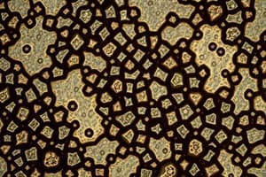
- The image depicts a cross-section of an organ, likely composed of epithelial tissue
- The tissue exhibits a pinkish-purple hue, indicating the presence of stained cells
- Numerous small, round to oval structures are observed, suggesting the presence of individual cells
- The tissue displays a layered structure, implying the possibility of stratified epithelial tissue
- Specific regions of the tissue exhibit characteristics suggestive of glandular tissue, potentially containing secretory cells
- Black arrows serve as pointers to different cell groups within the tissue
Microscopic Tissue Analysis
- The image presents a microscopic view of a tissue sample, likely a cross-section of an organ.
- The tissue exhibits a pinkish-purple color, a characteristic of stained biological specimens.
- The tissue contains numerous small, rounded to oval structures, indicating the presence of cells.
- Several labeled areas within the image pinpoint distinct layers or cell types.
- Black arrows within the image serve as points of reference for different cell groupings.
- The overall appearance of the tissue suggests an epithelial tissue, which is known for its lining function in various organs.
- The layered structure in specific regions points towards stratified epithelial tissue, characterized by multiple cell layers.
- The potential presence of secretory cells within the tissue suggests it may be a type of glandular tissue, specialized for producing and secreting substances.
Tissue Sample Analysis
-
The image depicts a microscopic view of a tissue sample.
-
The tissue exhibits a pinkish-purple coloration.
-
The tissue contains numerous small, round to oval structures, which are likely cells.
-
Labeled areas with blank boxes highlight different layers or cell types within the tissue.
-
Black arrows indicate points of reference for various cell groupings.
-
The overall appearance suggests epithelial tissue.
-
The layered structure in specific regions indicates stratified epithelial tissue.
-
The presence of secretory cells points to the possibility of glandular tissue.
Studying That Suits You
Use AI to generate personalized quizzes and flashcards to suit your learning preferences.


