Podcast
Questions and Answers
Which of the following is a characteristic of endospores?
Which of the following is a characteristic of endospores?
- They are resistant to heat (correct)
- They are reproductive structures
- They are easily stained
- They increase a cells innocence
What is the role of vegetative cells in endospore formation?
What is the role of vegetative cells in endospore formation?
- They form endospores through sporulation (correct)
- They are formed by budding
- They prevent endospore formation
- They directly become endospores
What is the purpose of sporulation in bacteria?
What is the purpose of sporulation in bacteria?
- To increase cell size
- To reproduce
- To survive harsh conditions (correct)
- To increase metabolic activity
Which of the following diseases is caused by a spore-forming bacterium?
Which of the following diseases is caused by a spore-forming bacterium?
What color are vegetative cells stained with safranin in the endospore staining procedure?
What color are vegetative cells stained with safranin in the endospore staining procedure?
What is the primary stain used in endospore staining?
What is the primary stain used in endospore staining?
What is the function of water in the endospore staining process?
What is the function of water in the endospore staining process?
Which of the following is a function of the bacterial capsule?
Which of the following is a function of the bacterial capsule?
What is the main purpose of a capsule stain?
What is the main purpose of a capsule stain?
What appearance do capsules have after a capsule stain?
What appearance do capsules have after a capsule stain?
Which of the following best defines a biofilm?
Which of the following best defines a biofilm?
Why is heat-fixing not required in capsule staining?
Why is heat-fixing not required in capsule staining?
Which staining method uses dyes to stain the background, leaving the capsule unstained and visible?
Which staining method uses dyes to stain the background, leaving the capsule unstained and visible?
Which of the following genera are spore-forming?
Which of the following genera are spore-forming?
After performing the spore stain, endospores will appear:
After performing the spore stain, endospores will appear:
After performing the spore stain, vegetative will appear:
After performing the spore stain, vegetative will appear:
During spore staining, what is the mordant?
During spore staining, what is the mordant?
During spore staining, what is the decolorizer?
During spore staining, what is the decolorizer?
Which of the following is an external structure of bacteria?
Which of the following is an external structure of bacteria?
The cell wall of bacteria is made of:
The cell wall of bacteria is made of:
What is the glycocalyx composed of?
What is the glycocalyx composed of?
Why is a capsule considered a virulence factor?
Why is a capsule considered a virulence factor?
Which of the following is a function of pili?
Which of the following is a function of pili?
What is contained within bacterial microcompartments?
What is contained within bacterial microcompartments?
What is the structure of bacterial ribosomes?
What is the structure of bacterial ribosomes?
Why is it important to indicate whether cells are Gram-positive or Gram-negative when giving Gram stain results?
Why is it important to indicate whether cells are Gram-positive or Gram-negative when giving Gram stain results?
What is the first thing that needs to be done for gram staining?
What is the first thing that needs to be done for gram staining?
The decolorizer in gram staining is:
The decolorizer in gram staining is:
What does it mean if a bacteria is acid-fast?
What does it mean if a bacteria is acid-fast?
What is Monday's biology tutoring time?
What is Monday's biology tutoring time?
Which of the following is a characteristic of bacterial endospores that contributes to their survival in extreme conditions?
Which of the following is a characteristic of bacterial endospores that contributes to their survival in extreme conditions?
What is the primary purpose of staining bacterial samples before observing them under a microscope?
What is the primary purpose of staining bacterial samples before observing them under a microscope?
Following Gram staining, Gram-positive bacteria appear what color under a microscope?
Following Gram staining, Gram-positive bacteria appear what color under a microscope?
What specific type of microorganism is targeted by acid-fast staining?
What specific type of microorganism is targeted by acid-fast staining?
In the context of bacterial staining, what does 'negative staining' refer to??
In the context of bacterial staining, what does 'negative staining' refer to??
Which staining protocol uses malachite green, often with heat, to force the stain into a particularly resistant bacterial structure?
Which staining protocol uses malachite green, often with heat, to force the stain into a particularly resistant bacterial structure?
What is the function of a culture's capsule?
What is the function of a culture's capsule?
There is a biofilm made up of millions of bacterial cells. Where are these films found?
There is a biofilm made up of millions of bacterial cells. Where are these films found?
Which of these options is the best for bacterial capsule visualization?
Which of these options is the best for bacterial capsule visualization?
What color do bacterial cells with a capsule appear against a dark background in a capsule stain?
What color do bacterial cells with a capsule appear against a dark background in a capsule stain?
What is the function of glycocalyx?
What is the function of glycocalyx?
What does the mordant do in the spore stain?
What does the mordant do in the spore stain?
What color is used as a primary stain in endospore staining?
What color is used as a primary stain in endospore staining?
Which genera of bacteria is NOT able to produce endospores?
Which genera of bacteria is NOT able to produce endospores?
Flashcards
What is Gram staining?
What is Gram staining?
A method used to distinguish between different types of bacterial cells based on their cell wall structure.
What are Gram-positive bacteria?
What are Gram-positive bacteria?
Bacteria that retain the crystal violet stain and appear purple under a microscope.
Bacillus and Clostridium
Bacillus and Clostridium
The major spore-forming genera of bacteria.
What is an endospore?
What is an endospore?
Signup and view all the flashcards
What is sporulation?
What is sporulation?
Signup and view all the flashcards
Endospore staining difficulty
Endospore staining difficulty
Signup and view all the flashcards
What is Schaeffer-Fulton stain?
What is Schaeffer-Fulton stain?
Signup and view all the flashcards
What is Malachite Green?
What is Malachite Green?
Signup and view all the flashcards
What is Safranin?
What is Safranin?
Signup and view all the flashcards
What are Fimbriae?
What are Fimbriae?
Signup and view all the flashcards
What are Pili?
What are Pili?
Signup and view all the flashcards
What is a Glycocalyx?
What is a Glycocalyx?
Signup and view all the flashcards
What is a Capsule?
What is a Capsule?
Signup and view all the flashcards
What is a Slime layer?
What is a Slime layer?
Signup and view all the flashcards
What are Biofilms?
What are Biofilms?
Signup and view all the flashcards
What are capsules?
What are capsules?
Signup and view all the flashcards
What is Negative staining?
What is Negative staining?
Signup and view all the flashcards
What is Nigrosin?
What is Nigrosin?
Signup and view all the flashcards
What is virulence?
What is virulence?
Signup and view all the flashcards
Prepare a Smear
Prepare a Smear
Signup and view all the flashcards
What is mordant
What is mordant
Signup and view all the flashcards
Spore resistance
Spore resistance
Signup and view all the flashcards
Hostile environment
Hostile environment
Signup and view all the flashcards
Endospore release
Endospore release
Signup and view all the flashcards
Clostridioides difficile
Clostridioides difficile
Signup and view all the flashcards
How to identify the capsule using the capsule/negative stain?
How to identify the capsule using the capsule/negative stain?
Signup and view all the flashcards
Study Notes
- Tutoring is available:
- Mondays: 10:00 AM - 2:00 PM
- Wednesdays: 10:00 AM - 1:00 PM & 1:30 PM - 5:00 PM
- Thursdays: 10:00 AM - 1:00 PM & 1:30 PM - 5:00 PM
- Written Test 1 : 02/27 (2 weeks)
- Practical test 1: 03/04-03/11 (2 sessions) covering Gram stain and streaking on 5 media types (BA, MS, MSA, PEA, NA)
- Lecture exam 1: 03/04 (lectures 1-4)
Smear Prep
- Prepare smear and let it dry
- Turn on hot plates for steaming
- Prepare smear with 1/3 tap water
Different Staining Types
- Endospore staining
- Capsule/negative staining
Gram Staining
- Used to determine if cells are Gram-positive or Gram-negative, also gives shape and arrangement
- Gram-positive cells (e.g., Staphylococcus) appear as cocci in clusters
- They exhibit clusters, chains, diplococci, and tetrads
- Gram-negative cells appear as bacilli in single arrangements
- They exhibit single, diplobacilli, chains, and palisades arrangements
Acid-Fast Stain (ZN Method)
- Carbol Fushsin: Primary stain
- Heat
- Acid-alcohol: Decolorizer
- Methylene Blue: Counter stain
- Acid-fast positive results in red
- Acid-fast negative results in blue
Bacterial Characterization Sessions
- Session 1: Microscopy
- Session 2: Inoculation and transfer techniques aseptically
- Session 3: Preparation and staining slides for characteristics
- Further characterization includes differential media, biochemical tests, and other future tests
Bacterial Cell Structures
- All bacteria contain a cell membrane, bacterial chromosome/nucleoid, ribosomes, and cytoplasm
- Some bacteria have S layers, fimbriae, cell walls, outer membranes, cytoskeleton, pili, glycocalyx, inclusions/granules, microcompartments, nanotubes/nanowires, plasmids, endospores, intracellular membranes, flagellum
External Structures of Bacteria
- Flagella enables movement and tumbling/running
- Fimbriae facilitates attachment
- Pili enables conjugation and exchange of plasmid DNAs
- Nanotubes/nanowires facilitate the transport of amino acids or smaller molecules
- S-layer is a shield of repeated proteins produced in hostile environments
- Glycocalyx is a polysaccharide layer that could be organized as a slime layer or capsule
Cell Envelope
- Cell membrane: All bacteria have it
- Cell wall presents in some bacteria; made of peptidoglycan
- Outer membrane found in Gram-negative bacteria only
Internal Structures
- Cytoplasm
- Bacterial chromosomes and plasmids
- Ribosomes (70S; unique to bacteria)
- Cytoskeleton
- Inclusion bodies, for food storage
- Microcompartments, for storing enzymes
- Endospores which are dormant bodies resistant to extreme conditions
Endospores
- Internal structures are present inside the cell
- Resistant structures formed by bacterial species
- Resistant to heat, radiation, desiccation, and chemical disinfection
- Formed by vegetative cells through sporulation
- Not for reproduction, but for survival
- Sporulation occurs under harsh conditions
- Increase the virulence of cells
Sporulation Process in Bacillus Species
- Hostile environment sensing: Vegetative cell begins to be depleted of nutrients
- DNA replication: Chromosome is duplicated and separated
- Forespore and cell separation: Cell is septated into a sporangium and forespore
- Engulfment of forespore: Sporangium engulfs forespore for further development
- Endospore layer synthesis: Sporangium begins to actively synthesize endospore layers around forespore
- Cortex, Endospore coat, Chromosome begin to be involved
- Layer deposition: Cortex and outer coat layers are deposited
- Endospore coats involved: Exosporium, Endospore coat, Cortex, Core
- Endospore ready: Mature endospore
- Free endospore: Free endospore is released with the loss of the sporangium
- Germination: Endospore swells and releases vegetative cell
Endospores
- Spores produced by Bacillus, Clostridium, and Sporosarcina
- Vegetative cell are metabolically active
- Sporulation induced by certain environmental conditions
- Endospores resist extremes of heat, drying, freezing, radiation, and chemicals that would kill vegetative cells
Medical Significance of Bacterial Endospores
- Bacillus anthracis can cause anthrax
- Clostridium tetani can cause tetanus
- Clostridium perfringens can cause gas gangrene
- Clostridium botulinum can cause botulism
- Clostridium difficile: "C. diff," causes a gastrointestinal disease (nosocomial)
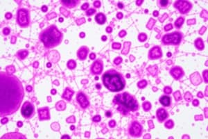
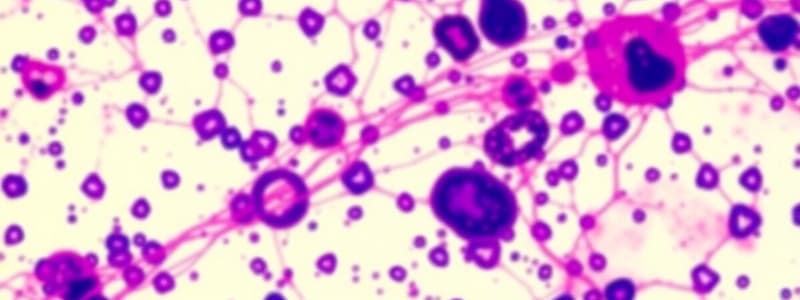
Endospore Staining
- These endospores are difficult to stain and can often be seen as clear bodies within bacterial cells
- Major spore-forming Bacillus and Clostridium
- Location of spore within the vegetative cell helps identify species within those genera
- Staining technique heats the primary stain (malachite green) into the endospores
- Once stained, endospores are difficult to decolorize
- Vegetative cells are then stained with a counterstain, like safranin
- Types of endospores: terminal, sub-terminal and central
Endospore Staining Steps
- Apply malachite green (primary stain), apply heat (mordant), apply water (decolorizer), apply safranin (counterstain)
- Malachite Green for 5 minutes and Safranin for one minute
Today’s Activities: Endospore Staining
- Bacillus subtilis( 48 hrs slant)
- Bacillus cereus (48 hrs slant)
- Requires staining tray, slide holder, forceps, water bottle, hot plate, newspaper, can containing water, Bunsen burner, loop, Malachite Green (primary), Safranin
Endospores
- Endospores are resistant to antibiotics, most disinfectants, and physical agents such as radiation, boiling, and drying
- Bacterial endospores can be destroyed at high temperatures in their spore state
- Otherwise by triggering endospore germination back to vegetative cells to be easily killed through traditional sterilization
External Structures
- Flagella for motility
- Pili for transfer of genetic material
- Fimbriae for attachment
- Glycocalyx as a slim layer or capsule, aiding virulence
Glycocalyx
- Glycocalyx is a capsule type that protects the cell against immune cells and enables attachment
- There is also the slime layer which enable attachment and aggregation of bacterial cells
Capsules
- Protect against white blood cells (phagocytes) by way of its glycocalyx
- Biofilms prevent bacteria from becoming dislodged (IE plaque on teeth)
- Responsible for persistent colonization on devices like catheters, IUDs, and pacemakers
Capsule Stain
- Capsule is polysaccharide-containing structure on the outside of cell
- Composed of fibrous polysaccharides or globular glycoproteins Capsule is in a tight matrix, firmly attached to wall, slime layer less organized/easily deformed
- Enables protection and can lead to biofilms (ecosystem with millions bacteria)
- Example organisms include Strep. pneumoniae, C. perfringens, Klebsiella pneumoniae, E. coli
- Bacterial capsules are non-ionic, so no acidic or basic stain will adhere
- Best way to visualize is with acidic stain
- It is recommended to have bacteria that are old
- No heat-fix is required, as it can distort capsules
- Structures can help increase bacteria's virulence
Capsule/Negative Stain
- Negative staining is a technique where the dark background makes the cell itself visible
- Heat-fixing not used
- Bacteria used for this stain are Staphylococcus aureus and Micrococcus luteus
- https://www.youtube.com/watch?v=avveXgPWVJ8
- Capsules formed by Klebsiella pneumoniae, Bacillus spp., Staphylococcus and Streptococcus spp., E. coli, Pseudomonas aeruginosa and Neisseria spp
Studying That Suits You
Use AI to generate personalized quizzes and flashcards to suit your learning preferences.