Podcast
Questions and Answers
What is the main difference between a light microscope and an electron microscope?
What is the main difference between a light microscope and an electron microscope?
- Light microscopes use a single lens while electron microscopes use multiple lenses
- Light microscopes use visible light while electron microscopes use a beam of electrons (correct)
- Light microscopes have a higher resolution than electron microscopes
- Light microscopes require a vacuum environment while electron microscopes do not
What is the formula used to calculate the magnification of an image in a microscope?
What is the formula used to calculate the magnification of an image in a microscope?
- Magnification = size of image x size of real object
- Magnification = size of real object / size of image
- Magnification = resolution of microscope / size of real object
- Magnification = size of image / size of real object (correct)
What is the main purpose of the vacuum environment in an electron microscope?
What is the main purpose of the vacuum environment in an electron microscope?
- To prevent the electrons from being deflected out of the beam alignment (correct)
- To increase the resolution of the microscope
- To reduce the amount of energy required to power the microscope
- To allow the use of a beam of electrons instead of visible light
What is the primary difference between a transmission electron microscope (TEM) and a scanning electron microscope (SEM)?
What is the primary difference between a transmission electron microscope (TEM) and a scanning electron microscope (SEM)?
What is the relationship between the resolution of a microscope and the wavelength of the light or electrons used?
What is the relationship between the resolution of a microscope and the wavelength of the light or electrons used?
What is the main limitation of light microscopes compared to electron microscopes?
What is the main limitation of light microscopes compared to electron microscopes?
What is the main purpose of cell fractionation?
What is the main purpose of cell fractionation?
Which of the following is a key limitation of using electron microscopes?
Which of the following is a key limitation of using electron microscopes?
What is the purpose of the homogenization step in cell fractionation?
What is the purpose of the homogenization step in cell fractionation?
What is the main difference between a Transmission Electron Microscope (TEM) and a Scanning Electron Microscope (SEM)?
What is the main difference between a Transmission Electron Microscope (TEM) and a Scanning Electron Microscope (SEM)?
What is the purpose of the buffered solution used in the cell fractionation process?
What is the purpose of the buffered solution used in the cell fractionation process?
Which of the following is a key advantage of using a Transmission Electron Microscope (TEM) over a light microscope?
Which of the following is a key advantage of using a Transmission Electron Microscope (TEM) over a light microscope?
Flashcards are hidden until you start studying
Study Notes
Electron Microscopes
- Transmission Electron Microscope (TEM):
- A beam of electrons passes through a thin section of a specimen
- Areas that absorb electrons appear darker on the electron micrograph
- Scanning Electron Microscope (SEM):
- A beam of electrons passes across the surface and scatters
- The pattern of scattering builds up a 3D image depending on the contours of the specimen
- Limitations of Electron Microscopes:
- The whole system must be in a vacuum, making it impossible to observe living specimens
- A complex staining process is required, which may introduce artefacts into the image
- Specimens have to be very thin, particularly for TEM
- SEM has a lower resolving power than TEM, but both have greater resolving power than a light microscope
Cell Fractionation and Ultracentrifugation
- Cell fractionation is the process of separating different parts and organelles of a cell to study them in detail
- The most common method of cell fractionation is differential centrifugation
- Steps of differential centrifugation:
- Homogenization: blending cells in an homogeniser to form a homogenate
- Centrifugation: spinning the homogenate at a slow speed to sediment the heaviest organelle (nuclei)
- Removing the supernatant and transferring it to another tube
- Spinning at a slightly faster speed to sediment the next heaviest organelle (mitochondria)
- Repeating the process to separate out other organelles
Cell Structure
- All living organisms are made up of cells, with various types of cells sharing common features
- Eukaryotic cells, found in humans, contain a nucleus and membrane-bound organelles
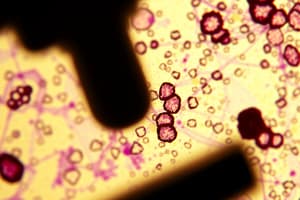
Microscopes
- Light Microscopes:
- Use convex glass lenses to resolve images 0.2um apart
- Limited by the wavelength of light, restricting resolution
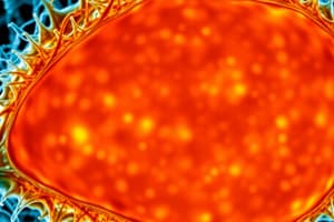
- Electron Microscopes:
- Can distinguish between items 0.1nm apart
- Allow for higher resolution than light microscopes
Studying That Suits You
Use AI to generate personalized quizzes and flashcards to suit your learning preferences.