Podcast
Questions and Answers
Which statement best describes the function of the hydrophilic head of phospholipids in the cell membrane?
Which statement best describes the function of the hydrophilic head of phospholipids in the cell membrane?
- It repels water molecules and stabilizes the membrane structure.
- It attracts water molecules and participates in cell signaling. (correct)
- It forms the bilayer's hydrophobic core to isolate the cell.
- It serves as a binding site for extracellular substances.
What characteristic of the cell membrane allows it to be visible only through electron microscopy?
What characteristic of the cell membrane allows it to be visible only through electron microscopy?
- The membrane thickness is minimal, measuring between 7.5 to 10 nm. (correct)
- It has a dynamic structure that changes rapidly.
- The presence of microvilli alters its visibility.
- Its chemical composition is complex and dense.
Which of the following correctly describes the organization of the cell membrane?
Which of the following correctly describes the organization of the cell membrane?
- Formed by a single layer of phospholipids arranged with heads inward.
- Incorporates only proteins with no lipid components.
- Composed of a trilaminar structure featuring two electron-dense lines and an electron-lucent layer. (correct)
- Consists solely of carbohydrate chains forming a protective layer.
Which organelle is characterized by a non-membranous structure and is involved in protein synthesis?
Which organelle is characterized by a non-membranous structure and is involved in protein synthesis?
What is the primary role of cholesterol in the phospholipid bilayer?
What is the primary role of cholesterol in the phospholipid bilayer?
What feature distinguishes the structure of the cytoskeleton from other cytoplasmic organelles?
What feature distinguishes the structure of the cytoskeleton from other cytoplasmic organelles?
Which type of protein is characterized by its firm embedding in the lipid bilayer and is not easily extractable?
Which type of protein is characterized by its firm embedding in the lipid bilayer and is not easily extractable?
What type of endocytosis involves the engulfing of solid particles by the cell?
What type of endocytosis involves the engulfing of solid particles by the cell?
Which of the following best describes the function of the cell coat or glycocalyx?
Which of the following best describes the function of the cell coat or glycocalyx?
What is true regarding the variability of mitochondria in terms of size and shape?
What is true regarding the variability of mitochondria in terms of size and shape?
Flashcards
Cell Membrane Function
Cell Membrane Function
The cell membrane acts as the outer boundary of the cell, controlling what enters and exits.
Cell Membrane Structure
Cell Membrane Structure
The cell membrane has a trilaminar appearance with two dense layers separated by a less dense layer.
Phospholipid Structure
Phospholipid Structure
Phospholipids have a hydrophilic head and hydrophobic tail, arranged in a bilayer.
Cell Membrane Composition
Cell Membrane Composition
Signup and view all the flashcards
Cell Membrane Component: Phospholipid Bilayer
Cell Membrane Component: Phospholipid Bilayer
Signup and view all the flashcards
Phospholipid bilayer
Phospholipid bilayer
Signup and view all the flashcards
Fluid Mosaic Model
Fluid Mosaic Model
Signup and view all the flashcards
Integral proteins
Integral proteins
Signup and view all the flashcards
Passive diffusion
Passive diffusion
Signup and view all the flashcards
Mitochondria
Mitochondria
Signup and view all the flashcards
Study Notes
Medicine (Semester 1) - Foundations of Normal Human Structure (BMS111) - Cell Structure (1)
- Objectives:
- Demonstrate the structure of the cell membrane.
- Determine the functions of the cell membrane and cell coat.
- Recognize the classification of cell organelles.
- Demonstrate routine histology staining methods.
- Determine the structure and functions of mitochondria.
- Recognize the structure, types, and functions of ribosomes.
Cell Structure
- Components:
- Cell membrane
- Nucleus
- Cytoplasm
- Membranous organelles (e.g., mitochondria, endoplasmic reticulum, Golgi apparatus, lysosomes)
- Non-membranous organelles (e.g., ribosomes)
- Cytoskeleton
- Cell inclusions
Cell Membrane
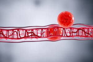
- Description: The outer limiting membrane surrounding the cell. Also known as the plasma membrane or plasmalemma.
- Thickness: 7.5-10 nm, visible only by electron microscopy (EM).
- EM Appearance: Trilaminar appearance with two electron-dense lines (black) separated by an electron-lucent line (white).
Molecular Structure of Cell Membrane
- Components:
- Lipids (phospholipids, cholesterol)
- Proteins (integral, peripheral)
- Carbohydrates (glycolipids, glycoproteins)
Phospholipids
- Structure: Composed of a hydrophilic head (water-attracting) and a hydrophobic tail (water-repelling).
- Arrangement: Organized into a double layer (bilayer) with hydrophobic tails facing inward and hydrophilic heads facing outward.
Cholesterol
- Location: Between phospholipid molecules.
- Function: Regulates the fluidity and stabilizes the phospholipid bilayer.
Proteins (Cell Membrane)
- Integral Proteins: Firmly embedded in the lipid bilayer, difficult to extract. Many are transmembrane proteins spanning the entire bilayer.
- Peripheral Proteins: Loosely attached to the outer or inner surfaces, easily extracted.
Carbohydrates (Cell Membrane)
- Glycolipids and Glycoproteins: Project from the external surface of the membrane, forming the cell coat or glycocalyx.
Functions of Cell Membrane
- Exchange of Materials:
- Passive diffusion (for gases and ions).
- Active transport (for amino acids, glucose, fatty acids).
- Selective transport (for hormones, drugs, bacteria).
- Endocytosis: Cell taking in substances.
- Phagocytosis (for solid particles).
- Pinocytosis (for fluids).
- Receptor-mediated endocytosis (for large molecules).
- Exocytosis: Cell expelling substances.
Mitochondria
- Definition: Membranous organelles specialized for energy production (ATP).
- Size and Shape: Vary in size and shape (elongated, rod-shaped, spherical).
- Number: Highly variable depending on cell activity (e.g., liver cells have many, lymphocytes have few).
- Location: Mobile, located where energy needs are high (e.g., between myofibrils in cardiac muscle).
- Structure: Double membrane, inner membrane with cristae (folds), intermembrane space, matrix (contains mitochondrial DNA and ribosomes).
Ribosomes
- Definition: Non-membranous organelles; protein factories of the cell.
- Size: Very small (20-30 nm in diameter).
- Light Microscopy (LM): Individual ribosomes are too small to be seen. Clusters lead to cytoplasm basophilia (due to rRNA).
- Electron Microscopy (EM): Appear as small electron-dense particles, composed of two subunits (small and large) formed of rRNA & proteins.
- Types:
- Free ribosomes/Polyribosomes/Polysomes: Synthesize proteins for use within the cell (cytosol and cytoskeleton).
- Attached ribosomes/Rough Endoplasmic Reticulum (RER): Synthesize proteins for secretion outside the cell, or remaining as primary lysosomes.
Staining (Hematoxylin and Eosin)
- Hematoxylin (H): Basic dye, stains acidic structures (DNA in nucleus) basophilic.
- Eosin (E): Acidic dye, stains basic structures (mitochondria) acidophilic.
Studying That Suits You
Use AI to generate personalized quizzes and flashcards to suit your learning preferences.


