Podcast
Questions and Answers
Match the following medical imaging techniques or technologies with their descriptions:
Match the following medical imaging techniques or technologies with their descriptions:
X-ray = First medical imaging technology invented in 1895 MRI = Developed in the 1970s using nuclear magnetic relaxation times CT scan = Developed in the 1970s for detailed internal imaging Sonar = Originally used in World War Two, later adapted for medical use
Match the following decades with their notable advancements in medical imaging:
Match the following decades with their notable advancements in medical imaging:
1900s = Use of pharmaceutical contrast agents to enhance organ visibility 1950s = Utilization of nuclear medicine for pathology diagnosis 1960s = Introduction of sonar for medical purposes 1970s = Development of CT scans and MRI technology
Match the following individuals or inventions with their historical significance:
Match the following individuals or inventions with their historical significance:
Wilhelm Rontgen = Invented the X-ray Nuclear medicine = Started being utilized in the 1950s Computed tomography = Also known as CT scan, developed in the 1970s Contrast agents = Used in the early 1900s for enhanced imaging
Match the following imaging techniques with their main principles:
Match the following imaging techniques with their main principles:
Match the following milestones in medical imaging with their corresponding years:
Match the following milestones in medical imaging with their corresponding years:
Who invented the X-ray?
Who invented the X-ray?
What technique began to be used in the 1960s for medical imaging?
What technique began to be used in the 1960s for medical imaging?
What is required to produce X-rays?
What is required to produce X-rays?
Computed Tomography (CT scan) was developed in the 1950s.
Computed Tomography (CT scan) was developed in the 1950s.
X-ray machines can be broadly classified into fixed, portable, and ______.
X-ray machines can be broadly classified into fixed, portable, and ______.
What does fluoroscopy enable physicians to examine?
What does fluoroscopy enable physicians to examine?
What is an OPG scan primarily used for?
What is an OPG scan primarily used for?
Dental X-rays use high levels of radiation to capture images.
Dental X-rays use high levels of radiation to capture images.
What is a bone density scan also known as?
What is a bone density scan also known as?
Which component is NOT part of the main components of an X-ray machine?
Which component is NOT part of the main components of an X-ray machine?
Flashcards are hidden until you start studying
Study Notes
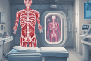
Medical Imaging Timeline
- Wilhelm Rontgen invented the X-ray in 1895, laying the foundation for medical imaging.
- Pharmaceutical contrast agents were introduced in the early 1900s, enabling visualization of organs and blood vessels.
- Nuclear medicine emerged as a diagnostic tool in the 1950s.
- Sonar, initially used in World War II, began being used for medical purposes in the 1960s.
- The 1970s saw the development of computed tomography (CT scan) for detailed anatomical imaging.
- Magnetic Resonance Imaging (MRI), based on nuclear magnetic relaxation times, was also developed in the 1970s.
History of Medical Imaging
- X-rays were invented in 1895 by Wilhelm Rontgen, a German physicist.
- Contrast agents were introduced in the early 1900s, allowing visualization of organs and blood vessels.
- Nuclear medicine emerged in the 1950s as a diagnostic tool.
- Sonar, previously used in warfare, began to be used for medical purposes in the 1960s.
- Computed Tomography (CT scan) was developed in the 1970s.
- Magnetic Resonance Imaging (MRI) was also developed in the 1970s, based on nuclear magnetic relaxation times.
X-ray Properties
- Short wavelength of the electromagnetic spectrum.
- Capable of traveling in a vacuum.
- Travel in a straight line
- Do not carry an electric charge.
- Requires high voltage to produce.
- Used to capture human skeleton defects.
X-ray Machine Parts
Main Components
-
X-Ray Tube: Consists of a cathode (electron source), anode (target), vacuum, and glass tube.
-
Operating Console: Controls the x-ray tube current and voltage, ensuring proper beam quantity and quality.
-
High Frequency Generator: Powers the x-ray tube; high frequency generators are preferred for stable operation and reduced voltage ripples.
Secondary Components
- Collimator: Restricts the field of view, minimizing scatter radiation. Lead shutters are used to further refine the beam.
- Grid: Similar to the collimator but positioned after the patient. Filters scattered radiation, improving image clarity.
- X-Ray Film: Turns black when exposed to x-rays, remaining white in areas where x-rays are absorbed, creating an image with varying shades of black, gray, and white.
Types of X-ray Machines Based on Movement
- Fixed X-ray machines: Large, powerful devices with transformers requiring dedicated electrical connections. Primarily used in teaching and research settings.
- Portable X-ray machines: Compact and lightweight, with smaller transformers. Designed for easy operation and often interface with other technologies, allowing for image transfer and analysis.
- Mobile X-ray machines: More powerful than portable machines due to larger transformers, mounted on wheels. Designed for easy movement within radiology departments.
Fluoroscopy
- A technique for visualizing moving body structures.
- Uses a continuous x-ray beam passed through the body, with images displayed on a monitor.
- Allows physicians and technologists to examine various body systems, including skeletal, digestive, urinary, respiratory, and reproductive systems.
- Can be used to monitor the movement of organs during procedures, such as swallowing studies, cardiac catheterization, and spinal or joint injections.
C-Arm X-ray
- A C-shaped arm connecting the x-ray source and detector, providing fluoroscopic intraoperative imaging.
- Primarily used for surgical, orthopedic, and emergency care procedures.
OPG X-Ray
- Orthopantomogram (OPG): A scan providing a panoramic view of the jaw and teeth.
- Used for dental assessments, including identifying dental problems, bone loss, and mandible trauma.
Dental X-rays
- Images of teeth produced using low-dose radiation.
- Help dentists diagnose problems like cavities, tooth decay, and impacted teeth.
Mammography
- A specialized breast imaging technique using low-dose x-rays.
- Employs two plates to compress the breast, spreading tissue for better visualization.
Bone Density Scan
- Also known as bone densitometry or Dual-energy X-ray Absorptiometry (DXA or DEXA).
- Measures bone density, used to assess bone health and diagnose conditions like osteoporosis.
Studying That Suits You
Use AI to generate personalized quizzes and flashcards to suit your learning preferences.