Podcast
Questions and Answers
What is the primary function of gamma cameras in SPECT imaging?
What is the primary function of gamma cameras in SPECT imaging?
- To perform anatomical imaging in real-time
- To produce images exclusively of the brain
- To detect gamma rays emitted by the tracer (correct)
- To provide high-resolution functional images
Which imaging modality combines functional and anatomical imaging for precision?
Which imaging modality combines functional and anatomical imaging for precision?
- CT Scan
- MRI
- PET/CT (correct)
- Ultrasound
What is a significant disadvantage of MRI for certain patients?
What is a significant disadvantage of MRI for certain patients?
- It provides too much radiation exposure
- It requires the use of contrast agents
- It may not be suitable for patients with claustrophobia (correct)
- It cannot image soft tissues effectively
What is a common use for Positron Emission Tomography (PET)?
What is a common use for Positron Emission Tomography (PET)?
Which factor is NOT a key consideration in selecting an imaging modality?
Which factor is NOT a key consideration in selecting an imaging modality?
What is the term used to describe the difference in charge between the anode and cathode in an x-ray tube?
What is the term used to describe the difference in charge between the anode and cathode in an x-ray tube?
How does increasing the milliAmpererage (mA) affect the x-ray beam?
How does increasing the milliAmpererage (mA) affect the x-ray beam?
What unit is the tube current measured in?
What unit is the tube current measured in?
What effect does increasing exposure time have in projection radiography?
What effect does increasing exposure time have in projection radiography?
What happens to patient radiation dose when the milliAmperage increases?
What happens to patient radiation dose when the milliAmperage increases?
Which of the following correctly follows the concept of the Inverse Square Law in x-ray imaging?
Which of the following correctly follows the concept of the Inverse Square Law in x-ray imaging?
What is the primary result of using a high kilovoltage peak (kVp) in an x-ray tube?
What is the primary result of using a high kilovoltage peak (kVp) in an x-ray tube?
Which statement is true regarding the relationship between exposure time and radiation dose?
Which statement is true regarding the relationship between exposure time and radiation dose?
What is a major limitation of a standard x-ray?
What is a major limitation of a standard x-ray?
How can the localization of lesions be improved in an x-ray examination?
How can the localization of lesions be improved in an x-ray examination?
What technique helps achieve better tissue differentiation in x-rays?
What technique helps achieve better tissue differentiation in x-rays?
What is a characteristic feature of mammography equipment compared to standard x-ray?
What is a characteristic feature of mammography equipment compared to standard x-ray?
What impact does the proximity of an organ to the image receptor have on x-ray imaging?
What impact does the proximity of an organ to the image receptor have on x-ray imaging?
In terms of radiation energy, what is the typical range used in mammography?
In terms of radiation energy, what is the typical range used in mammography?
Which of the following is NOT a structural element that might be visible in a chest x-ray?
Which of the following is NOT a structural element that might be visible in a chest x-ray?
What is a common misconception regarding the depth information provided by a general x-ray?
What is a common misconception regarding the depth information provided by a general x-ray?
What is the primary principle of general X-ray imaging?
What is the primary principle of general X-ray imaging?
Which component among the following is NOT part of the general X-ray system?
Which component among the following is NOT part of the general X-ray system?
What is the effect of increasing the kilovoltage (kVp) in X-ray imaging?
What is the effect of increasing the kilovoltage (kVp) in X-ray imaging?
In X-ray imaging, which of the following tissues would absorb the most X-rays?
In X-ray imaging, which of the following tissues would absorb the most X-rays?
What happens to the image quality when kVp is increased excessively?
What happens to the image quality when kVp is increased excessively?
Which of the following accurately describes transmission in the context of X-ray imaging?
Which of the following accurately describes transmission in the context of X-ray imaging?
The interaction of X-rays with tissues primarily depends on what characteristic?
The interaction of X-rays with tissues primarily depends on what characteristic?
What would be a distinct disadvantage of using general X-rays?
What would be a distinct disadvantage of using general X-rays?
What frequency range is typically used in ultrasound to visualize superficial structures?
What frequency range is typically used in ultrasound to visualize superficial structures?
What does 'gain' refer to in ultrasound imaging?
What does 'gain' refer to in ultrasound imaging?
Which of the following structures can be better visualized using lower frequency ultrasound?
Which of the following structures can be better visualized using lower frequency ultrasound?
What primarily distinguishes radionuclide imaging from techniques like CT or MRI?
What primarily distinguishes radionuclide imaging from techniques like CT or MRI?
What type of radiation do radionuclide tracers primarily emit?
What type of radiation do radionuclide tracers primarily emit?
What is a potential disadvantage of using lower frequency ultrasound?
What is a potential disadvantage of using lower frequency ultrasound?
In what way does radionuclide imaging contribute to understanding physiological processes?
In what way does radionuclide imaging contribute to understanding physiological processes?
What are the common projections used in radiography?
What are the common projections used in radiography?
Which imaging technique is utilized to track the absorption of radiopharmaceuticals for physiological imaging?
Which imaging technique is utilized to track the absorption of radiopharmaceuticals for physiological imaging?
Which feature distinguishes Fluoroscopy from conventional Projection Radiography?
Which feature distinguishes Fluoroscopy from conventional Projection Radiography?
What is a limitation of mobile X-ray equipment compared to static Projection Radiography equipment?
What is a limitation of mobile X-ray equipment compared to static Projection Radiography equipment?
What principle drives the generation of images in a CT scan?
What principle drives the generation of images in a CT scan?
Why is CT preferred over traditional Projection Radiography in some cases?
Why is CT preferred over traditional Projection Radiography in some cases?
What is one challenge of using conventional Projection Radiography?
What is one challenge of using conventional Projection Radiography?
How does the imaging capability of Fluoroscopy compare to that of CT?
How does the imaging capability of Fluoroscopy compare to that of CT?
What factor contributes to the innovation of CT technology?
What factor contributes to the innovation of CT technology?
Flashcards
X-ray imaging principle
X-ray imaging principle
X-rays are emitted from an X-ray tube and pass through the body. Different tissues interact with the rays differently based on their density. X-rays passing through less dense tissues are transmitted, while denser tissues absorb more X-rays.
Kilovoltage (kVp)
Kilovoltage (kVp)
The penetrating power of the X-ray beam is determined by the kilovoltage (kVp). Higher kVp results in more penetrating X-rays.
kVp effect on image contrast & dose
kVp effect on image contrast & dose
Higher kVp leads to increased penetration, resulting in more X-rays passing through the body. This reduces the contrast in the image, making it more black, but also reduces the radiation dose to the patient.
X-ray imaging components
X-ray imaging components
Signup and view all the flashcards
X-ray interaction with tissues
X-ray interaction with tissues
Signup and view all the flashcards
Tissue attenuation in X-ray imaging
Tissue attenuation in X-ray imaging
Signup and view all the flashcards
X-ray applications in bone imaging
X-ray applications in bone imaging
Signup and view all the flashcards
What is X-ray imaging?
What is X-ray imaging?
Signup and view all the flashcards
Tube potential
Tube potential
Signup and view all the flashcards
Kilovolts (kVp)
Kilovolts (kVp)
Signup and view all the flashcards
High kVp effect
High kVp effect
Signup and view all the flashcards
Milliamperage (mA)
Milliamperage (mA)
Signup and view all the flashcards
mA effect on intensity
mA effect on intensity
Signup and view all the flashcards
mA effect on radiation dose
mA effect on radiation dose
Signup and view all the flashcards
Exposure time
Exposure time
Signup and view all the flashcards
Exposure time effect
Exposure time effect
Signup and view all the flashcards
Why X-ray lacks depth information?
Why X-ray lacks depth information?
Signup and view all the flashcards
How to improve depth perception in X-RAY?
How to improve depth perception in X-RAY?
Signup and view all the flashcards
What is tissue attenuation in X-ray?
What is tissue attenuation in X-ray?
Signup and view all the flashcards
How does magnification affect X-ray images?
How does magnification affect X-ray images?
Signup and view all the flashcards
What is unique about the X-ray tube in Mammography?
What is unique about the X-ray tube in Mammography?
Signup and view all the flashcards
Projection Radiography
Projection Radiography
Signup and view all the flashcards
Fluoroscopy
Fluoroscopy
Signup and view all the flashcards
CT Scan
CT Scan
Signup and view all the flashcards
Tissue Attenuation
Tissue Attenuation
Signup and view all the flashcards
Contrast Resolution
Contrast Resolution
Signup and view all the flashcards
Superimposition
Superimposition
Signup and view all the flashcards
Mobile Radiography
Mobile Radiography
Signup and view all the flashcards
Real-Time Imaging
Real-Time Imaging
Signup and view all the flashcards
What is Ultrasound Imaging?
What is Ultrasound Imaging?
Signup and view all the flashcards
How does frequency affect ultrasound image depth?
How does frequency affect ultrasound image depth?
Signup and view all the flashcards
What is gain in ultrasound?
What is gain in ultrasound?
Signup and view all the flashcards
How does radionuclide imaging (RNI) work?
How does radionuclide imaging (RNI) work?
Signup and view all the flashcards
Why are tracers used in RNI?
Why are tracers used in RNI?
Signup and view all the flashcards
What makes RNI unique compared to CT or MRI?
What makes RNI unique compared to CT or MRI?
Signup and view all the flashcards
What are the advantages and disadvantages of RNI?
What are the advantages and disadvantages of RNI?
Signup and view all the flashcards
SPECT (Single Photon Emission Computed Tomography)
SPECT (Single Photon Emission Computed Tomography)
Signup and view all the flashcards
PET (Positron Emission Tomography)
PET (Positron Emission Tomography)
Signup and view all the flashcards
PET/CT (Positron Emission Tomography/Computed Tomography)
PET/CT (Positron Emission Tomography/Computed Tomography)
Signup and view all the flashcards
X-ray Imaging
X-ray Imaging
Signup and view all the flashcards
Choosing an Imaging Modality
Choosing an Imaging Modality
Signup and view all the flashcards
Study Notes
Imaging Modalities
- Medical imaging modalities are used to visualize internal body structures.
- An introduction to medical imaging was presented.
- This course is titled HSMI 1211.
- The objectives included historical development, principles of modalities (radiography, CT, MRI, US, RNI), imaging parameters, and advantages/disadvantages of each modality.
Historical Development
- X-rays were discovered in 1895 by Wilhelm Conrad Roentgen.
- The first use of diagnostic imaging and medicine occurred in the 1950s.
- Magnetic Resonance Imaging (MRI) was developed in the 1980s, using a magnetic field without ionizing radiation.
- Digital imaging and artificial intelligence integration became prominent in the 2000's.
- Computed Tomography (CT) was developed in the 1970s, by Godfrey Hounsfield & Allan Cormack.
- SPECT and PET scans appeared in the 1990s.
General X-ray
- X-rays interact with tissues, either by transmission (passing through less dense tissues like air or soft tissue) or absorption (being absorbed by denser tissues like bone).
- X-ray beams are generated by an X-ray tube, pass through the patient, and interact with tissues.
- The image is formed by the varying amount of X-rays absorbed or transmitted.
- X-rays are composed of three components: X-ray tube, patient, and image receptor.
Imaging Parameters (kVp)
- Kilovoltage (kV) determines the penetrating power of the radiation beam.
- Higher kV results in more penetrating beams (leading to overpenetration) decreasing contrast, and increasing blackness.
- Higher kV results in less radiation being absorbed by tissues, lowering patient radiation dose.
- The anode is positively charged and the cathode is negatively charged, thus electrons move quickly leading to higher energy X-rays
Imaging Parameters (mA)
- Milliamperage (mA) denotes the current in the X-ray tube.
- Higher mA increases the intensity of the X-ray beam; causing the image to be darker and resulting in higher exposure.
- Increasing mA increases the radiation intensity.
- Increasing mA increases the radiation dose to the patient.
Imaging Parameters (Seconds)
- Measured in seconds, exposure time relates to the duration of radiation exposure.
- Longer exposure times result in more radiation reaching the patient, leading to a darker image.
- Increasing exposure time increases radiation dose to the patient.
Imaging Parameters (SID)
- Source-to-Image Receptor Distance (SID) is the distance between the X-ray source and the image receptor.
- Following the inverse square law, an increase in distance reduces the intensity of radiation, leading to underexposure.
Imaging Parameters (Image Receptor)
- The image receptor records the image.
- The speed of the image receptor system affects the amount of radiation needed to produce an image.
- Faster systems need less radiation for the same image density.
Imaging Parameters (Filtration)
- Filtration in X-ray tubes uses filters to remove low-energy X-rays.
- This reduces the intensity of radiation reaching the image receptor, making the image less black (underexposed).
- Increase in filtration reduces radiation dose to the patient.
Imaging Parameters (OID)
- Object-to-Image Receptor Distance (OID) is the distance between the patient and the image receptor.
- Increasing OID results in less radiation reaching the image receptor, giving rise to underexposure.
- OID also magnifies the image, decreasing the detail of the image.
General X-ray Advantages and Limitations
- Lack of depth information, structures superimposed
- Limited tissue differentiation compared to other modalities.
- False size due to magnification by image distance
- Advantages include simplicity, cost-effectiveness and accessibility.
Mammography
- Mammography uses a specific anode in the X-ray tube (molybdenum or molybdenum/tungsten alloy) enabling the use of lower kVp (28-32kV) when compared to conventional X-ray (50-120kV).
- Medio-lateral oblique (MLO) and cranio-caudal (CC) projections are commonly employed.
Fluoroscopy
- Fluoroscopy is an extension of radiography, providing real-time imaging.
- It shows the anatomy dynamically.
- Key improvements from conventional radiography lie in the ability to show changes in real-time.
Mobile X-ray
- Mobile X-ray allows for imaging in places outside of dedicated radiology departments.
- Image quality potentially less than in a fixed facility, due to lower specifications of the equipment.
Computed Tomography (CT) Scan
- CT generates cross-sectional images using multiple X-ray projections and computer processing.
- This overcomes the limitations of conventional X-ray (i.e., lack of depth information).
- This allows the user to see various perspectives through CT scans in different planes (e.g., AP, PA, lateral)
CT Imaging Parameters
- Key parameters like Kilovoltage (kV), Milliamperes (mA), exposure time, collimation (beam width), slice thickness, and more determine image quality and patient dose.
- Multi-planar reformation enhances image visualization.
CT System Components
- The CT system includes components for data acquisition, processing, display, archive, and communication.
- These include the control system, data acquisition system, data processing system, data display, data storage and communication systems.
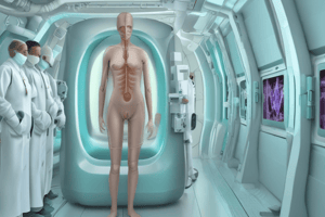
Magnetic Resonance Imaging (MRI)
- MRI uses a powerful magnetic field and radio waves to produce detailed images of the body.
- This contrasts sharply with ionizing X-rays in medical imaging in that it does not use ionizing radiation.
- Key strengths include detailed tissue discrimination in different planes.
- Common imaging parameters include repetition time (TR), echo time (TE), inversion time (TI), flip angle (FA), and spin-spin relaxation time (T2).
MRI Imaging Parameters
- Key imaging parameters used in MRI are repetition time (TR), echo time (TE), Inversion time (TI), Flip angle (FA), spin-lattice relaxation time (T1). and spin-spin relaxation time (T2).
MRI Advantages & Limitations
- MRI offers superior tissue differentiation.
- It doesn't utilize ionizing radiation, minimizing potential risks.
- Images can be acquired in multiple planes without repositioning the patient.
- However, the procedure can be time-consuming & uncomfortable, sensitive to motion, and potentially dangerous with metallic implants.
Ultrasound
- Ultrasound utilizes sound waves to create images of internal body structures.
- Important parameters include frequency and gain to improve the image for the user.
- Advantages include portability, real-time imaging, and non-ionizing radiation.
Ultrasound Imaging Parameters
- The frequency is a crucial component of ultrasound imaging, where higher frequencies provide better resolution for shallower structures; lower frequencies penetrate deeper but have poorer resolution.
- Gain is another vital parameter that controls the amplification of attenuated echoes to create a better contrast and visualization of the structures and their differences from a darker background.
Ultrasound Advantages & Limitations
- Ultrasound offers portability, real-time imaging, and doesn't use ionizing radiation.
- However, it can't penetrate bone or air-filled structures, and image quality depends on the operator's skills and experience.
Radionuclide Imaging (RNI)
- Radioactive tracers, or radiopharmaceuticals, are introduced into the body, and their radiation is detected to visualize metabolic and functional processes.
- This includes SPECT for 3D imaging and PET, which combines metabolic function with anatomical information.
RNI Imaging Parameters
- SPECT uses gamma cameras, while PET combines metabolic information with CT.
- This provides functional images for physiological processes.
- Specific radionuclides like fluorine-18 (FDG), are critical tracers in PET imaging for oncology.
RNI Advantages & Limitations
- RNI yields unique information on physiological processes.
- RNI detects disease at early stages and allows whole-body scans.
- However, RNI has factors like limited accuracy due to signal limitations and prolonged scanning duration from tracer absorption and detection.
Choice of Imaging Modality
- The choice of imaging modality is based on the intended information and patient factors.
- Factors include the intended information, patient factors (e.g., claustrophobia; trauma), and required examination time.
- MRI and ultrasound may not demonstrate bony pathology.
Studying That Suits You
Use AI to generate personalized quizzes and flashcards to suit your learning preferences.