Podcast
Questions and Answers
What is the primary benefit of multi slice CT scanners?
What is the primary benefit of multi slice CT scanners?
- They are smaller in size than traditional scanners.
- They produce color images.
- They allow for rapid collection of projection data. (correct)
- They require less radiation than single slice scanners.
Cone beam CT (CBCT) increases the size of the scanner while decreasing radiation dosage.
Cone beam CT (CBCT) increases the size of the scanner while decreasing radiation dosage.
False (B)
Who was the leader in the development of the first CT scanner introduced in 1971?
Who was the leader in the development of the first CT scanner introduced in 1971?
Sir Godfrey Hounsfield
CT scanners produce ___ dimensional images.
CT scanners produce ___ dimensional images.
Match the following components of a CT scanner with their functions:
Match the following components of a CT scanner with their functions:
Which of the following imaging modalities is NOT commonly used in oncology?
Which of the following imaging modalities is NOT commonly used in oncology?
X-rays provide functional information about the body.
X-rays provide functional information about the body.
What is a common use of contrast agents in imaging?
What is a common use of contrast agents in imaging?
The intensity of the emerging primary beam in X-ray imaging provides information about the interactions that have occurred within the _____ .
The intensity of the emerging primary beam in X-ray imaging provides information about the interactions that have occurred within the _____ .
Match the imaging modalities with their description:
Match the imaging modalities with their description:
What is an important limitation of X-rays?
What is an important limitation of X-rays?
Digital imaging has largely replaced film-based systems in medical imaging.
Digital imaging has largely replaced film-based systems in medical imaging.
How does the atomic number of tissues affect X-ray imaging?
How does the atomic number of tissues affect X-ray imaging?
What is the purpose of Image Guided Radiotherapy (IGRT)?
What is the purpose of Image Guided Radiotherapy (IGRT)?
Online treatment verification involves comparisons made after the treatment has been delivered.
Online treatment verification involves comparisons made after the treatment has been delivered.
What imaging technique allows direct visualization of the radiotherapy target volume?
What imaging technique allows direct visualization of the radiotherapy target volume?
The __________ compares images taken with reference images at a point after treatment has been delivered.
The __________ compares images taken with reference images at a point after treatment has been delivered.
Which of the following statements about DRR's is true?
Which of the following statements about DRR's is true?
Match the imaging technique with its description:
Match the imaging technique with its description:
Further treatment is never required after the initial radiotherapy session.
Further treatment is never required after the initial radiotherapy session.
What is one purpose of follow-up in radiotherapy?
What is one purpose of follow-up in radiotherapy?
What type of x-ray beam is used in second and third generation CT scanners?
What type of x-ray beam is used in second and third generation CT scanners?
Fourth generation CT scanners have detectors that are movable.
Fourth generation CT scanners have detectors that are movable.
What is the duration of a scan on a first generation CT scanner?
What is the duration of a scan on a first generation CT scanner?
MRI uses a strong magnetic field and _____ pulses to produce images.
MRI uses a strong magnetic field and _____ pulses to produce images.
Match the following CT generations with their key characteristics:
Match the following CT generations with their key characteristics:
Which of the following advantages does MRI have over CT?
Which of the following advantages does MRI have over CT?
Dual energy CT scanning is a feature of first generation CT technology.
Dual energy CT scanning is a feature of first generation CT technology.
What does T2 weighted MRI images reveal about cerebrospinal fluid (CSF)?
What does T2 weighted MRI images reveal about cerebrospinal fluid (CSF)?
What is a significant characteristic that differentiates cancerous cells from healthy tissue?
What is a significant characteristic that differentiates cancerous cells from healthy tissue?
PET imaging provides detailed anatomical information about the tissues.
PET imaging provides detailed anatomical information about the tissues.
What imaging technique has become the standard for localization in radiotherapy?
What imaging technique has become the standard for localization in radiotherapy?
PET is often fused with ______ to enhance anatomical detail.
PET is often fused with ______ to enhance anatomical detail.
Match the imaging techniques with their primary uses:
Match the imaging techniques with their primary uses:
Which of the following modalities is known for its ability to reveal distant metastases?
Which of the following modalities is known for its ability to reveal distant metastases?
Volumetric data from CT scans are solely 2D images.
Volumetric data from CT scans are solely 2D images.
What kind of data is generated for physics planning in radiotherapy?
What kind of data is generated for physics planning in radiotherapy?
What is one major advantage of using MRI for imaging?
What is one major advantage of using MRI for imaging?
Patients with non-ferromagnetic metal implants can be scanned using MRI.
Patients with non-ferromagnetic metal implants can be scanned using MRI.
Name one disadvantage of ultrasound imaging.
Name one disadvantage of ultrasound imaging.
PET imaging utilizes _____ that are administered to patients to gather information about physiological function.
PET imaging utilizes _____ that are administered to patients to gather information about physiological function.
Match the imaging technique with its main characteristic:
Match the imaging technique with its main characteristic:
What is a typical frequency range used in ultrasound imaging?
What is a typical frequency range used in ultrasound imaging?
Contrast-enhanced MRI is ineffective for diagnosis and staging.
Contrast-enhanced MRI is ineffective for diagnosis and staging.
What is the most common radiopharmaceutical used in PET scans?
What is the most common radiopharmaceutical used in PET scans?
MRI images can be fused with _____ data for better treatment planning.
MRI images can be fused with _____ data for better treatment planning.
What is one of the advantages of radionuclide imaging?
What is one of the advantages of radionuclide imaging?
Flashcards
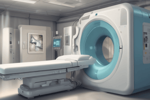
Computerized Tomography (CT)
Computerized Tomography (CT)
A medical imaging technique that uses X-rays to create detailed cross-sectional images of the body.
How CT scanner works?
How CT scanner works?
The CT scanner uses a rotating X-ray tube to create multiple images of the target area.
Evolution of CT scanners
Evolution of CT scanners
CT scanners have evolved over time, with increases in the number of detectors and decreases in scan time.
Multi-slice CT
Multi-slice CT
Signup and view all the flashcards
Cone Beam CT (CBCT)
Cone Beam CT (CBCT)
Signup and view all the flashcards
What is medical imaging?
What is medical imaging?
Signup and view all the flashcards
What is an image?
What is an image?
Signup and view all the flashcards
What are X-rays?
What are X-rays?
Signup and view all the flashcards
How does the amount of absorption of X-rays work?
How does the amount of absorption of X-rays work?
Signup and view all the flashcards
What are contrast agents?
What are contrast agents?
Signup and view all the flashcards
What are the limitations of X-rays?
What are the limitations of X-rays?
Signup and view all the flashcards
What is CT scan?
What is CT scan?
Signup and view all the flashcards
What is an MRI scan?
What is an MRI scan?
Signup and view all the flashcards
First Generation CT Scanners
First Generation CT Scanners
Signup and view all the flashcards
Second Generation CT Scanners
Second Generation CT Scanners
Signup and view all the flashcards
Third Generation CT Scanners
Third Generation CT Scanners
Signup and view all the flashcards
Fourth Generation CT Scanners
Fourth Generation CT Scanners
Signup and view all the flashcards
Helical CT
Helical CT
Signup and view all the flashcards
Dual-Energy CT (DECT)
Dual-Energy CT (DECT)
Signup and view all the flashcards
MRI
MRI
Signup and view all the flashcards
Advantages of MRI
Advantages of MRI
Signup and view all the flashcards
What is MRI?
What is MRI?
Signup and view all the flashcards
What are the applications of MRI?
What are the applications of MRI?
Signup and view all the flashcards
What are the limitations of MRI?
What are the limitations of MRI?
Signup and view all the flashcards
What are the advantages of modern MRI?
What are the advantages of modern MRI?
Signup and view all the flashcards
What is Ultrasound?
What is Ultrasound?
Signup and view all the flashcards
What are the applications of Ultrasound?
What are the applications of Ultrasound?
Signup and view all the flashcards
What are the advantages of Ultrasound?
What are the advantages of Ultrasound?
Signup and view all the flashcards
What are the disadvantages of Ultrasound?
What are the disadvantages of Ultrasound?
Signup and view all the flashcards
What is Radionuclide Imaging?
What is Radionuclide Imaging?
Signup and view all the flashcards
How does Radionuclide Imaging work?
How does Radionuclide Imaging work?
Signup and view all the flashcards
What is PET imaging?
What is PET imaging?
Signup and view all the flashcards
Why is PET often combined with CT or MRI?
Why is PET often combined with CT or MRI?
Signup and view all the flashcards
What are the advantages of combining PET and CT?
What are the advantages of combining PET and CT?
Signup and view all the flashcards
What are the roles of CT and MRI in oncology?
What are the roles of CT and MRI in oncology?
Signup and view all the flashcards
Why is CT used for radiotherapy localization?
Why is CT used for radiotherapy localization?
Signup and view all the flashcards
What are Digitally Reconstructed Radiographs (DRRs)?
What are Digitally Reconstructed Radiographs (DRRs)?
Signup and view all the flashcards
How are CT images used in radiation therapy setup?
How are CT images used in radiation therapy setup?
Signup and view all the flashcards
Can other imaging modalities be used with CT for radiotherapy planning?
Can other imaging modalities be used with CT for radiotherapy planning?
Signup and view all the flashcards
Image-Guided Radiotherapy (IGRT)
Image-Guided Radiotherapy (IGRT)
Signup and view all the flashcards
Treatment Verification
Treatment Verification
Signup and view all the flashcards
Online Treatment Verification
Online Treatment Verification
Signup and view all the flashcards
Offline Treatment Verification
Offline Treatment Verification
Signup and view all the flashcards
KV Imaging
KV Imaging
Signup and view all the flashcards
Follow-up
Follow-up
Signup and view all the flashcards
Magnetic resonance imaging (MRI)
Magnetic resonance imaging (MRI)
Signup and view all the flashcards
Study Notes
Imaging Principles/Imaging in Oncology
- The lecture aims to explain the principles of different imaging techniques and their role in the oncology patient pathway.
- The session will cover the understanding of the principles of different imaging techniques and the role of imaging in the oncology patient pathway.
Types of Imaging
- Different imaging modalities are used in oncology, such as X-rays, Computed Tomography (CT), Magnetic Resonance Imaging (MRI), Ultrasound, and Radionuclide Imaging.
- The images have specific applications, including X-rays used to demonstrate spatial relationships within the body, while CT scans produce 3-dimensional cross-sectional images in standard anatomical planes.
- Images from various imaging modalities can be combined (fused) for comprehensive analysis.
X-rays
- X-rays are created in an X-ray tube.
- The X-ray beam travels in straight lines and interacts with body tissue, where they are either absorbed or scattered.
- The intensity of the emerging beam contains information on tissue interactions.
- This beam is detected by an image intensifier to produce an X-ray image.
- The amount of absorption depends on the atomic number of the tissues and the energy of the X-rays.
- Contrast agents like iodine or barium enhance visual clarity by having different absorption characteristics than surrounding tissue.
- X-rays have limitations, including difficulty visualizing low contrast tissues and only providing a 2D image.
Computerised Tomography (CT)
- CT uses X-rays to produce 3-dimensional images .
- The images are cross-sectional in the 3 standard anatomical planes.
- CT scanners can be categorized into different generations based on detectors and methodologies.
- There are multi-slice CT scanners and cone beam CT.
- CT scans have different image configurations to meet various imaging needs.
Magnetic Resonance Imaging (MRI)
- MRI uses a strong magnetic field and radiofrequency pulses to create images of hydrogen distribution in the body.
- It provides visualization of the body in slices, similar to CT.
- MRI utilizes hydrogen atom properties and produces T1 or T2 weighted images for different tissue contrasts.
- T1 weighted images show fat and white matter brighter than CSF. CSF shows brighter on T2 weighted images.
- MRI has advantages like non-ionizing radiation and exceptional soft tissue visualization .
- MRI has a disadvantage where patients with metallic implants cannot be scanned.
Ultrasound
- Ultrasound uses high-frequency sound waves to create images of the internal body structures.
- The frequency range of the waves is beyond human hearing.
- Ultrasound can differentiate various tissues, like solid masses and fluid-filled cysts.
- It's used in various applications, including breast cancer assessment, uterus, ovary, prostate, kidney scans.
- Ultrasound is often used in combination with fine-needle biopsy.
- Ultrasound image advantages are being relatively cheap, safe, and demonstrating real-time images.
- Ultrasound image disadvantages are being subjective and difficult to interpret.
Radionuclide Imaging
- Radionuclide imaging primarily focuses on the physiological function of organs and tissues.
- It utilizes unsealed radiopharmaceuticals and isotopes to investigate and treat diseases.
- Radiopharmaceuticals are coupled with a radionuclide, which spontaneously decays and emits energy.
- This emission is detected to create an image.
- Radiolabeled compounds are detected using a gamma camera that creates an image of the compound distribution and function.
Positron Emission Tomography (PET)
- PET is focused on physiological function and uses positron emission from radioactive isotopes injected into the patient.
- Radioactive markers are attached to glucose typically (e.g. 18FDG) to target cells that metabolize glucose rapidly.
- PET itself lacks anatomical detail, usually paired with CT or MRI to provide full assessment.
CT and MRI Fusion
- This method integrates data of both CT and MRI to offer a more comprehensive visualization of the body.
Role of Imaging in Oncology
- Imaging plays a crucial role in the patient pathway, from diagnosis and staging to treatment planning and follow-up.
- The patient's pathway includes diagnosis, CT planning, treatment planning, treatment verification, treatment delivery, and follow-up steps.
Radiotherapy Localisation
-
Localisation in radiotherapy aims to accurately target the tumour and avoid sensitive structures.
-
Imaging methods, such as CT planning scans and virtual simulations, are necessary to create treatment plans.
-
Other imaging data can be integrated with CT data for complete assessment.
-
Methods for verification include comparing images to reference images, digitally reconstructed radiographs (DRR), portal imaging (MV), and kV imaging.
-
Online and Offline imaging are used to verify imaging, including image-guided radiotherapy (IGRT), and cone-beam CT (CBCT).
Follow-up
- Follow-up is essential to monitor tumour response and the outcome of treatment.
- Monitoring for signs of disease recurrence, spread, or treatment effectiveness is vital.
- Assessments are carried out to determine if the treatment was successful.
- Follow up considers treatment curative status, disease extent, if further treatment is required, and possible metastatic disease symptoms.
Further Reading
- NICE Pathways is an interactive toolkit for health professionals that provides access to NICE guidance and other resources.
- There are available on target documents for Radiotherapy.
Studying That Suits You
Use AI to generate personalized quizzes and flashcards to suit your learning preferences.