Podcast
Questions and Answers
What is the shape of the upper surface of the disc?
What is the shape of the upper surface of the disc?
- Concavoconvex (correct)
- Convexoconcave
- Flat
- Convex
What is the characteristic of the retrodiscal tissue?
What is the characteristic of the retrodiscal tissue?
- Fibrous and poorly innervated
- Avascular and poorly innervated
- Vascular and highly innervated (correct)
- Cartilaginous and avascular
What connects the articular disc to the tympanic plate of the temporal bone?
What connects the articular disc to the tympanic plate of the temporal bone?
- Synovial membrane
- Inferior retrodiscal layer
- Superior retrodiscal layer (correct)
- Articular capsule
Which nerve branch supplies the TMJ?
Which nerve branch supplies the TMJ?
What is the main blood supply to the TMJ?
What is the main blood supply to the TMJ?
What type of movement can occur in the TMJ?
What type of movement can occur in the TMJ?
What is the function of the synovial membrane?
What is the function of the synovial membrane?
What is the venous drainage of the TMJ?
What is the venous drainage of the TMJ?
What is the primary function of the lateral temporomandibular ligament?
What is the primary function of the lateral temporomandibular ligament?
What type of joint is the temporomandibular joint?
What type of joint is the temporomandibular joint?
Where is the sphenomandibular ligament attached above?
Where is the sphenomandibular ligament attached above?
What is the function of the fibrous bands that attach the articular disc to the head of the mandible?
What is the function of the fibrous bands that attach the articular disc to the head of the mandible?
What is the articular disc composed of?
What is the articular disc composed of?
What is the function of the stylomandibular ligament?
What is the function of the stylomandibular ligament?
Where is the capsule of the temporomandibular joint attached above?
Where is the capsule of the temporomandibular joint attached above?
What is the attachment of the articular disc in front?
What is the attachment of the articular disc in front?
What is the primary function of the lateral pterygoid muscle during depression of the mandible?
What is the primary function of the lateral pterygoid muscle during depression of the mandible?
Which muscles are responsible for elevation of the mandible?
Which muscles are responsible for elevation of the mandible?
What happens to the head of the mandible during protrusion of the mandible?
What happens to the head of the mandible during protrusion of the mandible?
Which muscle is responsible for pulling the articular disc backward during elevation of the mandible?
Which muscle is responsible for pulling the articular disc backward during elevation of the mandible?
What is the primary movement of the mandible during depression?
What is the primary movement of the mandible during depression?
Which muscle is responsible for pulling the head of the mandible backward during elevation of the mandible?
Which muscle is responsible for pulling the head of the mandible backward during elevation of the mandible?
What is the primary function of the fibroelastic tissue during depression of the mandible?
What is the primary function of the fibroelastic tissue during depression of the mandible?
During which movement of the mandible does the articular disc move backward into the mandibular fossa?
During which movement of the mandible does the articular disc move backward into the mandibular fossa?
What is the primary function of the posterior fibers of the temporalis muscle?
What is the primary function of the posterior fibers of the temporalis muscle?
Which muscle is responsible for elevating the mandible and also plays a role in retracting the mandible?
Which muscle is responsible for elevating the mandible and also plays a role in retracting the mandible?
What is the origin of the anterior belly of the digastric muscle?
What is the origin of the anterior belly of the digastric muscle?
Which nerve supplies the masseter muscle?
Which nerve supplies the masseter muscle?
What is the function of the lateral pterygoid muscle?
What is the function of the lateral pterygoid muscle?
What is the insertion of the medial pterygoid muscle?
What is the insertion of the medial pterygoid muscle?
Which structure is located anterior to the temporomandibular joint?
Which structure is located anterior to the temporomandibular joint?
What is the primary function of the geniohyoid muscle?
What is the primary function of the geniohyoid muscle?
What structure lies immediately in front of the external auditory meatus?
What structure lies immediately in front of the external auditory meatus?
What is the clinical significance of the lateral temporomandibular ligament?
What is the clinical significance of the lateral temporomandibular ligament?
What is the result of the articular disc becoming partially detached from the capsule?
What is the result of the articular disc becoming partially detached from the capsule?
What is the movement that causes the head of the mandible and the articular disc to move forward until they reach the summit of the articular tubercle?
What is the movement that causes the head of the mandible and the articular disc to move forward until they reach the summit of the articular tubercle?
What is the position of the head of the mandible in bilateral cases of dislocation?
What is the position of the head of the mandible in bilateral cases of dislocation?
What is the method used to reduce the dislocation of the temporomandibular joint?
What is the method used to reduce the dislocation of the temporomandibular joint?
What muscles are overcome by the downward pressure during the reduction of the dislocation?
What muscles are overcome by the downward pressure during the reduction of the dislocation?
What is the structure that the parotid gland lies laterally to?
What is the structure that the parotid gland lies laterally to?
Study Notes
Temporomandibular Joint
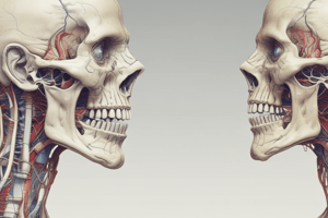
- The temporomandibular joint is a synovial joint that articulates between the articular tubercle and the anterior portion of the mandibular fossa of the temporal bone above and the head (condyloid process) of the mandible below.
- The articular surfaces are covered with fibrocartilage.
- The joint is divided into upper and lower cavities by the articular disc.
Capsule and Ligaments
- The capsule surrounds the joint and is attached above to the articular tubercle and the margins of the mandibular fossa and below to the neck of the mandible.
- The lateral temporomandibular ligament strengthens the lateral aspect of the capsule and limits the movement of the mandible in a posterior direction, protecting the external auditory meatus.
- The sphenomandibular ligament lies on the medial side of the joint and represents the remains of the first pharyngeal arch in this region.
- The stylomandibular ligament lies behind and medial to the joint and is a band of thickened deep cervical fascia that extends from the apex of the styloid process to the angle of the mandible.
Articular Disc
- The articular disc is an oval plate of fibrocartilage that is attached circumferentially to the capsule.
- The disc is also attached in front to the tendon of the lateral pterygoid muscle and by fibrous bands to the head of the mandible.
- These bands ensure that the disc moves forward and backward with the head of the mandible during protraction and retraction of the mandible.
Retrodiscal Tissue
- The retrodiscal tissue is vascular and highly innervated, making it a major contributor to the pain of Temporomandibular Disorder (TMD) when there is inflammation or compression within the joint.
- The retrodiscal zone (bilaminar zone) of the TMJ is located between the posterior band of the articular disc and the posterior part of the articular capsule.
- The superior retrodiscal layer of the TMJ joint is a lamina composed of connective tissue and elastic fibers that continues the posterior part of the articular disk and connects the disc to the tympanic plate of the temporal bone.
- The inferior retrodiscal layer of the TMJ joint is a lamina composed of collagen fibers that continues the posterior part of the articular disk and inserts into the condylar neck.
Nerve and Vascular Supply
- The TMJ is supplied by the auriculotemporal and masseteric branches of the mandibular nerve.
- The TMJ is supplied mainly by three arteries: the deep auricular artery (from the maxillary artery), the superficial temporal artery (a terminal branch of the external carotid artery), and the anterior tympanic artery (a branch of the maxillary artery).
- The venous drainage of the TMJ is via the superficial temporal vein and the maxillary vein.
Movements
- The mandible can be depressed or elevated, protruded or retracted, and rotated.
- Depression of the mandible is brought about by contraction of the digastrics, geniohyoids, and mylohyoids, with the lateral pterygoids playing an important role in pulling the mandible forward.
- Elevation of the mandible is brought about by contraction of the temporalis, masseter, and medial pterygoids.
- Protrusion of the mandible is brought about by contraction of the lateral pterygoid muscles of both sides, assisted by both medial pterygoids.
- Retraction of the mandible is brought about by contraction of the posterior fibers of the temporalis.
Muscles
- Masseter: originates from the zygomatic arch, inserts into the lateral surface of the ramus of the mandible, and elevates the mandible to occlude teeth.
- Temporalis: originates from the floor of the temporal fossa, inserts into the coronoid process of the mandible, and elevates the mandible while posterior fibers retract the mandible.
- Digastric: has two bellies, posterior and anterior, which depress the mandible or elevate the hyoid bone.
- Mylohyoid: originates from the mylohyoid line of the mandible, inserts into the body of the hyoid bone, and depresses the mandible or elevates the floor of the mouth and hyoid bone.
- Geniohyoid: originates from the inferior mental spine of the mandible, inserts into the body of the hyoid bone, and depresses the mandible or elevates the hyoid bone.
- Lateral pterygoid: originates from the greater wing of the sphenoid and lateral pterygoid plate, inserts into the neck of the mandible and articular disc, and pulls the neck of the mandible forward.
- Medial pterygoid: originates from the tuberosity of the maxilla and lateral pterygoid plate, inserts into the oblique line on the lamina of the thyroid cartilage, and elevates the mandible.
Clinical Notes
- The temporomandibular joint lies immediately in front of the external auditory meatus.
- The great strength of the lateral temporomandibular ligament prevents the head of the mandible from passing backward and fracturing the tympanic plate when a severe blow falls on the chin.
- Dislocation of the temporomandibular joint can occur when the mandible is depressed, and the joint is unstable.
- Reduction of the dislocation can be achieved by pressing the gloved thumbs downward on the lower molar teeth and pushing the jaw backward.
Studying That Suits You
Use AI to generate personalized quizzes and flashcards to suit your learning preferences.
Description
This quiz covers the movement of the mandible and jaw closure, including the roles of the articular disc, lateral pterygoid muscle, and surrounding structures.


