Podcast
Questions and Answers
What is the primary function of the heart in the cardiovascular system?
What is the primary function of the heart in the cardiovascular system?
- To store blood for later use
- To create pressure head to push blood through blood vessels (correct)
- To transport oxygen throughout the body
- To filter toxins from the blood
Where is the heart located in the human body?
Where is the heart located in the human body?
- In the abdominal cavity
- In the throat
- In the thoracic cavity above the diaphragm (correct)
- In the pelvic region
How does oxygen exchange occur in the pulmonary capillaries?
How does oxygen exchange occur in the pulmonary capillaries?
- Oxygenated blood enters and carbon dioxide diffuses into the blood
- Oxygen diffuses from blood to tissue
- Oxygen diffuses from the air directly into red blood cells
- Deoxygenated blood enters and oxygen diffuses from tissue to blood (correct)
What happens during the contraction phase of the cardiac cycle?
What happens during the contraction phase of the cardiac cycle?
What role does pressure difference play in blood flow within the cardiovascular system?
What role does pressure difference play in blood flow within the cardiovascular system?
How long is the action potential in a skeletal muscle cell?
How long is the action potential in a skeletal muscle cell?
What effect do Ca2+ channel antagonists like verapamil and diltiazem have on myocardial cells?
What effect do Ca2+ channel antagonists like verapamil and diltiazem have on myocardial cells?
Why is tetany prevented in cardiac muscle?
Why is tetany prevented in cardiac muscle?
What effect does an increased influx of Ca2+ have on muscle contraction?
What effect does an increased influx of Ca2+ have on muscle contraction?
What is the predominant type of Ca2+ channel in cardiac muscle?
What is the predominant type of Ca2+ channel in cardiac muscle?
What is the primary function of the Purkinje fibers in the heart?
What is the primary function of the Purkinje fibers in the heart?
Which cells in the heart typically generate action potentials at the fastest rate?
Which cells in the heart typically generate action potentials at the fastest rate?
What is characteristic of action potentials in autorhythmic cells?
What is characteristic of action potentials in autorhythmic cells?
What is the role of extracellular Ca2+ during myocardial contraction?
What is the role of extracellular Ca2+ during myocardial contraction?
In which phase of the cardiac action potential does early repolarization occur?
In which phase of the cardiac action potential does early repolarization occur?
What happens to K+ channels during repolarization of a myocardial cell?
What happens to K+ channels during repolarization of a myocardial cell?
Which phase of the cardiac action potential corresponds to the plateau phase?
Which phase of the cardiac action potential corresponds to the plateau phase?
What would happen if the sinoatrial (SA) node is destroyed?
What would happen if the sinoatrial (SA) node is destroyed?
What initiates the contraction of cardiac muscle cells?
What initiates the contraction of cardiac muscle cells?
How does blood flow from the atria to the ventricles during cardiac contraction?
How does blood flow from the atria to the ventricles during cardiac contraction?
What role do gap junctions have in myocardial cells?
What role do gap junctions have in myocardial cells?
What is the conduction velocity through the atrioventricular (AV) node?
What is the conduction velocity through the atrioventricular (AV) node?
What is the primary function of the sinoatrial (SA) node?
What is the primary function of the sinoatrial (SA) node?
Which structure conducts impulses between the right and left atrium?
Which structure conducts impulses between the right and left atrium?
What property describes the contraction of cardiac muscle cells?
What property describes the contraction of cardiac muscle cells?
Which characteristic distinguishes cardiac muscle from skeletal muscle?
Which characteristic distinguishes cardiac muscle from skeletal muscle?
At what point in the cardiac cycle does atrial pressure exceed ventricular pressure?
At what point in the cardiac cycle does atrial pressure exceed ventricular pressure?
What confirms the syncytium property of cardiac muscle?
What confirms the syncytium property of cardiac muscle?
Flashcards
Cardiac Muscle
Cardiac Muscle
Muscular pump that creates pressure to push blood through vessels.
Blood Vessels
Blood Vessels
Conduits that carry blood from heart to body and back.
Thoracic Cavity
Thoracic Cavity
Where the heart is located, separated from the abdominal cavity by the diaphragm
Heart Size
Heart Size
Signup and view all the flashcards
Pulmonary Circulation
Pulmonary Circulation
Signup and view all the flashcards
Systemic Circulation
Systemic Circulation
Signup and view all the flashcards
Cardiac Cycle
Cardiac Cycle
Signup and view all the flashcards
Pressure Gradient
Pressure Gradient
Signup and view all the flashcards
Atrial Contraction
Atrial Contraction
Signup and view all the flashcards
Ventricular Contraction
Ventricular Contraction
Signup and view all the flashcards
Purkinje Fibers
Purkinje Fibers
Signup and view all the flashcards
Conduction Velocity (Purkinje)
Conduction Velocity (Purkinje)
Signup and view all the flashcards
Autorhythmic Cells
Autorhythmic Cells
Signup and view all the flashcards
SA Node
SA Node
Signup and view all the flashcards
AV Node
AV Node
Signup and view all the flashcards
Fast Response AP
Fast Response AP
Signup and view all the flashcards
Slow Response AP
Slow Response AP
Signup and view all the flashcards
Phase 0 (AP)
Phase 0 (AP)
Signup and view all the flashcards
Phase 1 (AP)
Phase 1 (AP)
Signup and view all the flashcards
Phase 2 (AP)
Phase 2 (AP)
Signup and view all the flashcards
Phase 3 (AP)
Phase 3 (AP)
Signup and view all the flashcards
Phase 4 (AP)
Phase 4 (AP)
Signup and view all the flashcards
Effective Refractory Period (ERP)
Effective Refractory Period (ERP)
Signup and view all the flashcards
Relative Refractory Period (RRP)
Relative Refractory Period (RRP)
Signup and view all the flashcards
Myocardial Contractile Cells
Myocardial Contractile Cells
Signup and view all the flashcards
Myocardial Cell Contraction
Myocardial Cell Contraction
Signup and view all the flashcards
Skeletal Muscle Cell AP
Skeletal Muscle Cell AP
Signup and view all the flashcards
Myocardial Cell AP
Myocardial Cell AP
Signup and view all the flashcards
Plateau Phase
Plateau Phase
Signup and view all the flashcards
L-type Ca2+ Channels
L-type Ca2+ Channels
Signup and view all the flashcards
Ca2+ Influx and Contraction
Ca2+ Influx and Contraction
Signup and view all the flashcards
Ca2+ Channel Antagonists
Ca2+ Channel Antagonists
Signup and view all the flashcards
Blood flow
Blood flow
Signup and view all the flashcards
Cardiac Cycle Pressure
Cardiac Cycle Pressure
Signup and view all the flashcards
Atrial Contraction
Atrial Contraction
Signup and view all the flashcards
Atrial-Ventricular Pressure
Atrial-Ventricular Pressure
Signup and view all the flashcards
Simultaneous Atrial Ventricular Flow
Simultaneous Atrial Ventricular Flow
Signup and view all the flashcards
Ventricular Contraction
Ventricular Contraction
Signup and view all the flashcards
Cardiac Muscle
Cardiac Muscle
Signup and view all the flashcards
Myocardial Cell Structure
Myocardial Cell Structure
Signup and view all the flashcards
Intercalated Discs
Intercalated Discs
Signup and view all the flashcards
Gap Junctions
Gap Junctions
Signup and view all the flashcards
Cardiac Muscle Syncytium
Cardiac Muscle Syncytium
Signup and view all the flashcards
Cardiac Muscle Contraction Trigger
Cardiac Muscle Contraction Trigger
Signup and view all the flashcards
Autorhythmic Cells
Autorhythmic Cells
Signup and view all the flashcards
Sinoatrial (SA) Node
Sinoatrial (SA) Node
Signup and view all the flashcards
SA node impulse spread
SA node impulse spread
Signup and view all the flashcards
Cardiac Conduction System
Cardiac Conduction System
Signup and view all the flashcards
Atrial Conduction
Atrial Conduction
Signup and view all the flashcards
Atrioventricular (AV) Node
Atrioventricular (AV) Node
Signup and view all the flashcards
AV Node Delay
AV Node Delay
Signup and view all the flashcards
Ventricular Conduction
Ventricular Conduction
Signup and view all the flashcards
Study Notes
Lecture 4: Cardiac Muscle & Electrical Activity I
- The cardiovascular system has three main components: heart, blood vessels, and blood.
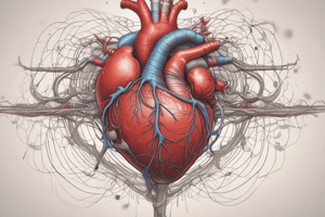
- The heart is a muscular pump that creates pressure to push blood through blood vessels.
- Blood vessels are conduits that allow blood flow from the heart to cells and back to the heart (e.g., arteries, arterioles, capillaries, venules, veins).
- Blood is the fluid carried through the system.
- The heart is located in the thoracic cavity, with the diaphragm separating it from the abdominal cavity.
- The heart weighs approximately 250-350 grams and is roughly the size of a fist.
Pulmonary and Systemic Circulations
- Pulmonary capillaries involve deoxygenated blood entering the lungs.
- Oxygen diffuses from tissues into the blood in the lungs.
- Blood leaving the lungs is oxygenated.
- Systemic capillaries involve oxygenated blood entering tissues.
- Oxygen diffuses from the blood into tissues.
- Blood leaving tissues is deoxygenated.
Cardiac Muscle
- Cardiac muscle cells contain actin and myosin arranged in sarcomeres, similar to skeletal muscle cells.
- Cardiac muscle cells contract via sliding filament mechanism.
- Myocardial cells are short, branched, and connect to neighboring cells creating a complex network.
- Adjacent myocardial cells are joined by intercalated discs containing gap junctions.
Myocardial Cells
- Gap junctions are fluid-filled channels allowing action potentials to spread rapidly from cell to cell, making the myocardial cells simultaneously contract.
- Cardiac muscle is a syncytium.
- The property is "all or nothing".
Cardiac Muscle Contraction
- Skeletal muscle contraction requires external stimulation by somatic motor nerves, while cardiac muscle contraction is triggered by specialized noncontractile myocardial cells, also known as autorhythmic cells or pacemaker cells.
- Cell membranes spontaneously depolarize to generate action potentials that spread to surrounding contractile myocardial cells.
Sinoatrial Node
- Autorhythmic cells are concentrated in the sinoatrial (SA) node in the right atrium near the superior vena cava opening.
- Spontaneous depolarizations in the SA node spread to surrounding myocardial cells, triggering contractions.
- Atrial myocardial cells contract first, followed by a pause, then the Ventricular myocardial cells.
- Atrial and ventricular syncytium complete the process.
Cardiac Conduction System
- Spontaneous action potentials (APs) generated by the SA node spread through the atrial contractile cells.
- The APs then spread to the ventricles through the cardiac conduction system.
Atrial Conduction
- Impulses travel down atrial fibres at a rate of 1 m/sec.
- Specialized fibres like Bachmann's bundle conduct the impulse from the right into the left atrium.
- Impulses continue to the AV node.
Atrioventricular Conduction
- The AV node is located at the base of the right atrium near the interatrial septum.
- It is the only conduction pathway from atria to ventricles.
- Conduction velocity slows to 0.05 m/sec, causing a delay between atrial and ventricular excitation.
- The delay allows optimal ventricular filling during atrial contraction.
Ventricular Conduction
- The AV node conducts to the bundle of His, which branches into Purkinje fibers that conduct the impulse into ventricles.
- Purkinje fibres have a conduction velocity of 1-4 m/sec, due to their larger cell size (80 μM) compared to myocytes (15 μM).
Autorhythmic Cells
- Location:
-
- Sinoatrial (SA) node in right atrium; most important location, APs spread to entire cardiac tissue (70-80 APs/min).
-
- Atrioventricular (AV) node in the right atrium; generated if SA node is destroyed, APs generated (40-60 APs/min).
-
- Pacemaker cells also in Purkinje fibers (30-40 AP/min). SA node APs normally inhibit autorhythmic activity in these cells.
Cardiac Action Potentials
- Two types of action potentials in cardiac tissue: fast response and slow response.
- Fast response is in atrial and ventricular myocytes (conductive or contractile cells).
- Slow response is in autorhythmic cells in the SA and AV nodes.
Action Potentials
- The Phases: Phase 0 (Upstroke), Phase 1 (Early repolarization), Phase 2 (Plateau), Phase 3 (Repolarization), and Phase 4 (Final Repolarization).
- ERP: Effective Refractory Period
- RRP: Relative Refractory Period
Myocardial Contractile Cells
- AP arrival opens voltage-gated Na+ channels, causing rapid depolarization.
- Voltage-gated Ca2+ channels open more slowly, prolonging depolarization.
- At +20 mV, Na+ channels close and K+ channels open, initiating repolarization.
- Slow inward Ca2+ diffusion balances the outward K+ diffusion, forming plateau phase.
- Ca2+ channels close and K+ channels complete repolarization.
Myocardial Cell Contraction
- Extracellular Ca2+ movement during depolarization opens Ca2+ channels on the sarcoplasmic reticulum.
- Increased intracellular [Ca2+] triggers contraction in an identical manner as skeletal muscle.
- Ca2+ is transported out of the cell during repolarization, leading to relaxation.
Excitation-Contraction Coupling
- Depolarization current spreads through gap junctions to contractile cells, allowing action potentials to travel along the plasma membrane and T-tubules.
- Ca2+ channels open in the plasma membrane and sarcoplasmic reticulum.
- Ca2+ induces Ca2+ release from the sarcoplasmic reticulum, causing exposure of myosin-binding sites.
- Crossbridge cycle begins, causing muscle contraction.
- Ca2+ is actively transported back into the sarcoplasmic reticulum and extracellular fluid, causing tropomyosin to block the myosin-binding sites, leading to muscle relaxation.
Cardiac Muscle Contraction
- Electrical current travels through the intercalated discs, connecting cells, and causing coordinated contraction.
Skeletal Muscle Contraction
- AP in skeletal muscle cell is fast (20 msec) and has a no-plateau phase.
- Repolarization occurs before contraction begins.
- The cell's responsiveness means further stimulation can occur during contraction, producing phenomena like summation and tetanus.
Myocardial Cell Contraction
- Myocardial cell AP duration (250 msec) is much longer than that of a skeletal muscle cell (20 msec).
- Plateau phase contributes to myocardial cell refractoriness.
- Summation of contraction and tetanus are prevented due to the long refractory periods of cardiac muscle cells.
Ca2+ Channels
- Cardiac muscle has various Ca2+ channels, but L-type Ca2+ channels (long-lasting) dominate.
- Opening these channels increases Ca2+ conductance due to their large concentration gradient.
- Ca2+ influx strengthens contraction.
In the Clinic
- Ca2+ channel antagonists (e.g., verapamil, diltiazem) decrease AP duration and contractility.
Studying That Suits You
Use AI to generate personalized quizzes and flashcards to suit your learning preferences.