Podcast
Questions and Answers
What is one of the main purposes of scleral indentation during examination?
What is one of the main purposes of scleral indentation during examination?
- To activate retinal tears
- To eliminate the need for optic coherence tomography
- To view peripheral fundus in profile (correct)
- To treat retinal breaks directly
Which of the following conditions is NOT a contraindication for performing scleral indentation?
Which of the following conditions is NOT a contraindication for performing scleral indentation?
- Recent penetrating injury
- Prior successful retinal detachment surgery (correct)
- Recent hyphema
- Recent intraocular surgery
In which patient demographic is lattice degeneration most commonly observed?
In which patient demographic is lattice degeneration most commonly observed?
- Patients with axial myopia (correct)
- Patients with hyperopia
- Patients with emmetropia
- Patients over 60 years of age
What characterizes snail track degeneration in the retina?
What characterizes snail track degeneration in the retina?
Which retinal condition is assessed using scleral indentation to enhance contrast?
Which retinal condition is assessed using scleral indentation to enhance contrast?
What is the significance of the phrase 'at the margins of the lattice degeneration'?
What is the significance of the phrase 'at the margins of the lattice degeneration'?
With what condition is a high axial myopia typically associated?
With what condition is a high axial myopia typically associated?
What unique feature characterizes retinal tears seen in posterior vitreous detachment?
What unique feature characterizes retinal tears seen in posterior vitreous detachment?
During a fundus examination, what is the role of optical coherence tomography (OCT)?
During a fundus examination, what is the role of optical coherence tomography (OCT)?
What describes the typical presentation of RPE hypertrophy as seen on examination?
What describes the typical presentation of RPE hypertrophy as seen on examination?
Which statement about scleral indentation during examinations is accurate?
Which statement about scleral indentation during examinations is accurate?
What is a distinguishing feature of pavingstone degeneration?
What is a distinguishing feature of pavingstone degeneration?
What common misconception about scleral indentation might be held by students?
What common misconception about scleral indentation might be held by students?
Which retinal degeneration is most often described as asymptomatic and usually found bilaterally?
Which retinal degeneration is most often described as asymptomatic and usually found bilaterally?
Which component is NOT included in the differential diagnosis of retinal degenerations?
Which component is NOT included in the differential diagnosis of retinal degenerations?
What is the estimated percentage of patients with lattice degeneration who develop retinal detachment?
What is the estimated percentage of patients with lattice degeneration who develop retinal detachment?
What is the primary mechanism by which retinal tears occur in lattice degeneration?
What is the primary mechanism by which retinal tears occur in lattice degeneration?
What is the estimated percentage of the general population that has snail track degeneration?
What is the estimated percentage of the general population that has snail track degeneration?
What is the characteristic appearance of snail track degeneration?
What is the characteristic appearance of snail track degeneration?
What is the primary difference between lattice degeneration and snail track degeneration?
What is the primary difference between lattice degeneration and snail track degeneration?
What is the recommended management for high-risk patients with lattice or snail track degeneration?
What is the recommended management for high-risk patients with lattice or snail track degeneration?
What is the characteristic appearance of laser spots after prophylactic laser intervention?
What is the characteristic appearance of laser spots after prophylactic laser intervention?
What is the estimated risk of retinal tear or hole in patients with vitreoretinal tuft?
What is the estimated risk of retinal tear or hole in patients with vitreoretinal tuft?
What is the characteristic appearance of vitreoretinal tuft?
What is the characteristic appearance of vitreoretinal tuft?
What is the recommended management for asymptomatic vitreoretinal tuft?
What is the recommended management for asymptomatic vitreoretinal tuft?
What is the primary difference between lattice degeneration and vitreoretinal tuft?
What is the primary difference between lattice degeneration and vitreoretinal tuft?
What is the estimated percentage of retinal detachments associated with lattice degeneration?
What is the estimated percentage of retinal detachments associated with lattice degeneration?
What is the primary mechanism by which retinal tears occur in snail track degeneration?
What is the primary mechanism by which retinal tears occur in snail track degeneration?
What is the recommended management for patients with lattice or snail track degeneration who have a strong family history of retinal detachment?
What is the recommended management for patients with lattice or snail track degeneration who have a strong family history of retinal detachment?
What is the characteristic appearance of retinal tears in lattice degeneration?
What is the characteristic appearance of retinal tears in lattice degeneration?
Flashcards are hidden until you start studying
Study Notes
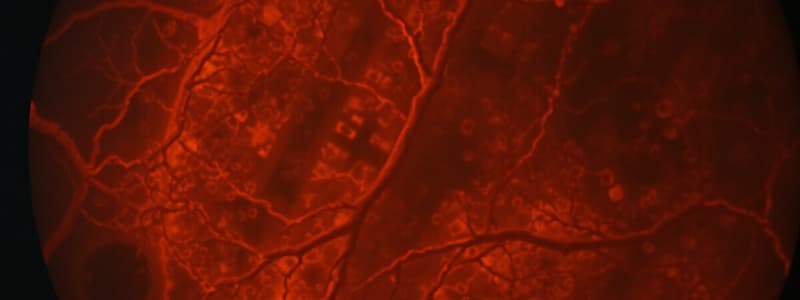
Lattice Degeneration
- Full-thickness missing retinal areas seen in OCT
- Can be stable or very slowly progressive, with a risk of retinal tears
- Approximately 1% of patients with lattice degeneration develop retinal detachment (RD), and ~20-30% of RDs are associated with lattice degeneration
Complications of Lattice Degeneration
- Retinal tears can occur with posterior vitreous detachment pulling on the retina
- Retinal degeneration is very rare in those with lattice degeneration
Snail Track Degeneration
- Degeneration of neural elements leading to atrophy of tissues with lipid deposits in the inner retinal surfaces
- Precursor of lattice degeneration
- Resembles the slime of a snail, with holes and focal thinning similar to lattice degeneration
- Can appreciate focal adhesions at the edges
Snail Track Degeneration Complications
- Similar to those of lattice degeneration, including retinal tears
Management of Lattice and Snail Track Degeneration
- Prophylactic laser intervention is debatable, but likely to treat high-risk patients, such as those with:
- Fellow eye with RD
- Strong family history of RD
- Symptomatic retinal breaks
- Highly myopic
- Aphakic
- Lattice in only seeing eye
Laser Prophylaxis
- White laser spots initially, becoming pigmented over time
Vitreoretinal Tuft
- Small foci of vitreous traction, slightly elevated, and may have a cystic appearance
- May be surrounded by hyperplastic RPE
- Low risk of retinal tear/hole/RD, and no treatment is required if asymptomatic
Scleral Indentation
- Also known as scleral depression, used to examine the peripheral fundus by displacing tissue inward
- Enhances contrast between normal and abnormal retina
- Indications include:
- Signs/symptoms of RD, retinal breaks in symptomatic patients with flashes or axial myopia
- History of blunt trauma
- High axial myopia
Scleral Indentation Technique
- Can be done over the eyelid or directly on the sclera
- Normal retina will be elevated towards the viewer when scleral indentation is applied
- Can help identify retinal breaks and detachments
Contraindications for Scleral Indentation
- Recent intraocular surgery
- Recent hyphema
- Recent penetrating injury
- Ruptured globe
Peripheral Retinal Degenerations
- Relatively common, with OCT playing a role in differential diagnosis
- Types include:
- Vitreoretinal degenerations (lattice degeneration, snail track degeneration, vitreoretinal tuft)
- Intraretinal degenerations (cystoid degeneration, retinoschisis, white without pressure)
- RPE/Chorioretinal degenerations (reticular degeneration, pavingstone degeneration)
Studying That Suits You
Use AI to generate personalized quizzes and flashcards to suit your learning preferences.


