Podcast
Questions and Answers
What percentage of glomerular filtered albumin is reabsorbed by the loop of Henle and distal tubule?
What percentage of glomerular filtered albumin is reabsorbed by the loop of Henle and distal tubule?
- 50%
- 71%
- 23% (correct)
- 3%
Dysfunction of albumin reabsorption in the proximal tubules may explain what condition in early-stage diabetes?
Dysfunction of albumin reabsorption in the proximal tubules may explain what condition in early-stage diabetes?
- Microalbuminuria (correct)
- Glomerulonephritis
- Macroalbuminuria
- Nephrotic syndrome
What is the estimated glomerular filtration rate of albumin in healthy human kidneys?
What is the estimated glomerular filtration rate of albumin in healthy human kidneys?
- 10 g/day
- 100 g/day
- 3.3 g/day (correct)
- 20 g/day
What is the reported size of the glomerular slit-pores, according to a recent electron tomography study?
What is the reported size of the glomerular slit-pores, according to a recent electron tomography study?
What is the molecular weight of albumin?
What is the molecular weight of albumin?
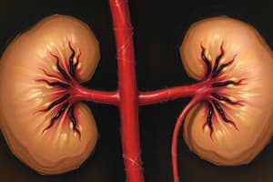
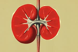
What is the main function of the kidneys?
What is the main function of the kidneys?
The glomerular filtration barrier is composed of how many layers?
The glomerular filtration barrier is composed of how many layers?
What charge does albumin have?
What charge does albumin have?
Damage to what part of the kidney can cause nonselective proteinuria?
Damage to what part of the kidney can cause nonselective proteinuria?
What is the role of megalin in the kidney?
What is the role of megalin in the kidney?
Flashcards
Sieving Coefficient
Sieving Coefficient
A measure of how much of a substance is filtered through the glomerulus compared to its concentration in the plasma.
Tubular Reabsorption
Tubular Reabsorption
The kidney's process of reclaiming filtered substances, like proteins, back into the bloodstream.
Microalbuminuria
Microalbuminuria
Small amounts of albumin in the urine, often an early sign of kidney issues in diabetes.
Glomerular Filtration Barrier
Glomerular Filtration Barrier
Signup and view all the flashcards
Podocytes
Podocytes
Signup and view all the flashcards
Slit Diaphragm
Slit Diaphragm
Signup and view all the flashcards
Endocytosis
Endocytosis
Signup and view all the flashcards
Albumin
Albumin
Signup and view all the flashcards
FcRn Receptors
FcRn Receptors
Signup and view all the flashcards
Transcytosis
Transcytosis
Signup and view all the flashcards
Study Notes
- Glomerular filtration occurs with an albumin sieving coefficient of 0.00062
- Approximately 3.3 g of albumin is filtered daily in human kidneys.
- The proximal convoluted tubule reabsorbs 71% of filtered albumin.
- The loop of Henle and distal tubule reabsorb 23% of filtered albumin.
- The collecting duct reabsorbs 3% of filtered albumin.
- This shows the kidney’s important role in protein metabolism.
- Dysfunction of albumin reabsorption in proximal tubules, caused by reduced megalin expression, leads to microalbuminuria in early-stage diabetes.
- Various disorders of the glomerular filtration barrier cause massive nonselective proteinuria.
- These disorders include:
- Podocyte detachment
- Glomerular basement membrane rupture
- And slit diaphragm dysfunction in focal segmental glomerulosclerosis, membranous nephropathy, and other glomerulonephritis
- Selective albuminuria linked to foot process effacement and tight junction-like slit alteration is seen in minimal-change nephrotic syndrome patients.
- Albumin uptake is enhanced in the podocyte cell body, which is potentially mediated by albumin receptors in the low-dose puromycin model.
- The role of enhanced podocyte albumin transport needs to be investigated to understand selective albuminuria in minimal-change disease.
Introduction
- The kidneys maintain body fluid homeostasis by regulating:
- Water balance
- Electrolyte balance
- Acid-base balance
- Excretion of uremic toxins
- The kidneys produce hormones like renin and erythropoietin and activate vitamin D3.
- The kidney's role in protein metabolism hasn't received much attention.
- Primitive urine contains many proteins smaller than albumin.
- Renal proximal tubules actively reabsorb these proteins and subsequently degrade them to amino acids in lysosomes and return them to the blood.
- Glomerular albumin filtration is restricted by size and charge barriers of the glomerular basement membrane and slit diaphragm pores.
- Smithies raised the question of the slit diaphragms not clogging with albumin if all filtered molecules pass through them.
- Glomerular albumin filtration occurs via diffusion across the GBM.
- It's unclear how albumin exits across effaced podocyte foot processes covering the basement membrane in minimal-change nephrotic syndrome.
- Discussed are the ultrastructural morphological changes of the glomerular filtration barrier in glomerular diseases.
- The discussion proposes a new mechanism of glomerular albumin filtration in minimal-change nephrotic syndrome.
Albumin Filtration Under Normal Conditions
- Albumin has three spherical domains, with a molecular weight of 69 kDa and a net charge of -15, and is a flexible, ellipsoid-shaped molecule, 3.8 nm in diameter and 15 nm long.
- The slit pore size was originally ≈ 40 by 140 angstroms in cross section and 70 angstroms long.
- Recent electron tomography study revealed that slit-pores are 35 angstroms in diameter with variation in size.
- Processing samples for electron microscopy causes a slight reduction in size, so the true size is likely larger.
- The effective Stock-Einstein radius of albumin is 35 angstroms (70 angstroms in diameter), so some molecules pass through the slit pores, due to their flexibility and ellipsoid shape.
- FITC-labeled albumin on the slit diaphragms between foot processes showed a small fraction of albumin goes through the slit pores in normal rats.
- Early micropuncture studies showed albumin concentration from 3 to 728 µg/mL in primitive rat urine in Bowman's capsule
Albumin Sieving Coefficient Comparisons:
- Fractional micropuncture showed 0.00062 in rats
- 131I-labeled BSA studies vary based on method and animal model.
- 125I-labeled human serum albumin measurements showed 0.00066 in rats when measuring kidney uptake and urinary excretion, and 0.0065 in rats with neutral human serum albumin.
- Urinary albumin excretion of congenital Fanconi syndrome patients showed 0.00008 in humans.
- Blockade of proximal tubular reabsorption by L-lysine studies showed 0.00033 to 0.0591 in puromycin aminonucleoside nephrotic rats.
- Inhibition of tubular function by cooling (8°C) studies showed 0.0019 in rats
- Urinary albumin excretion of megalin-knockout mice studies showed 0.00016 in megalin-knockout mice.
- Tritium-labeled albumin studies showed 0.074 in rats.
- Alexa-labeled albumin, confocal microscopy studies showed 0.0341 in rats.
- Alexa-labeled rat serum albumin, two-photon microscope with internal photodetectors studies showed 0.002 in Munich-Wistar rats.
- Isotope-labeled albumin clearance studies measure urinary excretion and tubular uptake, divided by the plasma isotope level, showing consistent values with fractional micropuncture data.
- Albumin clearance studies blocking proximal tubular reabsorption with L-lysine, low temperatures, or congenital abnormalities of tubular reabsorption showed smaller values than micropuncture data.
- Fractional excretion of albumin in Fanconi syndrome patients is 0.00008, roughly glomerular-sieving coefficient in the normal kidney.
- Nephron segments downstream of the proximal convoluted tubules reabsorb ~26% of glomerular filtrated albumin, if proximal tubular albumin reabsorption is impaired in Fanconi syndrome.
- Glomerular albumin-sieving coefficient may be greater than 0.00011 in humans.
- Large amounts of glomerular filtration have been reported.
- One study using tritium-labeled albumin demonstrated a sieving coefficient of 0.074
- Another with Alexa-labeled albumin observed by confocal microscopy found a sieving coefficient of 0.0341 (50-100x higher than previous studies).
- Technical limitations affect sensitivity, and the incomplete removal of unbound labeling molecules alters the glomerular filtration measurement.
- From the viewpoint of albumin metabolism, these values also seem unrealistically high.
Important Role of the Kidney in the Protein Metabolism
- Albumin concentration along the rat nephron was measured in fractional micropuncture studies.
- Renal tubules reabsorb about 3 g of albumin per day in humans.
- Isolated rabbit proximal tubule albumin reabsorption capacity measured at 99.9 × 10⁻³ ng/min/mm.
- Human kidneys can reabsorb 1.9g of albumin per day assuming a proximal tubule length of 6.5 mm.
- Nephron segments downstream of the proximal convoluted tubules further reabsorb ~26% of filtered albumin.
- The amount that the kidney reabsorbs comes to 2.6 g a day.
- Proximal tubule albumin molecules taken up into lysosomes in 6-15 minutes and then degraded into amino acids after 30-120 minutes.
- The kidney is an organ with important role in the protein metabolism
- Reported high sieving coefficient values suggest about 200g of albumin per day is filtered in the glomerulus and reabsorbed in the proximal tubule, which seems unlikely.
- The level of albumin cannot be measured in the normal kidney by immunostaining.
- Intact albumin cannot be transported by the proximal tubule in physiological conditions.
- Albumin filtration and metabolism in the kidney are higher than the liver’s daily production
- Low molecular weight proteins are almost all freely filtered at the glomerulus with a sieving coefficient of 0.987 ~9.6 g are reabsorbed per day.
- Hemodialysis cannot compensate for tubular dysfunction of protein metabolism in chronic renal failure; therefore, low molecular weight protein deposits (such as β2-microglobulin) cause amyloidosis in hemodialysis patients.
- The physiological role of protein metabolism in the kidney must be accounted for.
Mechanisms of Microalbuminuria in Diabetic Nephropathy
- Microalbuminuria may be an early marker of diabetic nephropathy caused by the increase in glomerular permeability and glomerular hyperfiltration.
- Proximal tubular albumin reabsorption decreases without glomerular albumin filtration increasing in early stages of streptozotocin-induced diabetic nephropathy.
- Tubular dysfunction may contribute to microalbuminuria in early-stage diabetes.
- Megalin is decreased in diabetic rats [27], and by measuring albumin clearance after blocking proximal reabsorption with lysine, using isotope labeled-bovine serum albumin [28].
- Brush border enzymes may degrade albumin in the proximal tubules the true amount of albuminuria may be larger than detected in the urine by measuring the intact albumin
- RAS inhibitors may restore megalin expression, which means it ameliorates the tubular dysfunction of albumin reabsorption and reduce albuminuria in diabetic rats.
- Reabsorbed albumin by receptor-mediated endocytosis into endosomes must be ligand-receptor dissociated in order to recycle the albumin-binding receptors back to the plasma membrane.
- Vesicular acidification by H+-ATPase, CLC-5, NHE-3 is functionally important for the pH-dependent dissociation between albumin and megalin, and effective albumin reabsorption.
- Angiotensin II blocks H+-ATPase [33], the acidification of endosomes may be reduced by inhibition of H+-ATPase by renal angiotensin II, the leading to decreased albumin reabsorption.
- RAS inhibitors prevent intraglomerular hypertension and glomerular permselectivity [34], but restore albumin metabolism in the proximal tubules.
Questions Pertaining to Glomerular Albumin Filtration through Slit Pores in the Nephrotic Syndrome
- The glomerular filtration barrier is made up of three layers.
- The fenestrated endothelium covered by a negatively charged glycocalyx.
- The glomerular basement membrane where a size barrier contains laminin and type IV collagen and a charge barrier generated by heparan sulfate as a coarse barrier.
- The slit diaphragm between foot processes is treated as a fine filter.
- Yamada identified by electron microscopy [36] by electron microscopy in 1955 [36].
- Rodewald and Karnovsky identified the zipper-like structure of the slit membrane [5]
- Albumin filters via the slit pores, similar to water because experiments using tracers have raised the controversy whether the GBM or slit diaphragm is more restrictive.
- Ferritin accumulates in the GBM, but not under the slit diaphragm [37].
- Horseradish peroxidase (HRP) is observed on the GBM [38, 39].
- The identification of nephrin at the slit diaphragm and its mutation in Finnish-type congenital nephrotic syndrome have lead to results that the slit membrane is the glomerular filtration barrier [40, 41].
- The importance of the GBM is for laminin β2 (LAMB2) knockout mice that show a severe disorganization of the GBM structure with accompanying proteinuria [42].
- The charge barrier of GBM needs to be further investigated by Heparan sulfate synthase deficient mice, which will not present signs of proteinuria.
- Reduction of the negative charge of the GBM and foot process effacement [43] shows that both the slit diaphragm and the GBM contribute to the a glomerular filtration barrier.
- Questions need to be answered before albumin filtration through slit pores is fully accepted.
- Albumin does not accumulate under the slit diaphragm in tracer studies.
- The albumin sieving coefficient is lower than similar-sized Dextran or Ficoll, which cannot be explained by molecular shape and charge differences [4, 44].
- The glomerular albumin coefficient should increase at high glomerular filtration rates.
- Reports have shown that the number of slit diaphragms decreases with a tight junction with structural change in minimal-change nephrotic syndrome.
- Reduction of what nephrin may cause shows that a slit pore enlargement can explain massive proteinuria but cannot have selective albuminuria in minimal-change.
Electron Microscopic Observation of Various Mechanisms of Proteinuria in Glomerular Diseases
- Observations of human renal biopsies in electron microscope images makes it possible to identify causes of proteinuria in different glomerular diseases
- Glomerulonephritis (IgA nephropathy, ANCA-related nephritis, acute glomerulonephritis, and lupus nephritis) damages the GBM via ruptures/holes caused by inflammatory cells.
- This results in a nonselective proteinuria with hematuria
- Podocyte detachment and apoptosis are observed in focal segmental glomerulosclerosis and membranous nephropathy.
- Proteins leak from the site of denuded GBM due to nonselective proteinuria Figures 3(b) and 3(c) This denuded GBM is known to adhere to the Bowman’s capsule.
- All of it leads to segmental sclerosis.
- Integrin-dependent signaling and oxidative stress from NADPH cause Podocyte detachment/apoptosis.
- Ultra structural morphological changes of the GBM may represent the shunt pathway assumed in mathematical of glomerular permselectivity.
- While these findings are often observed in the - high dose puromycin aminonucleoside nephrotic syndrome model [50] that show a shunt, and are rarely observed in both of these cases shows.
Possible Mechanisms of Selective Albuminuria in Minimal-Change Nephrotic Syndrome
- Common hypothesis is that that proteins leak from the pores that are due to decreased nephron expression, while podocyte ( detatchment is really not something we worry about).
- Podocytes that will show processes are going to completely widely cover and it must be in the capillary wall for most of the minimal change nephrotic syndrome, and podocyte pore density is decreased by 80% at most.
- Tight junction like structure half the time has Podocytes, the amount of proteins and what can cause questions.
- Podocytes show why the diaphragms cannot by way of Smith.
Conclusion: The nephrocytic syndromes cause proteinuria and the processes involved in them cause questions.
Studying That Suits You
Use AI to generate personalized quizzes and flashcards to suit your learning preferences.