Podcast
Questions and Answers
What is a key characteristic of a chronic condition resulting from repetitive trauma to a joint?
What is a key characteristic of a chronic condition resulting from repetitive trauma to a joint?
- The trauma leading to the condition does not have to be severe or catastrophic. (correct)
- It only affects weight-bearing joints.
- It always requires severe or catastrophic trauma as the initial cause.
- It primarily occurs in elderly individuals with pre-existing arthritis.
Which joints are commonly affected by chronic conditions resulting from repetitive trauma, particularly in service members?
Which joints are commonly affected by chronic conditions resulting from repetitive trauma, particularly in service members?
- Jaw, neck, and spinal joints
- Elbow, wrist, and finger joints
- Shoulder, hip, knee, and ankle joints (correct)
- Toe, clavicle, and rib joints
What aspect of a patient's history is MOST relevant when evaluating chronic or recurrent joint pain?
What aspect of a patient's history is MOST relevant when evaluating chronic or recurrent joint pain?
- Family history of autoimmune diseases.
- Dietary habits and supplement use.
- Detailed history of trauma and repetitive activities. (correct)
- Childhood illnesses and vaccinations.
During a physical examination for chronic joint pain, what assessment is crucial for detecting mechanical blocks?
During a physical examination for chronic joint pain, what assessment is crucial for detecting mechanical blocks?
When evaluating a knee joint, which finding would suggest a bucket handle meniscal tear rather than chronic joint pain from repetitive trauma?
When evaluating a knee joint, which finding would suggest a bucket handle meniscal tear rather than chronic joint pain from repetitive trauma?
What non-pharmacological treatment is recommended to reduce impact stress on joints in individuals with chronic joint pain?
What non-pharmacological treatment is recommended to reduce impact stress on joints in individuals with chronic joint pain?
When should a patient with chronic joint pain be referred for leg-EVAC/orthopedics consult?
When should a patient with chronic joint pain be referred for leg-EVAC/orthopedics consult?
Following the diagnosis of chronic joint pain, what timeline should prompt a patient to return for follow-up if their symptoms worsen?
Following the diagnosis of chronic joint pain, what timeline should prompt a patient to return for follow-up if their symptoms worsen?
What is a primary focus in managing lower extremity muscle strains?
What is a primary focus in managing lower extremity muscle strains?
When evaluating a lower extremity muscle strain, which historical detail is MOST important for determining the severity and course of treatment?
When evaluating a lower extremity muscle strain, which historical detail is MOST important for determining the severity and course of treatment?
In a physical exam for a suspected lower extremity muscle strain, which finding would be MOST indicative of a muscle injury?
In a physical exam for a suspected lower extremity muscle strain, which finding would be MOST indicative of a muscle injury?
During the physical exam, what is the significance of comparing tenderness with active range of motion (AROM) versus passive range of motion (PROM) in a suspected muscle strain?
During the physical exam, what is the significance of comparing tenderness with active range of motion (AROM) versus passive range of motion (PROM) in a suspected muscle strain?
What differential diagnosis should be considered in a patient presenting with lower extremity pain if they are unable to perform Active Range of Motion (AROM) if an avulsed tendon is suspected?
What differential diagnosis should be considered in a patient presenting with lower extremity pain if they are unable to perform Active Range of Motion (AROM) if an avulsed tendon is suspected?
In managing a lower extremity muscle strain, when would you recommend patients return for a follow-up visit?
In managing a lower extremity muscle strain, when would you recommend patients return for a follow-up visit?
According to the guidelines, when should a routine EVAC for PCM/orthopedics be considered for a patient with a lower extremity muscle strain?
According to the guidelines, when should a routine EVAC for PCM/orthopedics be considered for a patient with a lower extremity muscle strain?
What is the MOST immediate concern in a patient presenting with possible exertional rhabdomyolysis?
What is the MOST immediate concern in a patient presenting with possible exertional rhabdomyolysis?
Which of the following findings is MOST indicative of rhabdomyolysis rather than a typical muscle strain?
Which of the following findings is MOST indicative of rhabdomyolysis rather than a typical muscle strain?
In a patient suspected of having rhabdomyolysis from intense physical activity, what additional historical information would be MOST relevant?
In a patient suspected of having rhabdomyolysis from intense physical activity, what additional historical information would be MOST relevant?
What laboratory finding is MOST crucial in diagnosing rhabdomyolysis?
What laboratory finding is MOST crucial in diagnosing rhabdomyolysis?
Why is a urinalysis important in the evaluation of suspected rhabdomyolysis?
Why is a urinalysis important in the evaluation of suspected rhabdomyolysis?
In managing a patient with rhabdomyolysis, what is the initial approach to treatment focused on?
In managing a patient with rhabdomyolysis, what is the initial approach to treatment focused on?
What is the general recommendation for intravenous normal saline (NS) administration for a patient with rhabdomyolysis?
What is the general recommendation for intravenous normal saline (NS) administration for a patient with rhabdomyolysis?
What is the primary goal for urine output when managing a patient with rhabdomyolysis with IV fluids?
What is the primary goal for urine output when managing a patient with rhabdomyolysis with IV fluids?
When can a patient with rhabdomyolysis be treated as an outpatient with oral hydration?
When can a patient with rhabdomyolysis be treated as an outpatient with oral hydration?
Which of the following is a crucial element of a gradual return-to-duty protocol for a service member recovering from exertional rhabdomyolysis?
Which of the following is a crucial element of a gradual return-to-duty protocol for a service member recovering from exertional rhabdomyolysis?
During Phase 1 of a gradual return to duty protocol for exertional rhabdomyolysis, what is the recommended sleep duration?
During Phase 1 of a gradual return to duty protocol for exertional rhabdomyolysis, what is the recommended sleep duration?
During Phased return to duty after rhabdomyolysis, what is a component of phase 2?
During Phased return to duty after rhabdomyolysis, what is a component of phase 2?
If a service member's CK value remains >5X ULN and/or UA is persistently abnormal for 2 weeks after injury or hospitalization, what is the NEXT appropriate step?
If a service member's CK value remains >5X ULN and/or UA is persistently abnormal for 2 weeks after injury or hospitalization, what is the NEXT appropriate step?
For a patient diagnosed with Rhabdomyolysis, when should the patient return for reevaluation?
For a patient diagnosed with Rhabdomyolysis, when should the patient return for reevaluation?
What is the MOST critical factor when assessing compartment syndrome?
What is the MOST critical factor when assessing compartment syndrome?
What is a common early symptom in compartment syndrome?
What is a common early symptom in compartment syndrome?
What physical exam finding is MOST indicative of compartment syndrome in the lower leg?
What physical exam finding is MOST indicative of compartment syndrome in the lower leg?
In suspected compartment syndrome, what is the significance of diminished sensation distal to the injury site?
In suspected compartment syndrome, what is the significance of diminished sensation distal to the injury site?
When assessing a patient for compartment syndrome, which additional factor should be considered if circumferential burns are present?
When assessing a patient for compartment syndrome, which additional factor should be considered if circumferential burns are present?
What diagnostic tool is MOST directly useful in confirming a diagnosis of compartment syndrome?
What diagnostic tool is MOST directly useful in confirming a diagnosis of compartment syndrome?
What is the definitive treatment required for compartment syndrome?
What is the definitive treatment required for compartment syndrome?
For patients requiring a fasciotomy, what is a key management aspect after the procedure?
For patients requiring a fasciotomy, what is a key management aspect after the procedure?
What symptoms are categorized as 'red flags' and warrant immediate evacuation?
What symptoms are categorized as 'red flags' and warrant immediate evacuation?
In the context of musculoskeletal injuries, what does OPQRST primarily help assess?
In the context of musculoskeletal injuries, what does OPQRST primarily help assess?
What is the recommended initial urine output for patients being treated for rhabdomyolysis with intravenous fluids?
What is the recommended initial urine output for patients being treated for rhabdomyolysis with intravenous fluids?
What distinguishes compartment syndrome from a typical muscle strain?
What distinguishes compartment syndrome from a typical muscle strain?
In the management of rhabdomyolysis, what is the significance of monitoring serum electrolytes alongside creatinine kinase (CK) levels?
In the management of rhabdomyolysis, what is the significance of monitoring serum electrolytes alongside creatinine kinase (CK) levels?
When evaluating a patient for a lower extremity muscle strain, what is the significance of numbness or tingling?
When evaluating a patient for a lower extremity muscle strain, what is the significance of numbness or tingling?
What is the rationale for advising rest and avoiding aggravating activities in the initial management of a lower extremity muscle strain?
What is the rationale for advising rest and avoiding aggravating activities in the initial management of a lower extremity muscle strain?
What factors help differentiate a diagnosis of heat injury from exertional rhabdomyolysis?
What factors help differentiate a diagnosis of heat injury from exertional rhabdomyolysis?
When assessing limb swelling in a patient with suspected rhabdomyolysis, what additional condition should be considered if the patient reports pain out of proportion to the visible swelling?
When assessing limb swelling in a patient with suspected rhabdomyolysis, what additional condition should be considered if the patient reports pain out of proportion to the visible swelling?
If a patient is diagnosed with a lower extremity muscle strain and has not improved after 6 weeks of conservative management, what is the next step?
If a patient is diagnosed with a lower extremity muscle strain and has not improved after 6 weeks of conservative management, what is the next step?
A patient with a suspected lower extremity muscle strain is experiencing significant pain, and reports being unable to bear weight. Which aspect of their focused history is MOST crucial to consider in determining the next steps?
A patient with a suspected lower extremity muscle strain is experiencing significant pain, and reports being unable to bear weight. Which aspect of their focused history is MOST crucial to consider in determining the next steps?
What is the significance of asking about the history of supplement use in a patient presenting with possible rhabdomyolysis?
What is the significance of asking about the history of supplement use in a patient presenting with possible rhabdomyolysis?
In the context of exertional rhabdomyolysis, which of the following factors increases the risk in the military population?
In the context of exertional rhabdomyolysis, which of the following factors increases the risk in the military population?
What is the primary goal when using intravenous normal saline (NS) to manage a patient with rhabdomyolysis?
What is the primary goal when using intravenous normal saline (NS) to manage a patient with rhabdomyolysis?
A patient with suspected compartment syndrome reports that "the pain is worse than I would expect." What is an important component of the focused history to gather from the patient?
A patient with suspected compartment syndrome reports that "the pain is worse than I would expect." What is an important component of the focused history to gather from the patient?
What is the most appropriate next step for a service member whose CK value remains >5X ULN and/or UA is persistently abnormal for 2 weeks after injury or hospitalization due to Rhabdomyolysis?
What is the most appropriate next step for a service member whose CK value remains >5X ULN and/or UA is persistently abnormal for 2 weeks after injury or hospitalization due to Rhabdomyolysis?
During a physical exam for suspected compartment syndrome, what finding would raise the MOST immediate concern?
During a physical exam for suspected compartment syndrome, what finding would raise the MOST immediate concern?
What is the primary purpose of applying wet sterile dressings following a fasciotomy?
What is the primary purpose of applying wet sterile dressings following a fasciotomy?
During Phase 2 of a gradual return-to-duty protocol for exertional rhabdomyolysis, what type of physical activity is appropriate for the service member?
During Phase 2 of a gradual return-to-duty protocol for exertional rhabdomyolysis, what type of physical activity is appropriate for the service member?
What aspects of a patient's symptoms is categorized as a 'red flag' warranting immediate evacuation in the context of a musculoskeletal injury?
What aspects of a patient's symptoms is categorized as a 'red flag' warranting immediate evacuation in the context of a musculoskeletal injury?
What is PRIMARY presenting symptom for chronic joint pain?
What is PRIMARY presenting symptom for chronic joint pain?
What focused history questions are important to ask for chronic joint pain?
What focused history questions are important to ask for chronic joint pain?
What tools are helpful when confirming chronic joint pain?
What tools are helpful when confirming chronic joint pain?
What is an effective treatment for chronic joint pain that involves buoyancy?
What is an effective treatment for chronic joint pain that involves buoyancy?
How long should the patient follow up if they have worsening chronic joint pain?
How long should the patient follow up if they have worsening chronic joint pain?
How many muscles are there in the body capable of being injured through a variety of means?
How many muscles are there in the body capable of being injured through a variety of means?
What is recommended if a patient is unable to perform AROM if an avulsed tendon is suspected?
What is recommended if a patient is unable to perform AROM if an avulsed tendon is suspected?
What examination finding is expected during the assessment of a muscle strain?
What examination finding is expected during the assessment of a muscle strain?
What is considered if the mechanism suggests a possible fracture?
What is considered if the mechanism suggests a possible fracture?
When should a patient return for follow up care in weeks for lower extremity muscle strain?.
When should a patient return for follow up care in weeks for lower extremity muscle strain?.
What is a symptom of Rhabdomyolysis?
What is a symptom of Rhabdomyolysis?
What is the result of acute and excessive damage to skeletal muscle?
What is the result of acute and excessive damage to skeletal muscle?
What labs are useful when assessing possible rhabdomyolysis?
What labs are useful when assessing possible rhabdomyolysis?
What actions are important to consider for rhabdomyolysis?
What actions are important to consider for rhabdomyolysis?
Your patient has Compartment syndrome. Which statement is true?
Your patient has Compartment syndrome. Which statement is true?
What are late signs found with compartment syndrome?
What are late signs found with compartment syndrome?
What is the ultimate treatment for compartment syndrome?
What is the ultimate treatment for compartment syndrome?
What findings are expect after a patient had a fasciotomy?
What findings are expect after a patient had a fasciotomy?
There has been a report of “Red Flags”. Where should this patient be sent?
There has been a report of “Red Flags”. Where should this patient be sent?
A patient is experiencing neck pain from prolonged sitting looking at a computer. What cervical spine issue might this patient be experiencing?
A patient is experiencing neck pain from prolonged sitting looking at a computer. What cervical spine issue might this patient be experiencing?
Your patient has neck pain, you assess their posture as well as their ________?
Your patient has neck pain, you assess their posture as well as their ________?
What are helpful tools in the assessment of neck pain?
What are helpful tools in the assessment of neck pain?
How long does the recommendation include before EVACing or doing physical therapy?
How long does the recommendation include before EVACing or doing physical therapy?
Your team member is complaining of neck pain and needs battlefield acupuncture. What directive do you follow?
Your team member is complaining of neck pain and needs battlefield acupuncture. What directive do you follow?
Back pain in the lower back with or without ROM may be related to _______?
Back pain in the lower back with or without ROM may be related to _______?
What is the recommendation for a patient with suspected VA/DoD Clinical Practices Guidelines for low back pain?
What is the recommendation for a patient with suspected VA/DoD Clinical Practices Guidelines for low back pain?
Cauda Equina Syndrome has what specific symptoms?
Cauda Equina Syndrome has what specific symptoms?
To help strengthen your patients lower back do you recommend?
To help strengthen your patients lower back do you recommend?
When may you recommend bedrest?
When may you recommend bedrest?
A service member reports shoulder pain that worsens specifically when sleeping on the affected side. What condition is MOST likely indicated by this symptom?
A service member reports shoulder pain that worsens specifically when sleeping on the affected side. What condition is MOST likely indicated by this symptom?
When evaluating a patient for possible shoulder impingement syndrome, which historical detail would MOST strongly suggest this diagnosis?
When evaluating a patient for possible shoulder impingement syndrome, which historical detail would MOST strongly suggest this diagnosis?
During a physical examination for suspected shoulder impingement syndrome, which finding would be MOST indicative of the condition?
During a physical examination for suspected shoulder impingement syndrome, which finding would be MOST indicative of the condition?
A manual laborer reports shoulder pain that has not improved after several weeks of rest and NSAIDs. He notes pain with overhead activities. What is the MOST appropriate next step in management?
A manual laborer reports shoulder pain that has not improved after several weeks of rest and NSAIDs. He notes pain with overhead activities. What is the MOST appropriate next step in management?
A patient presents with shoulder and reports limited ROM, weakness, and a positive Hawkins test. Which of the following factors would warrant referral for orthopedic evaluation?
A patient presents with shoulder and reports limited ROM, weakness, and a positive Hawkins test. Which of the following factors would warrant referral for orthopedic evaluation?
Which of the following is MOST accurate regarding the typical mechanism of injury shoulder impingement?
Which of the following is MOST accurate regarding the typical mechanism of injury shoulder impingement?
A patient presents to your clinic and is diagnosed with an AC joint sprain. What in the history would suggest this diagnosis?
A patient presents to your clinic and is diagnosed with an AC joint sprain. What in the history would suggest this diagnosis?
A patient presents to your clinic and is diagnosed with an AC joint separation. What physical exam finding would suggest this diagnosis?
A patient presents to your clinic and is diagnosed with an AC joint separation. What physical exam finding would suggest this diagnosis?
A patient who fell directly onto his shoulder presents with a visible deformity at the distal end of the clavicle. What is the MOST appropriate next step?
A patient who fell directly onto his shoulder presents with a visible deformity at the distal end of the clavicle. What is the MOST appropriate next step?
Sling use for AC joint injuries should be implemented. How long should sling immobilization generally last?
Sling use for AC joint injuries should be implemented. How long should sling immobilization generally last?
A patient with low back pain presents with new onset bowel and bladder incontinence, saddle anesthesia. Which of the following is the MOST likely diagnosis?
A patient with low back pain presents with new onset bowel and bladder incontinence, saddle anesthesia. Which of the following is the MOST likely diagnosis?
What is the MOST common cause of lumbar radiculopathy (sciatica)?
What is the MOST common cause of lumbar radiculopathy (sciatica)?
A patient reports lower back pain radiating down the leg, worsening with sitting. Which of the following focused history questions is MOST relevant in differentiating between lumbar strain and disc herniation?
A patient reports lower back pain radiating down the leg, worsening with sitting. Which of the following focused history questions is MOST relevant in differentiating between lumbar strain and disc herniation?
During a focused history for a patient with low back pain radiating down the leg (Sciatica), what 'red flag' symptom requires further assessment?
During a focused history for a patient with low back pain radiating down the leg (Sciatica), what 'red flag' symptom requires further assessment?
A patient with low back pain and suspected lumbar radiculopathy has a positive straight leg raise test. What does this MOST likely indicate?
A patient with low back pain and suspected lumbar radiculopathy has a positive straight leg raise test. What does this MOST likely indicate?
For most patients with acute low back pain without 'red flag' symptoms, what is the MOST appropriate initial management strategy?
For most patients with acute low back pain without 'red flag' symptoms, what is the MOST appropriate initial management strategy?
The VA/DoD Clinical Practice Guidelines for low back pain recommends what?
The VA/DoD Clinical Practice Guidelines for low back pain recommends what?
A service member presents with new onset neck pain. On exam, you observe forward head posture. Beyond posture, what else should be assessed in the physical exam?
A service member presents with new onset neck pain. On exam, you observe forward head posture. Beyond posture, what else should be assessed in the physical exam?
A service member presents with neck pain. Their limited ROM is thought to be secondary to pain. What tools should be used to help diagnosis this patient?
A service member presents with neck pain. Their limited ROM is thought to be secondary to pain. What tools should be used to help diagnosis this patient?
A service member is seeking routine care for neck pain. You're a certified battlefield acupuncturist and would like to provide care. What directive should you follow?
A service member is seeking routine care for neck pain. You're a certified battlefield acupuncturist and would like to provide care. What directive should you follow?
Flashcards
Chronic joint pain
Chronic joint pain
A chronic condition from repetitive joint trauma.
Symptoms of Joint Pain
Symptoms of Joint Pain
Joint pain with movement and impact.
Focused History for Joint Pain
Focused History for Joint Pain
Trauma, occupation, and recreational activities.
Physical Exam findings in joint pain
Physical Exam findings in joint pain
Signup and view all the flashcards
Joint Pain Differential
Joint Pain Differential
Signup and view all the flashcards
Joint Pain Treatment Options
Joint Pain Treatment Options
Signup and view all the flashcards
When to Follow Up: Joint Pain
When to Follow Up: Joint Pain
Signup and view all the flashcards
Rhabdomyolysis
Rhabdomyolysis
Signup and view all the flashcards
Rhabdomyolysis Symptoms
Rhabdomyolysis Symptoms
Signup and view all the flashcards
Focused History: Rhabdomyolysis
Focused History: Rhabdomyolysis
Signup and view all the flashcards
Physical Exam findings: Rhabdomyolysis
Physical Exam findings: Rhabdomyolysis
Signup and view all the flashcards
Rhabdomyolysis Labs
Rhabdomyolysis Labs
Signup and view all the flashcards
Rhabdomyolysis Differential
Rhabdomyolysis Differential
Signup and view all the flashcards
Rhabdomyolysis Treatment
Rhabdomyolysis Treatment
Signup and view all the flashcards
Compartment Syndrome
Compartment Syndrome
Signup and view all the flashcards
Compartment Syndrome Findings
Compartment Syndrome Findings
Signup and view all the flashcards
Physical Exam: Compartment Syndrome
Physical Exam: Compartment Syndrome
Signup and view all the flashcards
Diagnostic Tools: Compartment Syndrome
Diagnostic Tools: Compartment Syndrome
Signup and view all the flashcards
Differential: Compartment Syndrome
Differential: Compartment Syndrome
Signup and view all the flashcards
Treatment: Compartment Syndrome
Treatment: Compartment Syndrome
Signup and view all the flashcards
"Red Flag" Back Symptoms
"Red Flag" Back Symptoms
Signup and view all the flashcards
Vertebral Column
Vertebral Column
Signup and view all the flashcards
Sternocleidomastoid Function
Sternocleidomastoid Function
Signup and view all the flashcards
Etiology of Neck Pain
Etiology of Neck Pain
Signup and view all the flashcards
Symptoms of Neck Pain
Symptoms of Neck Pain
Signup and view all the flashcards
Physical Exam: Neck Pain
Physical Exam: Neck Pain
Signup and view all the flashcards
Imaging for Neck Pain
Imaging for Neck Pain
Signup and view all the flashcards
Differential Diagnosis: Neck Pain
Differential Diagnosis: Neck Pain
Signup and view all the flashcards
Management of Neck Pain: Heat
Management of Neck Pain: Heat
Signup and view all the flashcards
What Not to Do
What Not to Do
Signup and view all the flashcards
Red Flag: Cauda Equina/Bowel
Red Flag: Cauda Equina/Bowel
Signup and view all the flashcards
Tear: Low Back Pain
Tear: Low Back Pain
Signup and view all the flashcards
Disk Rupture
Disk Rupture
Signup and view all the flashcards
Second-Pain in Injury: History
Second-Pain in Injury: History
Signup and view all the flashcards
Causes: Outside-Pain
Causes: Outside-Pain
Signup and view all the flashcards
Spinal Muscle Palpation- back tenderness
Spinal Muscle Palpation- back tenderness
Signup and view all the flashcards
Leg raise failure
Leg raise failure
Signup and view all the flashcards
X-ray limits
X-ray limits
Signup and view all the flashcards
Water retention /Core increase
Water retention /Core increase
Signup and view all the flashcards
Given Likely- Fluids/Pain
Given Likely- Fluids/Pain
Signup and view all the flashcards
Superior Pain
Superior Pain
Signup and view all the flashcards
Neurological Problems: C6
Neurological Problems: C6
Signup and view all the flashcards
Is less at arm/problems
Is less at arm/problems
Signup and view all the flashcards
Muscle Imbalance
Muscle Imbalance
Signup and view all the flashcards
Problems: Injury
Problems: Injury
Signup and view all the flashcards
Rotate and Stretch
Rotate and Stretch
Signup and view all the flashcards
Relieved Limbs- Acupuncture
Relieved Limbs- Acupuncture
Signup and view all the flashcards
Study Notes
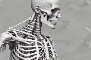
- Repetitive trauma to a joint can cause a chronic condition.
- Trauma does not have to be severe or catastrophic.
- Chronic or recurrent joint pain is commonly caused this way.
- Any joint can experience this condition.
- It is often seen in the shoulder, hip, knee, and ankle joints of service members.
- Which joints are impacted may depend on one's occupation and recreational activities.
Signs and Symptoms
- Pain is felt in the joint with movement, particularly impact.
- Intraarticular effusion may develop.
- Pain radiates along the joint line.
Focused History
- Enquire about any history of trauma.
- Ask about the nature of occupation and recreational activities.
- Note any loss of range of motion.
- Be mindful of instability or lack of confidence when weight-bearing.
Physical Exam
- There may be a decreased range of motion.
- If decreased in active range of motion, perform passive range of motion to assess for mechanical blocks.
- Palpation along the joint line can reveal tenderness.
- Swelling is occasionally noted.
Tools
- X-rays may help rule out fracture, but they may not be necessary.
Differential diagnoses
- Septic arthritis: Accompanied by fever, erythema, edema, and warmth of the joint. The knee is often held in slight flexion to maximize joint space.
- Bucket handle meniscal tear: prevents full extension of the leg.
- ACL injury: causes effusion of the knee.
- Fracture: Tenderness to palpation over bone.
- Soft tissue injuries: Maximal pain is localized away from the joint line.
Treatment
- PRICE (Protection, Rest, Ice, Compression, Elevation)
- NSAIDs for pain relief
- Topical or oral administration.
- Battlefield Acupuncture (BFA)
- Physical therapy can help.
- Range of motion exercises
- Strengthening the muscular support of the knee.
- “Aquatic therapy” can reduce impact stress on joints.
- If a patient is unable to straighten their leg, consider leg-EVAC/orthopedics consult (this may represent a “mechanical block").
Follow up Actions
- Re-evaluation is needed for increased pain, instability, inability to walk, or inability to fully extend the leg.
- Weight loss, physical therapy, and activity modification are vital for management.
- Chronic osteoarthritis management may be augmented by intraarticular injections of corticosteroids and/or hyaluronic acid.
- Return in 48-72 hours if symptoms worsen.
- Routine review by a preceptor IAW 44-103 is pertinent.
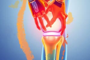
Lower Extremity Strains
- The body has more than 600 muscles that may be injured in various ways.
- Lower extremity strains are very common injuries that can be caused by trauma or overuse
Signs and Symptoms
- Pain is felt over the affected muscle.
- Pain increases with movement.
Focused History
- Ascertain the ability to bear weight if dealing with a lower extremity muscle injury.
- Inquire about activities of daily living (ADL's).
- Take note of numbness or tingling.
Physical Exam
- Palpation at the origin/insertion of the affected muscle reveals tenderness.
- Ecchymosis and swelling may be present.
- Tenderness occurs with AROM, but is reduced or absent with PROM.
- Tenderness is detectable with resisted ROM.
Tools
- X-rays are to be considered if the pain is over bone or if the mechanism suggests a possible fracture.
Differential Diagnosis
- Avulsion fracture: Patient is unable to perform AROM if the tendon is avulsed.
- Osteoarthritis: Chronic recurrent pain localized to a joint, possibly with swelling.
- Osteonecrosis of the associated joint: causes chronic dull ache.
- Bone tumor/cancer: pain at rest or at night, increased pain with weight bearing.
Treatment
- Avoid aggravating activities.
- Gentle stretching throughout ROM should begin once pain has started to resolve.
- Use PRICE (Protection, Rest, Ice, Compression, Elevation).
- NSAIDs.
- Battlefield Acupuncture (BFA).
Follow-up Actions
- Avoid aggravating activities.
- Manage expectations; it may take several weeks to fully resolve.
- Return if pain increases, if numbness or tingling develops, or if the patient is unable to bear weight or perform ADL's.
- Return in 2 weeks.
- If there is no improvement, consider physical therapy consult if available.
- Consider X-rays if available.
- If no improvement is seen after 6 weeks, consider routine EVAC for PCM/orthopedics.
- Routine review by a preceptor IAW 44-103, No BFA
- Battlefield Acupuncture (BFA)
Rhabdomyolysis
- Describes the acute and excessive damage to skeletal muscle.
- Damage results in necrosis and the release of intracellular myoglobin, creatinine kinase, and multiple electrolytes.
- Can be caused by extreme physical exertion, use of dietary supplements, and poor hydration, and is not an uncommon occurrance for military personnel.
- Members with Sickle Cell Disease or Sickle Cell Trait are at higher risk.
- Crush injuries, toxins, compartment syndrome, and prolonged immobilization are all common causes.
- Build up of myoglobin can potentially obstruct the renal tubular system, ultimately leading to renal failure.
Signs and Symptoms
- Severe and proximal muscle pain is usually present.
- Weakness.
- Limb swelling.
- Dark urine.
Focused History
- Consider physical activity.
- Enquire about injuries or trauma.
- Supplement use.
- Illicit drug use
- Recent illnesses.
Physical Exam
- Tenderness upon palpation of muscles.
- Weakness.
- Swelling of affected muscles.
- Evidence of trauma or compartment syndrome.
Labs
- Serum creatinine kinase (CK) will be at least 5 times the upper limit of normal (ULN usually ~200 U/L), often in the 10,000s initially.
- Peaks 24-72 hours after injury.
- Urinalysis (dipstick) exhibits hematuria.
- Includes myoglobin.
- Metabolic panel (monitor Na+, K+, Ca+, Phos, and Creatinine for renal function).
- Liver function tests (AST/ALT are usually elevated).
- Consider CBC to monitor for hemolysis or infection.
- Uric acid levels help to monitor for potential renal injury.
Differential Diagnoses
- Compartment syndrome: Pain is out of proportion (particularly with passive stretching of muscle tissue), woody texture or muscle, weakness, diminished sensation, decreased pulses distally.
- Muscle strain: Muscle pain is not out of proportion and is without lab and exam findings.
- Heat injury: Excessive activity resulting in elevated core body temperature and end organ damage (liver, renal, and muscle).
- Urinary tract infection: Hematuria, but without muscle pain.
Treatment
- Fluids (IV or oral) help to maintain a higher urine output to dilute myoglobin to forestall renal injury.
- CK < 20,000 U/L can often be treated in an outpatient setting with oral hydration.
- Recommend normal saline at 1-2L/hr initially if starting IV fluids.
- An adequate goal for urine output is > 200mL per hour or 1L every 6 hours.
- Monitor electrolytes and renal function frequently given high levels of fluids.
- Rest.
- Bed rest for 24-72 hours for outpatient treatment.
Gradual return to duty
- Phase 1:
- Strict light indoor duty for 72 hours, with encouragement for oral hydration and salting of food.
- No weight training.
- Seven to eight consecutive hours of sleep per night.
- Maintenance in a thermally controlled environment.
- Patients must follow-up in 24-72 hours for repeat CK/UA.
- Phase 2 may begin after the first 72 hours of limited duty if the CK value at 24-72 hours continues to be <5X ULN and UA continues to be normal.
- If CK value at the 24-72 hour follow-up is >5X ULN or UA is positive for blood with no RBC's, the Warfighter is considered a high-risk marker and inpatient versus continued outpatient follow up should be considered.
- If the clinician continues with outpatient management, the Warfighter is to continue on Phase 1 delineated above (Low Risk Rhabdomyolysis Profile) and followed in 24-72 hours with CK, creatinine and UA as per clinical judgment.
- Phase 2 may begin if the CK value is <5X ULN and UA has normalized. Remain in Phase 1 and return every 72 hours for repeat CK/UA until the indicated criteria are met.
- If CK remains >5X ULN or UA is persistently abnormal for 2 weeks after injury or hospitalization, refer for expert consultation.
- Phase 2:
- Light outdoor duty, with no strenuous physical activities.
- Lightweight resistance training.
- Supervised physical activity at own pace and distance (i.e., physical therapy, athletic trainer).
- Follow-up with a caregiver in one week.
- If clinical symptoms subside, begin Phase 3. Otherwise, remain in Phase 2 and revert to 1-week intervals. Progress to Phase 3 when there is no significant muscle weakness, swelling, pain, or soreness. Consider specialty evaluation to include psychiatry, if myalgia persists beyond 4 weeks with no objective findings.
- Phase 3:
- Return to regular outdoor duty and physical training.
- Follow-up with care provider as needed.
Additional Treatment
- Rest is essential.
- Monitor urine output (goal more than 200mL/hr), titrate fluids to reach goal.
- Seek medical evaluation with increasing pain, chest pain, shortness of breath, palpitations, and significantly increasing edema.
Follow up Actions
- Return in 24 hours for reevaluation and labs if outpatient treatment. Contact preceptor immediately, which may lead to a transfer or evacuation that depends on severity.
Compartment Syndrome
- Skeletal muscles are surrounded by connective tissue called fascia.
- The surrounding fascia is strong and non-compliant, separating muscles into sections or compartments
- The pressure in a compartment can rise if excessive fluid builds, possibly resulting in tissue/muscle damage and compromising circulation distal to the edema.
- Causes of trauma: crush injuries, penetrating injuries, long bone fractures, construction from circumferential burns, construction bandages or splints, bites and stings.
- The forearm and lower leg are frequent sites given their well defined compartments.
- Foot, thigh, and gluteal region may occur.
- Compartment syndrome can occur inside the abdomen as well when patients ar given large volumes of IV fluids.
- Both the pathophysiology and management are similar to compartment syndrome of the extremities.
- Compartment syndrome can occur inside the abdomen as well when patients ar given large volumes of IV fluids.
- Compartment syndrome can be intermittent (exercise induced) and mandates measurement of intracompartmental pressures during exercise to diagnose
Signs and Symptoms
- Pain is out of proportion
- Swelling
- Distally Decreased pulses
- Late signs are decreased sensation and paralysis
Focused History
- Recent trauma
- Recent physical activity
Physical Exam
- Pain with passive stretch of the muscle group
- Tense woody texture or the affected muscle group
- Edema or Swelling of limb
- Weakness
- Distal to the injury, decreased pulses
- Distal to the injury, diminished sensation
- Check for circumferential burns as they may act as a tourniquet
Tools
- Measurement of Intracompartmental pressures requires the services of Orthopedics.
- Labs are NOT necessary for diagnosis, but CK, Metabolic panel, LFTs, UA, and Uric acid may be requested as for Rhabdomyolysis.
Differential Diagnosis
- Rhabdomyolysis: Muscle pain and swelling without elevated intracompartmental pressures.
- Muscle strain: Muscle pain although not disproportionate and without addition lab and exam findings.
- Heat Injury can cause liver, renal, and muscle organ damage as a result of excessive activity that causes elevated body temperature.
Treatment
- Requires that the pressure be released.
- Fasciotomy requires the services of a trained provider.
- Pain management and risks for renal and hepatic toxicity has to be assessed.
- Fluids which could be considered include injuries to the muscle, and therefore potential rhabdomyolysis.
Hospital Care and Follow up
- Hospital admission and close monitoring are necessary, as fasciotomies should remain covered with frequently changed wet sterile dressings.
- A follow up action will be scheduled after hospital discharge.
- The patient may need skin grafts in order to cover fasciotomies.
Red Directive:
- Immediate Evacuation and contact the preceptor ASAP.
Red Flags
- Trauma
- Duration of pain >6 weeks
- Neurologic deficit (bowel/bladder incontinence, saddle anesthesia, lower extremity weakness)
- Fever
- Severe nocturnal or disabling pain
- Unexplained pain after age 55
- Unexplained weight loss
- Steroid use
Cervical/Thoracic region
- Consists of anterior and posterior ligaments.
- Anterior and posterior ligaments limit motion and provide support
- The region is divided into 5 areas
- Cervical, thoracic, lumbar, sacrum, coccyx
- Sternocleidomastoid (SCM):
- Rotate, forward flex Other head movements:
- Extend, bend, rotate - Splenius capitus - Semispinalis
- Etiology:
- Etiology is often multifactorial with the underlying disorder exacerbated by fatigue, physical deconditioning, muscle pain,poor posture, weakness of stabilizing muscles, decreased flexibility and sometimes psychosocial stress or psychiatric abnormality
- Pain can also be musculoskeletal in nature
- Secondary to poor posture, trauma (like whiplash), stress, tension, r apidly advancing an exercise program, etc
Signs and Symptoms
- Depending on the cause, the pain can manifest as stiffness, muscle tension, spasm, and tenderness
- May be accompanied by neurologic symptoms
- Pain (encourage patient to describe) and limited ROM
- Strength, sensation, and reflexes of the area innervated by that root may be impaired
- Strength, sensation, and reflexes may be impaired at the affected spinal cord level and all levels below, if the spinal cord is affected
Focused History
- OPQRST – Onset, Provocation/Palliation, Quality, Region or Radiation, Severity, Tim
- Presence of numbness, tingling, or weakness
- Assess for red flag symptoms
- History of back or neck problems
- Recent activity
- History of Fever -could indicate meningitis
Physical Exam
- Assess posture
- Limited ROM secondary to pain
- Motor and sensory dysfunction represent radicular symptoms
- Spurling test
- Specific for cervical radiculopathy
- Negative does not rule out radicular pain
Tools
- Most atraumatic neck pain does not require imaging (traumatic does)
Consider imaging: (UpToDate has a good algorithm)
- Progressive neurologic findings
- Constitutional symptoms
- Infectious risk with systemic symptoms
- History of malignancy
- Persistent moderate to severe neck pain lasting >6 weeks and affecting sleep or ADLs
Differential
- Shoulder problems: pain may be radiating from shoulder
- Peripheral nerve entrapment (CTS): positive Phalen, Tinel at elbow or wrist (pain radiating proximal to distal)
Cervical radiculopathy:
- Numbness or tingling in the UE
- Cervical sprain/strain: neck pain without associated symptoms
Treatment
- Rest, ice or heat (whichever feels better)
- Gentle ROM stretching
- NSAIDs
- if certified in Battlefield Acupuncture (BFA) and with preceptor concurrence, consider administering up to 10 treatment sessions as an alternative to NSAIDS as long as there is clinical benefit and no worsening of the patient's condition
- Muscle relaxants if the most likely diagnosis is spasm
- Cyclobenzaprine with preceptor guidance -Methocarbamol
- Avoid narcotics/steroids
Follow-up Actions
- Avoid aggravating activities
- Gentle stretching throughout ROM
- Modify posture
- Follow-up Actions:
- Return in 2 weeks if no improvement
- Consider physical therapy If gross motor weakness is present, then EVAC or transfer
- Blue Directive: Contact Preceptor immediately for approval if considering Battlefield Acupuncture (BFA)
- Green Directive: Routine review by preceptor IAW 44-103, No BFA
Lower Back Pain (LBP)
- is among the most common medical complaints although it is a symptom, not a disease
- The exact cause is often difficult to diagnose and is often multifactorial
- Because of the multifactorial nature, clinicians should determine whether pain has a spinal or extra spinal cause and whether the cause is a serious disorder
Upper/Lower Back Pain Symptoms
- Pain may occur in the lower back with or without ROM. Acute pain (pain may last <4 weeks)
- Subacute Pain lasts for 4-12 weeks
- Chronic pain lasts longer than 12 weeks
- There also may be redicular symptoms and radiate into the buttock region.
- Standing may prove difficult.
Upper/Lower Back Pain Focused History
- History of injury.
- PMHx of back pain.
- Numbness or tingling in LE.
- Difficulty controlling bowel or bladder function.
- Radiation of pain.
Upper/Lower Back Pain Physical Exam
- Tenderness of the lumbar paraspinal muscles, muscle spasms in the lumbar paraspinal, are palpable and often tender. -ROM is frequently limited due to pain, so it's important to differentiate this cause of limitation from a neurological issue.
- Perform a straight leg raise, it will be negative if there is no sciatic nerve involvement with lumbar strain -Motor and sensory deficits with a more ominous problem.
Tools
- If no “red flag” symptoms, X-rays are not appropriate unless the pain has been present for greater than 6 weeks.
Differential Diagnosis
- Cauda Equina Syndrome: This is evidenced by paralysis and bowel/bladder dysfunction.
- Disk herniation: Exhibits unilateral redicular symptoms that extend below the knee.
- Infection: Indicated by fever, chills, and night sweats.
- Consider Causes outside the spine such as: constipation, pancreatitis, kidney stones
- Fracture: Important to note the mechanism of injury
Treatment (if no red flag)
- Follow these symptoms, treat the patient rest, ice, and or heat (whichever feels better)
- Gentle ROM stretching
- NSAIDs
- Battlefield acupuncture
- Muscle relaxants
- Do not provide narcotics/steroids
If red flag symptoms or gross motor weakness is present: - EVAC or transfer immediately
Lower back pain Treatment
- It is acute and will resolve on its own within a few days with self-care and there is no residual loss of function
- Some cases may require months for symptoms to resolve.
- National Institute of Neurological Disorders and Stroke
Follow-up Actions:
- Return in 2 weeks if no improvement.
- Recommended physical therapy if available.
- If physical therapy unavailable continue stretching and or consider changing muscle relaxants or NSAIDs. Re-evaluate after 6 weeks if pain persists despite following conservative management.
Lower Limb disk herniation
- Prolapse of an intervertebral disk through a tear in the surrounding annulus fibrosus
- The tear causes pain when the disk impinges on an adjacent nerve root, a segmental radiculopathy with paresthesias and a weakness in the distribution of the affected root results
- C6 and C7 are most commonly affected in the cervical area
-
80% of disk ruptures affect L5 or S1 nerve roots in the lumbar area
Etiology
- Common cause is as humans age the nucleus Pulposus central part of the disk becomes less hydrated and weakens
- 2nd most common trauma.
- Other congenital tissue disorders connect it to such.
- Likely to occur posterolaterally and therefore more likely to compress the nerve root.
- (The symptomology associated with lumbar disk herniation is commonly referred to as Sciatica.) NCBI StatPearls 11/2020)
Lower Limb disk herniation - Signs and Symptoms
-
Back Pain
-
Potential for Nerve root compression (weakness in UE/LE).
-
Sciatica.
-
Pain worsening from sitting, coughing, sneezing, raising Arms over your head.
-
Focused History: Assess for Red flags and or possible injury
disk herniation - Physical Exam
- Tenderness to palpation at paraspinal Muscles
- Possible muscle spasm in the paraspinal
- ROM (ROM is in pain)
- Straight leg raise tests can provide information regarding sciatic nerve involvement
- Red flag motor and or sensory deficits
Tools
- No “Red Flags” symptoms means X-rays will not be appropriate unless symptoms last greater than 6 weeks
- https://www.youtube.com/watch?v=GfNLPgbnpAg
disk herniation - Differential Diagnosis
- Cauda Equina Syndrome: Evidence of bowel and or bladder disfunction
- Trochanteric bursitis pain: down lateral thigh, Exquisite tenderness to palpation over greater trochanter
- Fever: Chills and night sweats
- Outside of the spine such as Constipation and pancreatitis + kidney stones Fracture: Note mechanism of injury
disk herniation - Treatment: if no "red flags" symptoms
- Treat those symptoms
- Encourage rest ice or use heat, encourage gentle movements/Stretching 🧘♀️ Follow-up actions after 24 hours
Cauda Equina Syndrome
- Cauda Equina Syndrome occurs when the nerve roots at the caudal end of the cord, become compressed or damaged this causes disruption to the motor and sensory pathways to the Lower body with eventual bladder issues
- Most commonly this happens because of herniated disc in the lumbar section of spinal cord
- Congenital anomalies spinal cord infection spinal epidural abscess tumor or trauma it can occur from stenosis as well
- Although this is classified as very rear medical event it is emergent and is potentially caused by several back pain patients
- Symptoms can include but need not be limited to*
- Distal leg and or any type of leg weakness and also loss or change in both muscle Sensory and muscle responses
- Loss of Feeling of needing to defecate or urinate -Dysfuctionial bowels (late sign)
- Incontinence (late sign): Erection's or erections is a sign you may need immediate attention Loss over sensory or muscle feedback
CAUDA Equina -Focused history
-Check for all previous listed symptoms but also add: Recent incident or injury
History of spinal stenosis or herniated disc
- Ask the patient and inquire: Is there any bowel/bladder following symptoms?
Physical exam
   - Low back exam
   - Note: any loss of rectal tone
   - Unable to move from chair
   - Inability to feel or move toes
       - Check neurology and or sensory dysfunction and for any possible issues related to the Legs
Tools for Checkup
- If X-rays are available, lumbar films are May be appropriate if EVAC or transfer but have limited benefit
Differential exams
- Look for: intact reflexes in legs because its a very complex issue which requires immediate expert atnntion and consultation
-
- If suspicious:**
- Immediate transfer -Consider Foley Catheter -And to encourage and administer support well awaiting transportation
Shoulder Impingement
- Shoulder dislocation / Instability
- Adhesive capsulitis
- AC joint separation
- Brachial plexus injury
- Biceps tender rupture
The humerus will not be centered on the glenohumeral joint
AC (Separation of the Acromioclavicular Ligaments)
- AC: acromioclavicular
- Known as a shoulder separation this injury occurs most frequently when the patient has fallen on the shoulder will less if they have out stretched hand
- Severe strains tear the AC and coracoclavicular ligaments which can displace the distal clavicle superiorly or inferiorly from the acromion
Symptoms for AC joint and/or Separation
Symptoms include pain at the AC joint from range of motion of the shoulder as well as swelling that can be caused depending on the severity of injury
- Focused history:* Fall on shoulder from outstretched hand Hands domination and occupation
Physical exam :
Perform palpation to the a-c joint but look at the shoulders to confirm or examine any other injuries ( SC joint to clavicle as well as humerus) and then reduced the range of motion in the shoulder • Tools;Bilateral Calvi
- Use tools: Bilateral Clavicle X-ray if available, it May to be helpful in finding the grade of the sprain -Use the tools to determine what the symptoms are (Active range of motion to passive range motion)
- Tendenitis : look for history and or injuries • Shoulder and or joint dislocated or great deltoid range pain and can not perform
- Differential*: Range of motion will be reduced and passive movement be full/
- Treatment*
- Sling for7-10 days
- Gentle stretching * -PRICE NSAIDS and or BATTLEFIELD ACUPUNCTURE:
- if symptoms are showing you need to treat as if neuroCompromise or orthopedic as mentioned
Shoulder impingement:
- In the shoulder the ligaments from the deltoid to a-c to humerus there are often mechanisms of injury *
- Symptoms:* -Symptoms such as pain and loss of motion are all side effects of shoulder issues -Nocturnal pain with muscle fatigue
- Focused History: Note the patients' hand Dominance or injuries or sports
- *Physical exam: possible muscle waste; and possible inflammation
Tools
- Tools do need to be used unless there appears to be trauma
- Differentials; trauma and biceps injury
- If the goal is the gentle process of restoration you need to also protect the area by following the symptoms
Use the listed treatments; - The first seven to ten days after the injury you may begin stretching you can Ask them review a PT before you go
- PRICE the affected pain with non-steroidal and Battlefield -Acupuncture and or any other listed options
If needed and or if pain continues evaluate by calling the Preceptor for extra procedures / tests, or or to discuss orthopedic’s
Studying That Suits You
Use AI to generate personalized quizzes and flashcards to suit your learning preferences.