Podcast
Questions and Answers
What is the MOST accurate description of protein conformation?
What is the MOST accurate description of protein conformation?
- The linear sequence of amino acids linked by glycosidic bonds.
- The process of permanently unfolding a polypeptide chain.
- The disruption of a protein's native state due to covalent bond breakage.
- The spatial arrangement of atoms dependent on bond rotation. (correct)
Which characteristic is associated with fibrous proteins?
Which characteristic is associated with fibrous proteins?
- Dynamic flexibility allowing for hinge-like movements.
- High enzymatic activity due to a defined active site.
- Spherical shape that is soluble in aqueous environments.
- Structural rigidity, often found in structural proteins. (correct)
What determines the overall shape of a folded polypeptide?
What determines the overall shape of a folded polypeptide?
- The quantity of alpha helices present.
- The presence of motifs and domains.
- The location of beta sheets.
- The amino acid sequence. (correct)
Which statement accurately describes the role of hydrogen bonds in protein secondary structure?
Which statement accurately describes the role of hydrogen bonds in protein secondary structure?
What is required to determine a protein's primary structure?
What is required to determine a protein's primary structure?
What is a limitation of using Nuclear Magnetic Resonance (NMR) to determine protein structure?
What is a limitation of using Nuclear Magnetic Resonance (NMR) to determine protein structure?
What characteristic of alpha helices was discovered by Linus Pauling?
What characteristic of alpha helices was discovered by Linus Pauling?
Which amino acid residue would MOST likely destabilize an alpha helix within a protein?
Which amino acid residue would MOST likely destabilize an alpha helix within a protein?
Where are amphipathic helices commonly located in globular proteins that exist in aqueous solutions?
Where are amphipathic helices commonly located in globular proteins that exist in aqueous solutions?
What stabilizes beta sheets?
What stabilizes beta sheets?
In beta sheets, where do the side chains of the amino acids point?
In beta sheets, where do the side chains of the amino acids point?
What is a characteristic of protein motifs?
What is a characteristic of protein motifs?
What primarily stabilizes the tertiary structure of a globular protein?
What primarily stabilizes the tertiary structure of a globular protein?
Which statement BEST describes protein domains?
Which statement BEST describes protein domains?
What is the defining characteristic of a protein with quaternary structure?
What is the defining characteristic of a protein with quaternary structure?
What is the role of 'chaperones' in protein folding?
What is the role of 'chaperones' in protein folding?
When does a protein begin to fold?
When does a protein begin to fold?
Which action DOES NOT cause protein denaturation?
Which action DOES NOT cause protein denaturation?
What accounts for the tensile strength of collagen?
What accounts for the tensile strength of collagen?
What is a unique aspect of collagen's triple helix?
What is a unique aspect of collagen's triple helix?
What is required by enzymes that catalyze the hydroxylation of proline and lysine in collagen?
What is required by enzymes that catalyze the hydroxylation of proline and lysine in collagen?
What is the function of myoglobin?
What is the function of myoglobin?
Why does hemoglobin have a higher oxygen-binding capacity than myoglobin?
Why does hemoglobin have a higher oxygen-binding capacity than myoglobin?
What would be the effect of an increased concentration of 2,3-bisphospho-D-glycerate (BPG) concentration?
What would be the effect of an increased concentration of 2,3-bisphospho-D-glycerate (BPG) concentration?
Under physiological conditions, what promotes oxygen release from hemoglobin?
Under physiological conditions, what promotes oxygen release from hemoglobin?
Which level of protein structure is characterized by the linear sequence of amino acids?
Which level of protein structure is characterized by the linear sequence of amino acids?
What is the MAIN difference between globular and fibrous proteins?
What is the MAIN difference between globular and fibrous proteins?
Which of these methods would be MOST appropriate for determining the three-dimensional structure of a large protein complex in solution?
Which of these methods would be MOST appropriate for determining the three-dimensional structure of a large protein complex in solution?
What interaction is responsible for maintaining the alpha-helical structure of proteins.
What interaction is responsible for maintaining the alpha-helical structure of proteins.
Which characteristic distinguishes parallel and antiparallel beta-sheets?
Which characteristic distinguishes parallel and antiparallel beta-sheets?
What defines a protein domain?
What defines a protein domain?
Which force is LEAST important to stabilizing quaternary structures?
Which force is LEAST important to stabilizing quaternary structures?
What is the MOST important aspect of the 'cooperative folding' model of protein folding?
What is the MOST important aspect of the 'cooperative folding' model of protein folding?
In protein denaturation, what level of protein structure is MOST directly affected?
In protein denaturation, what level of protein structure is MOST directly affected?
What is the MAIN structural feature of collagen?
What is the MAIN structural feature of collagen?
What is the role of inter-chain hydrogen bonds in the collagen helix?
What is the role of inter-chain hydrogen bonds in the collagen helix?
What is a consequence of vitamin C deficiency on collagen structure?
What is a consequence of vitamin C deficiency on collagen structure?
What is the structure of myoglobin?
What is the structure of myoglobin?
What is the MAIN function of the heme prosthetic group in both myoglobin and hemoglobin?
What is the MAIN function of the heme prosthetic group in both myoglobin and hemoglobin?
Which factor contributes to myoglobin's ability to bind oxygen more tightly than hemoglobin at low oxygen concentrations?
Which factor contributes to myoglobin's ability to bind oxygen more tightly than hemoglobin at low oxygen concentrations?
During the binding of oxygen to hemoglobin, what conformational change promotes increased oxygen affinity.
During the binding of oxygen to hemoglobin, what conformational change promotes increased oxygen affinity.
Increased amounts of carbon dioxide and a decrease in pH facilitates what?
Increased amounts of carbon dioxide and a decrease in pH facilitates what?
Which statement about protein structure is NOT correct?
Which statement about protein structure is NOT correct?
Flashcards
What is conformation?
What is conformation?
Spatial arrangement of atoms that depend on bond rotation, not breaking covalent bonds.
What is native conformation?
What is native conformation?
The single, stable shape a protein assumes under physiological conditions.
Fibrous vs Globular Proteins
Fibrous vs Globular Proteins
Proteins are classified into two types depending on their structure.
Primary Structure
Primary Structure
Signup and view all the flashcards
Secondary Structure
Secondary Structure
Signup and view all the flashcards
Tertiary Structure
Tertiary Structure
Signup and view all the flashcards
Quaternary Structure
Quaternary Structure
Signup and view all the flashcards
What is X-ray crystallography?
What is X-ray crystallography?
Signup and view all the flashcards
What is NMR spectroscopy?
What is NMR spectroscopy?
Signup and view all the flashcards
What are Secondary Structures?
What are Secondary Structures?
Signup and view all the flashcards
What is the alpha helix?
What is the alpha helix?
Signup and view all the flashcards
Alpha helix hydrogen bonds
Alpha helix hydrogen bonds
Signup and view all the flashcards
What is alanine's role in helix?
What is alanine's role in helix?
Signup and view all the flashcards
What are beta sheets?
What are beta sheets?
Signup and view all the flashcards
What are beta barrels?
What are beta barrels?
Signup and view all the flashcards
What are random coils/loops?
What are random coils/loops?
Signup and view all the flashcards
What are motifs?
What are motifs?
Signup and view all the flashcards
Non-covalent interactions
Non-covalent interactions
Signup and view all the flashcards
What are protein domains?
What are protein domains?
Signup and view all the flashcards
What is quaternary structure?
What is quaternary structure?
Signup and view all the flashcards
Why quaternary structure?
Why quaternary structure?
Signup and view all the flashcards
What is protein folding?
What is protein folding?
Signup and view all the flashcards
What is denaturation?
What is denaturation?
Signup and view all the flashcards
Methods of protein denaturation
Methods of protein denaturation
Signup and view all the flashcards
What confirms protein folding?
What confirms protein folding?
Signup and view all the flashcards
What is collagen?
What is collagen?
Signup and view all the flashcards
What are Glycine, Proline & Hydroxyproline?
What are Glycine, Proline & Hydroxyproline?
Signup and view all the flashcards
Covalent Cross-Linking
Covalent Cross-Linking
Signup and view all the flashcards
Inter-chain hydrogen bonds
Inter-chain hydrogen bonds
Signup and view all the flashcards
What is Vitamin C?
What is Vitamin C?
Signup and view all the flashcards
What does Myoglobin do?
What does Myoglobin do?
Signup and view all the flashcards
What is a prosthetic group?
What is a prosthetic group?
Signup and view all the flashcards
What does prosthetic group of Heme need to be stabilized as?
What does prosthetic group of Heme need to be stabilized as?
Signup and view all the flashcards
What is Hemoglobin
What is Hemoglobin
Signup and view all the flashcards
What is Quaternary Structure of Hemoglobin?
What is Quaternary Structure of Hemoglobin?
Signup and view all the flashcards
What is positive cooperative binding?
What is positive cooperative binding?
Signup and view all the flashcards
What does Deoxyhemoglobin have?
What does Deoxyhemoglobin have?
Signup and view all the flashcards
Oxyhemoglobin
Oxyhemoglobin
Signup and view all the flashcards
Half saturation affinity
Half saturation affinity
Signup and view all the flashcards
What is 2,3-bisphospho-D-glycerate (BPG)
What is 2,3-bisphospho-D-glycerate (BPG)
Signup and view all the flashcards
What does hemoglobin do for O2
What does hemoglobin do for O2
Signup and view all the flashcards
Study Notes
Introduction to Proteins
- Proteins have varied functions arising from key structural features.
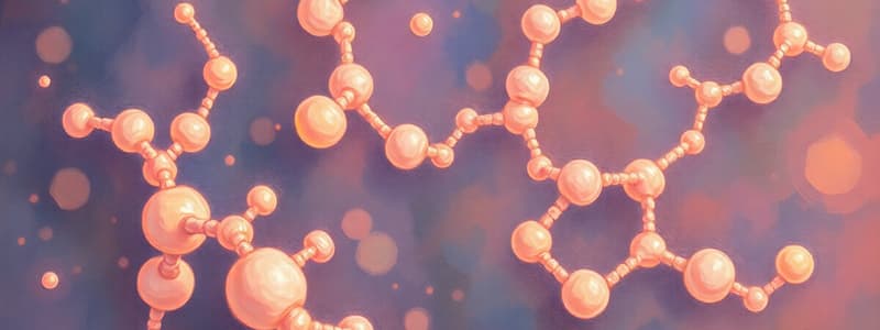
Linear Polymers of Amino Acids
- An unlimited order of amino acids allows for a vast variety of proteins
- Each protein is a biologically functional, unique sequence of amino acid residues folded into a unique and stable 3D structure
- These amino acids are linked by stable peptide bonds, and peptide bonds can last thousands of years without an enzyme
Functional Groups in Proteins
- Proteins contain a wide range of functional groups, including hydroxides, thiols, thioesters, carboxylic acids, amines, and phosphates
- The reactivity of these functional groups accounts for enzyme activity
Protein Interactions and Assemblies
- Proteins interact with each other and other biological molecules to form complex assemblies
- Molecular cooperation leads to molecular synergism
Polypeptide Chains
- A protein may consist of a single polypeptide chain or many chains
Conformation Importance
- 3D shape is critical to the biological processes of proteins
- The spatial arrangement of atoms that depends on rotation of bonds defines protein conformation
- Conformation does not break covalent bonds
- Polypeptides have numerous possible conformations.
- Native conformation is the single, stable shape a protein assumes under physiological conditions.
- A protein's biological function depends completely on its conformation
Genome Encoding
- The number of polypeptides encoded in a genome can be determined through genomic sequencing
- E. coli contains approximately 4,000 polypeptides
- D. melanogaster contains approximately 14,000 polypeptides.
- H. sapiens contains roughly 20,000 different polypeptides.
- Proteomics is the study of the entire complement of proteins in a cell
Protein Rigidity and Flexibility
- Some proteins are rigid, while others exhibit limited flexibility
- Rigid proteins are often structural proteins
- Flexibility is useful in making hinges, springs, and levers
Globular vs. Fibrous Proteins
- Proteins can be classified as either globular or fibrous, based on their structure
- Fibrinogen is a fibrous protein
- Superoxide dismutase exemplifies a globular protein
Primary Structure
- This is the linear sequence of amino acids held together by covalent forces (peptide bonds).
- The primary structure determines the overall shape of folded polypeptides and influences secondary, tertiary, and quaternary structures
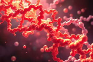
Secondary Structure
- This element involves local regions with repeating conformations like alpha helices, beta sheets, and loops
- Secondary structures are maintained by hydrogen bonds (H-bonds) between amide hydrogens and carbonyl oxygen
- Examples include alpha helix and beta sheets
Tertiary structure
- The completely folded and compacted polypeptide chain represents the tertiary structure.
- It is stabilized by the interactions of side chains of non-neighboring amino acid residues
Quaternary Structure
- This arrangement involves the association of two or more polypeptide chains into a multi-subunit protein
Protein Sequencing
- Primary structure is determined through DNA or sequencing
Determining 3-D Structure
- Elucidating 3-D native conformation is crucial for understanding protein function
- X-ray crystallography and nuclear magnetic resonance (NMR) imaging can determine 3-D conformation
X-Ray Crystallography Process
- A beam of parallel X-rays is directed at a protein crystal
- Electrons in the crystal diffract the X-rays
- The diffraction of X-rays by electrons is recorded on film or by an electronic detector
- Mathematical analysis of the diffraction pattern, along with knowledge of the primary structure, is used to deduce 3-D conformation
- The development of computers has aided this process
Max Perutz
- Determined the structure of hemoglobin
John Kendrew
- Determined the structure of myoglobin
Nobel Prize
- Perutz and Kendrew shared the Nobel Prize in 1962
Pros of X-Ray Crystallography
- Proteins retain their native conformation in crystal form
- Crystals contain 20-50% water molecules
- Researchers can add ligands, which diffuse into active sites
- Crystals retain catalytic activities, which shows the crystal represents active, in vivo conformation
Cons of X-Ray Crystallography
- Requires preparation of protein crystals
- It can be difficult and time-consuming
- Some proteins do not easily adopt a crystalline form
NMR Spectroscopy
- A sample of protein is placed in a magnetic field
- Electromagnetic radiation is applied, and certain atomic nuclei absorb it as the applied magnetic field varies
- Absorbance reveals interactions of atoms that are close together
- Data is combined with knowledge of the amino acid (aa) sequence to calculate a number of structures that satisfy the observed interactions
Bovine Ribonuclease A
- An NMR structure
Pros of NMR Spectroscopy
- Permits study of proteins in solution
- Does not require preparations of crystals
Cons of NMR Spectroscopy
- Better suited for use for small proteins
- Difficult to determine the structures of large, complex proteins
Discovery of the Alpha Helix
- Discovered by Linus Pauling in 1950
- This structure is the most frequently observed secondary structure
- The structure is virtually always found in nature as right-handed form
Alpha Helix Details
- An alpha helix generally has ~3.6 (3.5-3.7) residues per turn
- Pitch (advance per turn) is 0.54 nm
- Rise (advance per amino acid residue) is 0.15 nm
Alpha Helix Bonding and Location
- Each carbonyl oxygen (residue n) of the polypeptide backbone is H-bonded to the backbone amide hydrogen of the fourth residue toward the C-terminus (n + 4)
- The first three and last three residues lack H-bonding partners within the helix
- Each hydrogen bond closes a loop of 13 atoms
- Hydrogen bonds are nearly parallel to the long axis of the helix
- All carbonyl groups point toward the C-terminus
- Stabilized by hydrophobic regions.
Side Chain Impact
- The side chains of amino acids in an alpha helix point outwards from the cylinder of the helix
- Alanine, with its small, uncharged side chain, fits well within the helix
- It is often found in helices of globular proteins
- Tyrosine and asparagine are less common due to their bulky R-groups
- Glycine destabilizes helices because rotation around the alpha-carbon is unconstrained, disrupting the normal structure/rigidity of the peptide backbone
- Proline is never found in the interior of alpha-helices, due to the rigid cyclic side chain causing steric interference
- Proline also lacks amide hydrogen for H-bonding with carbonyl oxygen of other amino acids in the helix.
Termination
- Alpha helices usually end with structures known as the helix stop signal (or capping box)
- The helix stop signal is a specific pattern of amino acid residues at the beginning of an alpha helix
- The side chain of N-terminal residue (n) of helix interacts with backbone amide hydrogen of the fourth residue of helix, resulting in a conformation that is not compatible with formation of alpha helix
- Serine and threonine are usually found at initial positioning of the capping box, and glutamate is often the fourth residue
Alpha Helix Content
- Globular proteins vary in their alpha-helical content
- Myoglobin contains 75% alpha-helices
- Chymotrypsin has very little
- Average for globular proteins is ~26%
Amphipathic Helices
- These structures have hydrophilic amino acids on one face of the helix cylinder and hydrophobic amino acids on the opposite face
- They are often located on the surface of globular proteins
Beta Structure
- Portions of the polypeptide chain are almost fully extended in the beta structure
- It includes beta strands and beta sheets
- Beta sheets consist of multiple beta strands arranged in sheets
- Stabilized by H-bonding between carbonyl oxygens and amide hydrogens on adjacent beta strands (of same or different polypeptide)
Parallel vs. Antiparallel Beta Strands
- In beta sheets, side chains point alternately above and below the plane of the sheet
- Parallel strands are less stable because of disordered H-bonds
- Beta sheets may contain 2-15 strands and can form barrel-shaped structures
Beta-Sandwich
- Two antiparallel sheets stacked against each other
- One side of the sheet is hydrophilic, while the other is hydrophobic
- These sheets come together so that hydrophobic regions face each other in the center of the protein
- Hydrophilic regions face the surface
Random Coils/Loops and Turns
- These regions are non-repetitive, but are actually stable, highly-ordered structures
- They connect secondary structures and provide directional changes necessary for proper folding
- Examples include loops, turns, and hairpin loops
- Globular proteins can contain as many residues in non-repetitive conformations as in helices and strands
- The least conserved areas of protein e.g. in the variable domains of the antibody molecule that bind to antigens
Motifs
- Supersecondary structures
- Combinations of alpha helices and beta strands that appear in a number of different proteins can be recognized
- These motifs sometimes have a conserved function between different proteins
Tertiary Structure
- This structure results from the folding of a polypeptide into a closely packed, spherical shape
- Non-covalent interactions (mostly hydrophobic effects) between amino acid and side chains primarily stabilize the whole structure
- There is great variety in tertiary structures
Protein Domains
- Many globular proteins are comprised of independently folded globular units called domains.
- Combinations of motifs makeup domains, and these range in size from 30-300 amino acids
- Small proteins usually contain one domain
- Larger proteins may contain more than one domain
- Often each domain has a function
- In multifunctional enzymes, each catalytic activity is associated with a different domain
- Regions between domains form crevices, which sometimes serve as binding sites
- Some domain structures occur in many different proteins
Quaternary Structure
- This structure is limited to proteins with multiple subunits
- Each subunit is called a monomer
- A multisubunit protein is called an oligomer
- Subunits in an oligomeric protein always have a definite stoichiometry
- Monomers may be identical or different
- Dimers and tetramers are the most common when monomers are identical
- When the monomers differ, each subunit has a unique function
- When several metabolic reactions are catalyzed by an oligomer, it is called a multienzyme complex
- Oligomer subunits are usually held together by noncovalent forces - hydrophobic interactions
Purpose of Quaternary Structure
- More stable than dissociated subunits
- Active sites of some oligomers are formed by amino acids on adjacent polypeptides
- Likelihood of errors is greater for long polypeptides
- It is more efficient to synthesize several smaller subunits
- 3D-structure changes when proteins bind ligands - critical step in biological activity of many proteins
- Different proteins can share the same subunits
- Allows for sequential enzyme action
Protein Folding Process
- As it is translated, the peptide begins to fold
- The first few interactions begin subsequent interactions by assisting in the alignment of groups
- Cooperative folding refers to when the formation of one part of a structure leads to the formation of the remaining parts
- Protein folding and stabilization depend on non-covalent forces - hydrophobic effect, H-bonding, charge-charge interactions, and van der Waals forces
- Formation of disulfide bonds (covalent)
Native Conformation and Stability
- The native conformation of a protein is its most stable shape
- Protein folding is a spontaneous process
- The process is driven by an overall increase in entropy coupled with a decrease in free energy
- Sections of polar backbone that are forced into interior neutralized by H- bonding
Chaperones
- Chaperons assist in folding by ensuring the correct folding pattern
- Some proteins require chaperones in order to fold correctly
- Chaperones prevent incorrectly folded intermediates from arising in the first place
- Chaperone do not "direct" folding; rather, they make sure that the target proetin achieves maximum stability
Denaturation
- Denaturation refers to disruption in the native conformation of a protein, resulting in a loss of biological activity
- Altering very few interactions is sufficient to denature proteins
Protein Denaturation Via Heating
- Increased thermal energy causes more rapid vibrations of molecules
- Heating can break relatively weak hydrogen bonds and hydrophobic interactions
Protein Denaturation Via Changes in pH
- Acids and bases alter the ionization of amino acid side chains
- This alters/disrupts ionic bonds/salt bridges that stabilize protein structure
Protein Denaturation Via Chaotropic agents
- Molecules that can disrupt the hydrogen bonding network between water molecules ultimately disrupting hydrophobic interactions
- These agents compete for H-bonding partners within polypeptide backbones and side chains disrupting H-bonds stabilizing protein structures
Protein Denaturation Via Detergents
- Compete for ionic bonds partners and/or non-polar side chains in proteins
- Disrupts interactions that stabilize protein structures
Protein Denaturation Via Cleavage
- Breaking disulfide bonds using mercaptoethanol, a reducing agent
Ribonuclease
- Scientists have demonstrated that correct disulfide bonds only form after proteins fold into native conformation
Collagen
- A common fibrous protein that is 25-30% of total protein in animals
- Made by connective tissue cells and secreted to the extracellular matrix
- Gives high tensile strength to connective tissue, as in teeth, bones, skin, tendons, ligaments, blood vessels, and cartilage
- There exists a diverse form and function
- The collagen in tendons are stiff, rope-like fibers
- The collagen in skin creates loosely woven fibers that permit expansion in all directions
Collagen Composition
- Consists of 35% Gly, 11% Ala, 21% Pro & Hyp (hydroxyproline)
- Its primary structure consists of repeating tripeptide Gly-X-Y with e.g. Gly-Pro-Hyp
- Collagen fibrils consist of 3 left- handed helical polypeptides (alpha chains) wrapped into a right-handed triple helix (superhelix or tropocollagen)
- They are arranged head-to-head in parallel bundles.
Tropocollagen
- Covalent cross-links between molecules of tropocollagen are responsible for that rigidity and tensile strength of collagen fibers
Collagen Structure
- Collagen triple helix is stabilized by inter-chain H-bonds
- Glycine residues in each form H-bonds with X residue of adjacent chain
- No intrachain H-bonds in collagen helix
Vitamin C
- Hydroxylation of proline and lysine catalyzed by enzymes often require ascorbic acid (vitamin C)
- Vitamin C deficiency results in improper formation of collagen resulting in scurvy - Skin lesions, bleeding gums, loose teeth, fragile blood vessels, etc.
Myoglobin Function
- Myoglobin functions by selectively and reversibly binding to O2 in muscles
- It is a globular protein that contains a heme prothetic group
- The heme prosthetic group gives source of red color in muscles and blood
- The protein is a member of globin multigene family, which also includes hemoglobins and leghemoglobin from plants
- High concentrations of myoglobin in muscle cells allow organisms to hold their breath for a longer period of time
- Diving mammals such as whales and seals have muscles with particularly high abundance of myoglobin
Myoglobin Structure
- Consists of 8 alpha-helices
- Contains heme prosthetic group
- Protein-bound organic molecule
- Protein molecule activity depends essential
- Heme molecule: complexed to iron (Fe2+)
- Forms a complex with six ligands: 4 N of protoporphyrin ring, His-93, and O2
Heme Prosthetic Group
- Prosthetic group of myoglobin wedged into hydrophobic cleft
- No covalent bonds hold heme group
- Protein structure: essential for the reversible binding of oxygen
- Oxidation of heme prevents reversible interaction with oxygen
Hemoglobin Functions
- Functions by selectively and reversibly binding O2 in erythrocytes
- Transports O2 in the blood from gas-exchange organs to tissues
- Has 300 million/erythrocyte and is globular/tetrameric
- Is oligomeric A2B2 , with alpha and beta chains that are similar and each have an identical heme prosthetic group
- Unique oxygen-binding properties compared to myoglobin
- Acts as a dimer of alpha/ beta subunits
Oxygen-Binding and Saturation
- The extent of oxygen binding at equilibrium depends on the concentration of the protein and the concentration of oxygen
- Fraction of molecules that are oxygenated (Y) vs. partial pressure of gaseous oxygen (pO2)
- Y = % saturation
- Oxygen-binding curves help determine the relationship between myoglobin and hemoglobin
Hyperbolic vs Sigmoidal
- Myoglobin has a hyperbolic oxygen binding curve, while hemoglobin has a sigmoidal O2- binding curve
- Hyperbolic curves are indicative of single ligand binding
- Sigmoidal is indicative of more than ligand binding
- Each tetramer can bind up to 4 O2, 1 per heme group
Cooperative Binding
- Shape of curve suggests that binding of first O2 facilitates binding of O2 to other heme molecules
- The oxygen affinity of hemoglobin increases as each O2 is bound
- Illustrates positive cooperative binding
Hemoglobin States
- Cooperative binding is related to changes in protein confirmation
- Tense conformation- Deoxyhemoglobin/Low oxygen affinity
- Movement disrupts ion pairs within the protein
- Favors conformation with higher oxygen affinity
- RelaxedConformation-Oxyhemoglobin/High oxygen affinity
Oxygen Affinity
- The partial pressure at half-saturation (P50) is the measure of the affinity of the protein for O2
Globin Protein Binding Systems
- Differential affinities for O2 separate myoglobin and hemoglobin in terms of oxygen delivery
- At high po₂ the proteins become saturated
- At low pO2, hemoglobin loses O2
- Myogobin then picks up 02
Allosteric Protein
- Hemoglobin is an allosteric protein that contains 2,3-bisphospho-D-glycerate (BPG)
- BPG is a by-product of glycolysis and is produced by metabolically active tissues
- Binding of BPG to hemoglobin reduces affinity of hemoglobin to O2 by lowering its capacity to bind oxygen
Regulators of O2 Affinity
- Both H+ and CO2 also affects affinity of hemoglobin for O2
- In the Bohr Effect, this system affects the conformation of said protein
Regulation Summary
- An increased amount of H+/CO results in in a lower pH
- Both of these compounds lower the resulting overall affinity
High/Low Metabolism
- High metabolism=Reduced 02 affinity
- Low metabolism= Increased 02 affinity
Studying That Suits You
Use AI to generate personalized quizzes and flashcards to suit your learning preferences.