Podcast
Questions and Answers
What is the primary method for preparing tissue for microscopic examination?
What is the primary method for preparing tissue for microscopic examination?
- Staining and mounting
- Direct observation using light microscopy
- Tissue dissection
- Sectioning, staining, and mounting (correct)
What is the main purpose of staining in histological studies?
What is the main purpose of staining in histological studies?
- Visualizing cells under the microscope
- Enhancing contrast between different tissues and structures (correct)
- Making tissues more flexible for sectioning
- Preserving the structural integrity of tissues
Which of these is NOT a key application of histological studies?
Which of these is NOT a key application of histological studies?
- Diagnosing diseases
- Researching biological processes
- Identifying the chemical composition of cells (correct)
- Evaluating treatment responses
How can histological studies contribute to understanding development and function of organisms?
How can histological studies contribute to understanding development and function of organisms?
Which field benefits most from the application of histological studies?
Which field benefits most from the application of histological studies?
Which of the following is NOT a key characteristic of epithelial tissue?
Which of the following is NOT a key characteristic of epithelial tissue?
What type of epithelial tissue is found lining the alveoli in the lungs?
What type of epithelial tissue is found lining the alveoli in the lungs?
Which of the following connective tissues is specialized for support and flexibility?
Which of the following connective tissues is specialized for support and flexibility?
Which type of muscle tissue is responsible for voluntary movements?
Which type of muscle tissue is responsible for voluntary movements?
What is the primary function of neurons?
What is the primary function of neurons?
Which of the following steps is NOT involved in preparing tissue samples for microscopic analysis?
Which of the following steps is NOT involved in preparing tissue samples for microscopic analysis?
What is the function of dense irregular connective tissue?
What is the function of dense irregular connective tissue?
Which type of gland secretes its products directly into the bloodstream?
Which type of gland secretes its products directly into the bloodstream?
Which of the following best describes the interdependent relationship between cells and the extracellular matrix (ECM)?
Which of the following best describes the interdependent relationship between cells and the extracellular matrix (ECM)?
Why is the preparation of thin tissue sections essential for light microscopy?
Why is the preparation of thin tissue sections essential for light microscopy?
How do advances in other fields contribute to the understanding of histology?
How do advances in other fields contribute to the understanding of histology?
What is the primary focus of histology in the study of tissues?
What is the primary focus of histology in the study of tissues?
How does the extracellular matrix (ECM) influence cellular behavior and function?
How does the extracellular matrix (ECM) influence cellular behavior and function?
Which of the following is a critical consideration when preparing tissue samples for microscopic examination?
Which of the following is a critical consideration when preparing tissue samples for microscopic examination?
What is the role of the extracellular matrix (ECM) in relation to cells within tissues?
What is the role of the extracellular matrix (ECM) in relation to cells within tissues?
How do organs achieve their specific functions through the arrangement of tissues?
How do organs achieve their specific functions through the arrangement of tissues?
What is the fundamental principle behind the use of heavy metals in TEM sample preparation?
What is the fundamental principle behind the use of heavy metals in TEM sample preparation?
In cryofracture and freeze etching, why is the process carried out under a vacuum?
In cryofracture and freeze etching, why is the process carried out under a vacuum?
Why is a thin layer of heavy metal, such as gold, applied to specimens before SEM analysis?
Why is a thin layer of heavy metal, such as gold, applied to specimens before SEM analysis?
How does autoradiography enable the study of dynamic cellular processes, such as protein secretion?
How does autoradiography enable the study of dynamic cellular processes, such as protein secretion?
What is the key advantage of using cell and tissue culture for biological research?
What is the key advantage of using cell and tissue culture for biological research?
In autoradiography, what is the role of silver bromide crystals within the photographic emulsion?
In autoradiography, what is the role of silver bromide crystals within the photographic emulsion?
How does cryofracture specifically aid in the study of membrane structure under TEM?
How does cryofracture specifically aid in the study of membrane structure under TEM?
What is a critical consideration when preparing cells for cell culture to ensure successful growth and adherence?
What is a critical consideration when preparing cells for cell culture to ensure successful growth and adherence?
Why is scanning electron microscopy (SEM) particularly useful for visualizing the three-dimensional structure of specimens?
Why is scanning electron microscopy (SEM) particularly useful for visualizing the three-dimensional structure of specimens?
What specific information can be obtained using autoradiography with a radioactive precursor like tritium-labeled thymidine?
What specific information can be obtained using autoradiography with a radioactive precursor like tritium-labeled thymidine?
Which staining method is best suited for visualizing the distribution of glycogen granules within liver cells?
Which staining method is best suited for visualizing the distribution of glycogen granules within liver cells?
If a tissue sample exhibits reduced cytoplasmic basophilia after being pretreated with ribonuclease, which cellular component was most likely removed?
If a tissue sample exhibits reduced cytoplasmic basophilia after being pretreated with ribonuclease, which cellular component was most likely removed?
In a study of a metabolic disease, which staining technique would be most appropriate for identifying intracellular accumulations of cholesterol and phospholipids?
In a study of a metabolic disease, which staining technique would be most appropriate for identifying intracellular accumulations of cholesterol and phospholipids?
During the preparation of a tissue sample, which of these steps is most critical for preserving lipid-rich structures?
During the preparation of a tissue sample, which of these steps is most critical for preserving lipid-rich structures?
What is the fundamental principle behind fluorescence microscopy that allows for high-contrast imaging of specific cellular components?
What is the fundamental principle behind fluorescence microscopy that allows for high-contrast imaging of specific cellular components?
What is the primary advantage of virtual microscopy over traditional light microscopy for histology education?
What is the primary advantage of virtual microscopy over traditional light microscopy for histology education?
What is the function of the condenser in a bright-field microscope?
What is the function of the condenser in a bright-field microscope?
What is the effect of the eyepiece lens on resolution?
What is the effect of the eyepiece lens on resolution?
Which of the following describes resolving power?
Which of the following describes resolving power?
In fluorescence microscopy, what property of light is exploited to visualize specific cellular components?
In fluorescence microscopy, what property of light is exploited to visualize specific cellular components?
What does the Feulgen reaction allow a researcher to visualize?
What does the Feulgen reaction allow a researcher to visualize?
Which modification to standard staining procedures is used to identify specific components, such as ECM fibers, in nervous tissue?
Which modification to standard staining procedures is used to identify specific components, such as ECM fibers, in nervous tissue?
What change would improve the clarity of a bright-field microscopic image?
What change would improve the clarity of a bright-field microscopic image?
How does eosin contribute to the visualization of cellular structures, and why is it considered a counterstain?
How does eosin contribute to the visualization of cellular structures, and why is it considered a counterstain?
If the objective lens has a magnifying power of 40x and the ocular lens has a magnifying power of 10x, what is the total magnification of the microscope?
If the objective lens has a magnifying power of 40x and the ocular lens has a magnifying power of 10x, what is the total magnification of the microscope?
Why is it crucial to promptly place tissue samples in fixatives after removal from the body?
Why is it crucial to promptly place tissue samples in fixatives after removal from the body?
What is the primary chemical mechanism by which formalin and glutaraldehyde act as fixatives?
What is the primary chemical mechanism by which formalin and glutaraldehyde act as fixatives?
Why is osmium tetroxide used after glutaraldehyde in electron microscopy sample preparation?
Why is osmium tetroxide used after glutaraldehyde in electron microscopy sample preparation?
What is the purpose of 'clearing' in the context of tissue embedding?
What is the purpose of 'clearing' in the context of tissue embedding?
Why is plastic resin embedding preferred over paraffin embedding for certain histological applications?
Why is plastic resin embedding preferred over paraffin embedding for certain histological applications?
What biophysical principle underlies the staining of tissue sections with dyes?
What biophysical principle underlies the staining of tissue sections with dyes?
Under what circumstances would frozen sections be preferred over fixed tissue sections in histological studies?
Under what circumstances would frozen sections be preferred over fixed tissue sections in histological studies?
What is the purpose of using a cryostat in preparing tissue biopsies for rapid microscopic analysis?
What is the purpose of using a cryostat in preparing tissue biopsies for rapid microscopic analysis?
Which of the following best describes the function of the pinhole aperture in confocal microscopy?
Which of the following best describes the function of the pinhole aperture in confocal microscopy?
In the context of staining, what is the significance of a tissue component being termed 'basophilic'?
In the context of staining, what is the significance of a tissue component being termed 'basophilic'?
Why is vascular perfusion sometimes used to introduce fixatives, particularly in large organs?
Why is vascular perfusion sometimes used to introduce fixatives, particularly in large organs?
What is the principle behind how phase-contrast microscopy enhances the visibility of transparent specimens?
What is the principle behind how phase-contrast microscopy enhances the visibility of transparent specimens?
What considerations must be prioritized when preparing tissue for electron microscopy, compared to light microscopy?
What considerations must be prioritized when preparing tissue for electron microscopy, compared to light microscopy?
In polarizing microscopy, what property of highly organized biological structures allows them to be visualized?
In polarizing microscopy, what property of highly organized biological structures allows them to be visualized?
What is the role of ethanol in the preparation of tissues for embedding?
What is the role of ethanol in the preparation of tissues for embedding?
What is the key advantage of using electron microscopy over light microscopy for studying cellular structures?
What is the key advantage of using electron microscopy over light microscopy for studying cellular structures?
Which of the following best describes how Acridine orange functions as a fluorescent stain?
Which of the following best describes how Acridine orange functions as a fluorescent stain?
How does the process of freezing tissues aid in histochemical studies related to enzyme activity?
How does the process of freezing tissues aid in histochemical studies related to enzyme activity?
Why is the hematoxylin and eosin (H&E) staining method so commonly used in histology?
Why is the hematoxylin and eosin (H&E) staining method so commonly used in histology?
What distinguishes differential interference contrast (DIC) microscopy from standard phase-contrast microscopy?
What distinguishes differential interference contrast (DIC) microscopy from standard phase-contrast microscopy?
What is the function of electromagnetic "lenses" in a transmission electron microscope (TEM)?
What is the function of electromagnetic "lenses" in a transmission electron microscope (TEM)?
Why are biopsies fixed in formalin for processing and microscopic analysis in a pathology laboratory?
Why are biopsies fixed in formalin for processing and microscopic analysis in a pathology laboratory?
Which of the following describes a critical step in preparing samples for transmission electron microscopy (TEM)?
Which of the following describes a critical step in preparing samples for transmission electron microscopy (TEM)?
What does the term "birefringence" refer to in polarizing microscopy?
What does the term "birefringence" refer to in polarizing microscopy?
Why is it important that the point light source, the lens focal point, and the detector’s pinhole aperture are all optically conjugated in confocal microscopy?
Why is it important that the point light source, the lens focal point, and the detector’s pinhole aperture are all optically conjugated in confocal microscopy?
In electron microscopy, what causes some areas of a micrograph to appear darker or more electron-dense compared to others?
In electron microscopy, what causes some areas of a micrograph to appear darker or more electron-dense compared to others?
How do digital cameras enhance the utility of light microscopic methods?
How do digital cameras enhance the utility of light microscopic methods?
Which of the following is a primary advantage of using phase-contrast microscopy in cell culture laboratories?
Which of the following is a primary advantage of using phase-contrast microscopy in cell culture laboratories?
How do compounds like DAPI and Hoechst function in fluorescence microscopy?
How do compounds like DAPI and Hoechst function in fluorescence microscopy?
What is a primary reason for creating "optical sections" at a series of focal planes in confocal microscopy?
What is a primary reason for creating "optical sections" at a series of focal planes in confocal microscopy?
Flashcards
Sectioning
Sectioning
Cutting tissue samples into thin slices using a microtome.
Staining
Staining
Applying dyes to tissue samples to enhance visual contrast under a microscope.
Mounting
Mounting
Attaching stained tissue sections to microscope slides for observation.
Role of Histology
Role of Histology
Signup and view all the flashcards
Importance of Histological Studies
Importance of Histological Studies
Signup and view all the flashcards
Histology
Histology
Signup and view all the flashcards
Four Main Tissue Types
Four Main Tissue Types
Signup and view all the flashcards
Epithelial Tissue
Epithelial Tissue
Signup and view all the flashcards
Connective Tissue
Connective Tissue
Signup and view all the flashcards
Muscular Tissue
Muscular Tissue
Signup and view all the flashcards
Nervous Tissue
Nervous Tissue
Signup and view all the flashcards
Epithelial Classification
Epithelial Classification
Signup and view all the flashcards
Tissue Fixation
Tissue Fixation
Signup and view all the flashcards
What is Histology?
What is Histology?
Signup and view all the flashcards
Tissue Components
Tissue Components
Signup and view all the flashcards
ECM Function
ECM Function
Signup and view all the flashcards
ECM-Cell Interaction
ECM-Cell Interaction
Signup and view all the flashcards
Tissue Specialization
Tissue Specialization
Signup and view all the flashcards
Organ Formation
Organ Formation
Signup and view all the flashcards
Tools of Histology
Tools of Histology
Signup and view all the flashcards
Ideal Microscopic Prep
Ideal Microscopic Prep
Signup and view all the flashcards
Fixation
Fixation
Signup and view all the flashcards
Fixatives
Fixatives
Signup and view all the flashcards
Formalin
Formalin
Signup and view all the flashcards
Glutaraldehyde
Glutaraldehyde
Signup and view all the flashcards
Osmium tetroxide
Osmium tetroxide
Signup and view all the flashcards
Embedding
Embedding
Signup and view all the flashcards
Paraffin
Paraffin
Signup and view all the flashcards
Dehydration (Tissue)
Dehydration (Tissue)
Signup and view all the flashcards
Clearing (Tissue)
Clearing (Tissue)
Signup and view all the flashcards
Microtome
Microtome
Signup and view all the flashcards
Rapid Freezing (Biopsy)
Rapid Freezing (Biopsy)
Signup and view all the flashcards
Cryostat
Cryostat
Signup and view all the flashcards
Basophilic
Basophilic
Signup and view all the flashcards
Acidophilic
Acidophilic
Signup and view all the flashcards
H&E Staining
H&E Staining
Signup and view all the flashcards
Heavy Metal Staining (TEM)
Heavy Metal Staining (TEM)
Signup and view all the flashcards
Cryofracture and Freeze Etching
Cryofracture and Freeze Etching
Signup and view all the flashcards
Scanning Electron Microscopy (SEM)
Scanning Electron Microscopy (SEM)
Signup and view all the flashcards
Autoradiography
Autoradiography
Signup and view all the flashcards
Radiolabeled Metabolites
Radiolabeled Metabolites
Signup and view all the flashcards
Cell & Tissue Culture (in vitro)
Cell & Tissue Culture (in vitro)
Signup and view all the flashcards
Culture Media
Culture Media
Signup and view all the flashcards
Cell Harvesting
Cell Harvesting
Signup and view all the flashcards
Tritium-Labeled Thymidine
Tritium-Labeled Thymidine
Signup and view all the flashcards
Dynamic Events Analysis
Dynamic Events Analysis
Signup and view all the flashcards
Hematoxylin
Hematoxylin
Signup and view all the flashcards
Eosin
Eosin
Signup and view all the flashcards
Periodic Acid-Schiff (PAS)
Periodic Acid-Schiff (PAS)
Signup and view all the flashcards
Feulgen Reaction
Feulgen Reaction
Signup and view all the flashcards
Enzyme Digestion
Enzyme Digestion
Signup and view all the flashcards
Sudan Black
Sudan Black
Signup and view all the flashcards
Metal Impregnation
Metal Impregnation
Signup and view all the flashcards
Bright-Field Microscopy
Bright-Field Microscopy
Signup and view all the flashcards
Condenser (Microscopy)
Condenser (Microscopy)
Signup and view all the flashcards
Objective Lens
Objective Lens
Signup and view all the flashcards
Eyepiece (Ocular Lens)
Eyepiece (Ocular Lens)
Signup and view all the flashcards
Resolving Power
Resolving Power
Signup and view all the flashcards
Virtual Microscopy
Virtual Microscopy
Signup and view all the flashcards
Fluorescence
Fluorescence
Signup and view all the flashcards
Fluorescence Microscopy
Fluorescence Microscopy
Signup and view all the flashcards
Fluorescent Stains
Fluorescent Stains
Signup and view all the flashcards
Acridine Orange
Acridine Orange
Signup and view all the flashcards
DAPI and Hoechst
DAPI and Hoechst
Signup and view all the flashcards
Fluorescent Labeling
Fluorescent Labeling
Signup and view all the flashcards
Immunohistologic Staining
Immunohistologic Staining
Signup and view all the flashcards
Phase-Contrast Microscopy
Phase-Contrast Microscopy
Signup and view all the flashcards
Refractive Index Principle
Refractive Index Principle
Signup and view all the flashcards
Differential Interference Contrast Microscopy
Differential Interference Contrast Microscopy
Signup and view all the flashcards
Confocal Microscopy
Confocal Microscopy
Signup and view all the flashcards
Confocal Arrangement
Confocal Arrangement
Signup and view all the flashcards
Optical Sectioning
Optical Sectioning
Signup and view all the flashcards
Polarizing Microscopy
Polarizing Microscopy
Signup and view all the flashcards
Birefringence
Birefringence
Signup and view all the flashcards
Electron Microscopy
Electron Microscopy
Signup and view all the flashcards
Transmission Electron Microscopy (TEM)
Transmission Electron Microscopy (TEM)
Signup and view all the flashcards
Study Notes
- Histology involves studying tissues and their arrangement in organs, focusing on how cell structure and arrangement optimize organ-specific functions.
- Tissues comprise cells and the extracellular matrix (ECM).
- The ECM supports cells and facilitates nutrient transport and waste removal.
- Cells produce the ECM, and interactions between matrix components and cell surface receptors create a continuum for coordinated function.
- During development, cells and the ECM specialize, forming fundamental tissue types.
- Organs arise from the combination of these tissues, enabling organ and organismal function.
- Histology relies on microscopy and molecular methods due to the small size of cells and matrix components.
- Advances in various fields enhance the understanding of tissue biology.
Preparation of Tissues for Study
- Tissue preparation involves creating thin sections for light microscopy.
- The goal is to preserve tissue structure, although preparation can cause distortions.
Fixation
- Fixation preserves tissue structure and prevents degradation using stabilizing or cross-linking fixatives.
- Tissues are cut into small fragments to facilitate fixative penetration.
- Vascular perfusion can introduce fixatives to improve preservation in large organs.
- Formalin (buffered isotonic 37% formaldehyde solution) is a common fixative for light microscopy.
- Glutaraldehyde, used for electron microscopy, cross-links proteins and reinforces cell and ECM structures.
- Electron microscopy requires careful fixation to preserve ultrastructural detail, often involving glutaraldehyde followed by osmium tetroxide.
Embedding & Sectioning
- Fixed tissues are embedded in a firm material like paraffin or plastic resins for thin sectioning.
- Dehydration with increasing ethanol solutions is required before infiltration with embedding media.
- Clearing replaces ethanol with an organic solvent miscible with both alcohol and the embedding medium, rendering the tissue translucent.
- Paraffin embedding occurs in an oven at 52°-60°C.
- Plastic embedding avoids high temperatures, minimizing tissue distortion.
- The hardened block is trimmed and sectioned using a microtome.
- Paraffin sections are typically 3-10 μm thick for light microscopy.
- Electron microscopy needs sections less than 1 μm thick.
- Sections are placed on glass slides or metal grids for staining and examination.
- Micrometer (μm), nanometer (nm), and angstrom (Å) are spatial units used in microscopy.
Medical Applications
- Biopsies are tissue samples analyzed in pathology laboratories.
- Rapid freezing in liquid nitrogen is used for quick analysis during medical procedures, employing a cryostat.
- Freezing preserves enzyme activity for histochemical studies and is useful for studying lipids.
Staining
- Staining is essential because most cells and extracellular material are colorless.
- Dyes selectively stain tissue components based on their chemical properties.
- Basophilic components (nucleic acids) are anionic and have an affinity for basic dyes.
- Cationic components (proteins) are acidophilic and stain with acidic dyes.
- Basic dyes include toluidine blue, alcian blue, and methylene blue, while hematoxylin behaves like a basic dye.
- Acid dyes include eosin, orange G, and acid fuchsin.
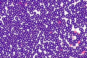
- Hematoxylin and eosin (H&E) staining is commonly used; hematoxylin stains DNA dark blue or purple, and eosin stains cytoplasm and collagen pink.
- Eosin acts as a counterstain.
- Trichrome stains provide greater distinction among extracellular tissue components.
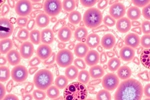
- The periodic acid–Schiff (PAS) reaction stains carbohydrate-rich structures purple or magenta.
- The Feulgen reaction specifically stains DNA.
- Enzyme digestion can further identify basophilic or PAS-positive material.
- Lipid-rich structures are revealed by avoiding heat and organic solvents and using lipid-soluble dyes like Sudan black.
- Metal impregnation techniques, often using silver salts, visualize certain ECM fibers and cellular elements in nervous tissue.
- Slide preparation takes 12 hours to 2½ days, concluding with mounting a coverslip.
Light Microscopy
- Light microscopy includes bright-field microscopy and specialized applications like fluorescence, phase-contrast, confocal, and polarizing microscopy.
- These are based on the interaction of light with tissue components.
Bright-Field Microscopy
- Bright-field microscopy uses ordinary light to examine stained tissue.
- The microscope includes a condenser, objective lens, and eyepiece.
- Total magnification is obtained by multiplying the magnifying power of the objective and ocular lenses.
- Resolving power is the smallest distance between two structures that can be seen as separate objects.
- The maximum resolving power of a light microscope is approximately 0.2 μm.
- Resolving power, image clarity and richness of detail, depends mainly on the quality of the objective lens.
- Virtual microscopy uses high-resolution digital images of stained tissues.
- It allows tissue study on a computer without a physical slide, scanned with a slide-scanning microscope.
Fluorescence Microscopy
- Fluorescence microscopy uses UV light to irradiate tissue sections, causing fluorescent substances to emit light.
- The fluorescent substances appear bright on a dark background.
- Fluorescent compounds with affinity for specific cell macromolecules can be used as fluorescent stains.
- Acridine orange binds both DNA and RNA.
- DAPI and Hoechst specifically bind DNA and stain cell nuclei blue under UV light.
- Fluorescein can be coupled to molecules that specifically bind to certain cellular components.
- Antibodies labeled with fluorescent compounds are extremely important in immunohistologic staining.
Phase-Contrast Microscopy
- Phase-contrast microscopy allows the study of unstained cells and tissue sections.
- It uses a lens system that produces visible images from transparent objects and can be used with living, cultured cells.
- It is based on the principle that light changes its speed when passing through structures with different refractive indices.
- Differential interference contrast microscopy with Nomarski optics produces a 3D image of living cells.
Confocal Microscopy
- Confocal microscopy uses a point of high-intensity light and a pinhole aperture to achieve high resolution and sharp focus.
- It avoids stray light.
- A computer-driven mirror system moves the point of illumination across the specimen.
- Digital images captured at many individual spots in a very thin plane of focus are reconstructed into a 3D image.
Polarizing Microscopy
- Polarizing microscopy identifies structures made of highly organized subunits.
- Tissue structures containing oriented macromolecules rotate the axis of light and appear as bright structures against a dark background.
- Birefringence is the ability to rotate the direction of vibration of polarized light.
- It is a feature of crystalline substances or substances containing highly oriented molecules, such as cellulose, collagen, microtubules, and actin filaments.
- Digital cameras extend the utility of light microscopic methods.
Electron Microscopy
- Transmission and scanning electron microscopes are based on the interaction of tissue components with beams of electrons.
- The wavelength in an electron beam is much shorter than that of light, allowing a 1000-fold increase in resolution.
Transmission Electron Microscopy
- The transmission electron microscope (TEM) has a resolution around 3 nm.
- It uses electromagnetic "lenses" to focus a beam of electrons through the tissue section.
- Regions of an electron micrograph correspond to tissue areas through which electrons passed readily (appearing brighter or electron-lucent) and areas where electrons were absorbed or deflected (appearing darker or more electron-dense).
- Compounds with heavy metal ions, such as osmium tetroxide, lead citrate, and uranyl compounds, improve contrast and resolution.
- Cryofracture and freeze etching allow TEM study of cells without fixation or embedding, useful in the study of membrane structure.
Scanning Electron Microscopy
- Scanning electron microscopy (SEM) provides a high-resolution view of the surfaces of cells, tissues, and organs.
- The beam of electrons does not pass through the specimen.
- The surface of the specimen is spray-coated with a thin layer of heavy metal that reflects electrons.
- Reflected electrons are captured to produce a black-and-white image.
Autoradiography
- Microscopic autoradiography localizes newly synthesized macromolecules in cells or tissue sections.
- Radioactively labeled metabolites are incorporated into specific macromolecules.
- Slides are coated with photographic emulsion containing silver bromide crystals to detect radiation.
- Silver grains indicate the locations of radiolabeled macromolecules in the tissue under the microscope or TEM.
- The radioactive precursor of DNA (such as tritium-labeled thymidine) can show cells in a tissue replicating DNA and preparing to divide.
Cell & Tissue Culture
- Live cells and tissues can be maintained and studied outside the body in culture (in vitro).
- Cell culture allows the direct observation of cellular behavior under a phase-contrast microscope.
- Cells and tissues are grown in complex solutions of known composition with serum or specific growth factors.
- Cells are dispersed and placed in a dish to which they adhere, usually as a single layer.
Studying That Suits You
Use AI to generate personalized quizzes and flashcards to suit your learning preferences.