Podcast
Questions and Answers
What is the primary function of the microscope?
What is the primary function of the microscope?
- To measure the dimensions of cells
- To enhance the color of stains
- To inspect objects too small for the naked eye (correct)
- To magnify objects beyond perception
Which of the following statements about resolution is correct?
Which of the following statements about resolution is correct?
- It is better in light microscopes than in electron microscopes.
- It is irrelevant if magnification is high.
- It is determined by the size of the specimen.
- It allows the distinction of fine details within a specimen. (correct)
What is the resolving power of an electron microscope?
What is the resolving power of an electron microscope?
- 1 nm (correct)
- 1 µm
- 0.2 µm
- 0.2 mm
Which type of microscope provides a three-dimensional image of surfaces?
Which type of microscope provides a three-dimensional image of surfaces?
How does magnification relate to resolution in microscopy?
How does magnification relate to resolution in microscopy?
What distinguishes a transmission electron microscope (TEM) from a scanning electron microscope (SEM)?
What distinguishes a transmission electron microscope (TEM) from a scanning electron microscope (SEM)?
Which property of the light microscope allows it to create clear images?
Which property of the light microscope allows it to create clear images?
What are the two main components of tissues as defined in histology?
What are the two main components of tissues as defined in histology?
What is the main purpose of using Sudan black dye in histological studies?
What is the main purpose of using Sudan black dye in histological studies?
Which type of microscope utilizes a beam of electrons for imaging?
Which type of microscope utilizes a beam of electrons for imaging?
What distinguishes the scanning electron microscope from other types of microscopes?
What distinguishes the scanning electron microscope from other types of microscopes?
Which staining method is primarily used to identify glycogen in histological samples?
Which staining method is primarily used to identify glycogen in histological samples?
What type of lens is characteristic of an electron microscope?
What type of lens is characteristic of an electron microscope?
In histology, why are thin stained tissue sections preferred for light microscopic examination?
In histology, why are thin stained tissue sections preferred for light microscopic examination?
Which statement describes the resolution of a microscope?
Which statement describes the resolution of a microscope?
Which of the following is NOT a type of staining method used in histology?
Which of the following is NOT a type of staining method used in histology?
What is the primary source of illumination used in Light Microscopy (LM)?
What is the primary source of illumination used in Light Microscopy (LM)?
Which of the following is NOT a characteristic of Electron Microscopy (EM)?
Which of the following is NOT a characteristic of Electron Microscopy (EM)?
What is the resolving power of Light Microscopy as compared to Electron Microscopy?
What is the resolving power of Light Microscopy as compared to Electron Microscopy?
Which type of staining method is primarily used for revealing general structure in tissue?
Which type of staining method is primarily used for revealing general structure in tissue?
Which of the following statements about Hematoxylin and Eosin (H&E) staining is false?
Which of the following statements about Hematoxylin and Eosin (H&E) staining is false?
What is the purpose of Sudan black dye in histological staining?
What is the purpose of Sudan black dye in histological staining?
Which of the following is a characteristic of staining methods termed as 'histochemical' and 'cytochemical'?
Which of the following is a characteristic of staining methods termed as 'histochemical' and 'cytochemical'?
What does the term 'acidophilic' refer to in the context of staining methods?
What does the term 'acidophilic' refer to in the context of staining methods?
Flashcards are hidden until you start studying
Study Notes
Introduction to Histology
- Histology is the study of the microscopic structure of cells, tissues, and organs.
- Tissues are composed of two interacting components: cells and extracellular matrix (ECM).
- The study of histology requires the use of microscopes due to the small size of cells.
Microscopes
- Microscopes are instruments with magnifying lenses for viewing objects too small for the human eye.
- They have two main functions:
- Resolution: The ability to distinguish fine details.
- Magnification: The ability to enlarge an image.
Resolution
- The resolving power of the human eye is 0.2 millimeters (mm).
- The resolving power of a light microscope (LM) is 0.2 micrometers (µm).
- The resolving power of an electron microscope (EM) is 1 nanometer (nm).
Magnification
- Magnification is only useful when accompanied by sufficient resolution.
- An LM with a resolving power of 0.2µm can produce clear images magnified 1000 times.
- An EM with a resolving power of 1 nm can produce clear images magnified 100,000 times.
Types of Microscopes
- There are two main types of microscopes:
- Light microscope (LM): Utilizes light passing through thin, stained tissue sections.
- Electron microscope (EM): Employs electron beams and electromagnetic lenses.
Electron Microscope (EM)
- There are two types of EM:
- Transmission electron microscope (TEM): Uses an electron beam that passes through ultrathin tissue sections to study fine tissue structures.
- Scanning electron microscope (SEM): The electron beam scans the surfaces of cells, tissues, and organs, providing a three-dimensional (3D) image.
Differences between LM and EM
| Feature | Light Microscope (LM) | Electron Microscope (EM) |
|---|---|---|
| Resolving Power | 0.2 µm | 1 nm |
| Magnification | 1000 times | 100,000 times |
| Illumination | Light (longer wavelength) | Electron Beam (shorter wavelength) |
| Lenses | Glass lenses | Magnetic lenses |
| Image | Colored | Black and white |
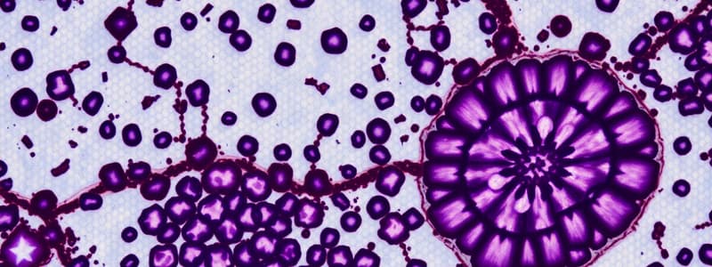
Tissue Preparation for Microscopic Examination
- Thin, stained tissue sections must be prepared for microscopic examination.
Types of Staining Methods
- There are three main types of staining methods:
- General staining methods: Reveal the general structure of the tissue.
- Hematoxylin and eosin (H&E) staining is the most common general stain.
- Hematoxylin is a blue basic dye that stains acidic cell components like nucleic acids (DNA & RNA). These components are termed basophilic.
- Eosin is a pink acidic dye that stains basic cell components like most cytoplasm proteins (e.g., mitochondria). These components are termed acidophilic.
- Special staining methods: Reveal specific tissue components.
- For example, Sudan black dye stains lipids.
- Histochemical and cytochemical staining methods: Employ chemical reactions to stain specific tissue components.
- The Periodic Acid Schiff (PAS) reaction stains carbohydrates (glycogen).
- General staining methods: Reveal the general structure of the tissue.
Studying That Suits You
Use AI to generate personalized quizzes and flashcards to suit your learning preferences.


