Podcast
Questions and Answers
What is the primary transport system in humans?
What is the primary transport system in humans?
- Lymphatic system
- Gastrointestinal system
- Nervous system
- Blood circulatory system (correct)
Which germ layer contributes to the development of the human transport system?
Which germ layer contributes to the development of the human transport system?
- Ectoderm
- Endoderm
- Mesoderm (correct)
- All of the above
The lymphatic system is referred to as the primary transport system.
The lymphatic system is referred to as the primary transport system.
False (B)
What is the function of the heart in the circulatory system?
What is the function of the heart in the circulatory system?
How much does a normal human heart weigh?
How much does a normal human heart weigh?
Name the three layers of the heart wall.
Name the three layers of the heart wall.
Where is the apex of the heart typically located?
Where is the apex of the heart typically located?
The _______ prevents the backflow of blood in the heart.
The _______ prevents the backflow of blood in the heart.
What is the purpose of the septum in the heart?
What is the purpose of the septum in the heart?
What sound is produced by the closure of the mitral and tricuspid valves?
What sound is produced by the closure of the mitral and tricuspid valves?
What do the pulmonary veins do?
What do the pulmonary veins do?
Match the following heart structures with their functions:
Match the following heart structures with their functions:
The left ventricle walls are thinner than the right ventricle walls.
The left ventricle walls are thinner than the right ventricle walls.
Flashcards are hidden until you start studying
Study Notes
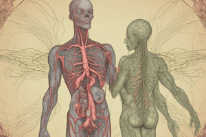
Transportation Systems in Humans
- Human transport systems involve the circulatory system, which includes the blood circulatory system and the lymphatic system.
- The blood circulatory system is known as the primary transport system, while the lymphatic system is referred to as the secondary transport system.
Development of Systems
- The human transport system develops from the mesodermal germ layer.
- Ectoderm contributes to the nervous system and integumentary system.
- Endoderm gives rise to the gastrointestinal tract, liver, and pancreas.
Blood Circulatory System
- Functions as the main transport fluid system, responsible for distributing blood.
- Comprises the heart and blood vessels which facilitate the flow of blood throughout the body.
- The heart acts as a double pump, circulating blood to the lungs for oxygenation and supplying oxygenated blood to the body.
Lymphatic System
- Acts as a compensatory system, involving lymphatic vessels that transport lymph fluid.
- Collects excess interstitial fluid from tissues and returns it to blood circulation.
- The lymphatic system consists of lymph nodes, lymph vessels, and lymph fluid.
Structure and Function of the Human Heart
- The heart is conical in shape, appearing reddish, and functions as a muscular organ.
- The heart generates its own nerve impulses, making it myogenic, meaning it does not require external signals from the brain to contract.
- Normal heart weight ranges from 250 to 350 grams, comparable in size to a fist.
Heart Location and Orientation
- Positioned slightly left within the thoracic cavity, located between the first and second rib space.
- Apex of the heart lies in the fifth intercostal space, typically 9 cm away from the sternum.
Layers of the Heart
- Heart wall is composed of three layers:
- Pericardium: Outer protective double-layered membrane surrounding the heart.
- Myocardium: Thick muscular layer responsible for contraction.
- Endocardium: Inner layer made up of simple squamous epithelium.
Blood Flow through the Heart
- Deoxygenated blood enters the heart through the superior and inferior vena cavae into the right atrium.
- The right atrioventricular (tricuspid) valve opens to allow blood to enter the right ventricle.
- From the right ventricle, blood is pumped into the pulmonary trunk towards the lungs for oxygenation.
- Oxygenated blood from the lungs returns to the left atrium via pulmonary veins.
- Blood flows from the left atrium to the left ventricle through the bicuspid (mitral) valve, then to the aorta for distribution throughout the body.
Important Heart Structures
- Septum: Separates the chambers of the heart to prevent mixing of oxygenated and deoxygenated blood.
- Chambers:
- Atria: Upper chambers with thinner walls.
- Ventricles: Lower chambers with thicker walls for powerful contraction.
- Valves: Control blood flow direction between different chambers (e.g., tricuspid valve, bicuspid valve).
Cardiac Muscle Cells
- Cardiac muscle cells are branched and interconnected via intercalated discs, allowing synchronized contractions.
- These disc structures are crucial for efficient heart function and coordination during heartbeat cycles.
Key Concepts
- The heart's pumping action is vital for maintaining circulation and providing oxygen to aerobic tissues in the body.
- Understanding the layout and functionalities of the heart and its associated systems is essential for grasping human physiology.### Heart Structure and Function
- Heart functions as a pump, controlling blood flow to various body parts.
- The internal structure of the heart includes valves critical for proper blood circulation.
Key Valves
- Tricuspid Valve: Found between the right atrium and right ventricle, allows blood flow into the ventricle.
- Chordae Tendineae: Thin strings connecting the papillary muscles to the valves, preventing backflow of blood.
- Bicuspid (Mitral) Valve: Located between the left atrium and left ventricle, manages oxygen-rich blood flow.
Blood Flow Pathways
- Inferior and Superior Vena Cava: Return deoxygenated blood to the right atrium.
- Pulmonary Arteries: Carry deoxygenated blood from the right ventricle to the lungs for oxygenation.
- Pulmonary Veins: Return oxygenated blood from the lungs to the left atrium.
- Aorta: Distributes oxygen-rich blood from the left ventricle to the rest of the body.
Ventricular Differences
- Left ventricle walls are thicker than right ventricle walls, necessary to generate higher pressure to pump blood throughout the body.
Heart Sounds
- Two main heart sounds:
- Lub: Produced by closure of the mitral and tricuspid valves when the ventricles contract.
- Dub: Produced by closure of the pulmonary and aortic valves when the ventricles relax.
Circulatory Pathways
- Pulmonary Circulation: Moves carbon dioxide-rich blood from the heart to the lungs and returns oxygenated blood back to the heart.
- Systemic Circulation: Distributes oxygenated blood from the heart to the body and returns deoxygenated blood back to the heart.
Blood Types
- The heart receives both oxygenated blood from pulmonary veins and deoxygenated blood from venae cavae.
- It does not receive arterial blood directly; arteries carry blood away from the heart.
Vascular Structures
- Arteries: Generally carry oxygenated blood away from the heart to tissues (exceptions include pulmonary and umbilical arteries).
- Factors affecting blood supply to various organs:
- Carotid Arteries: Supply blood to the brain.
- Renal Arteries: Supply blood to the kidneys.
- Coronary Arteries: Supply blood to the heart muscle itself.
Important Terminology
- Lumen: The cavity within a tubular structure, such as blood vessels.
- Epicardium: Innermost layer of pericardium, thin layer covering the heart.
- Endocardium: Innermost layer of the heart, lining the chambers.
Overall Importance
- Understanding the heart’s structure and function is crucial for comprehension of the cardiovascular system and its role in overall health.
- Each component has a specific role in maintaining efficient blood circulation throughout the body.
Transportation Systems in Humans
- Circulatory system includes two main systems: the blood circulatory system (primary) and the lymphatic system (secondary).
Development of Systems
- Human transport systems originate from the mesoderm, with the ectoderm forming the nervous and integumentary systems, and the endoderm developing the gastrointestinal tract, liver, and pancreas.
Blood Circulatory System
- Main transport system distributing blood, encompassing the heart and blood vessels for overall flow in the body.
- The heart functions as a double pump: circulating deoxygenated blood to the lungs for oxygenation and sending oxygen-rich blood to body tissues.
Lymphatic System
- A secondary transport system, consisting of lymphatic vessels that transport lymph fluid and help maintain fluid balance by collecting excess tissue fluid.
Structure and Function of the Human Heart
- The heart is a conically shaped muscular organ, reddish in color, capable of generating its own nerve impulses.
Heart Location and Orientation
- Located slightly left within the thoracic cavity, between the first and second rib space, with the apex positioned in the fifth intercostal space, roughly 9 cm from the sternum.
Layers of the Heart
- Pericardium: The outer protective double-layered membrane.
- Myocardium: The thick muscular layer responsible for contraction.
- Endocardium: The inner layer composed of simple squamous epithelium.
Blood Flow through the Heart
- Deoxygenated blood enters through the superior and inferior vena cavae into the right atrium, then flows to the right ventricle via the tricuspid valve.
- Blood is pumped into the pulmonary trunk towards the lungs and returns oxygenated to the left atrium via pulmonary veins.
- Blood moves from the left atrium to the left ventricle through the bicuspid valve and then to the aorta for systemic circulation.
Important Heart Structures
- Septum: Separates heart chambers to prevent mixing blood types.
- Atria: Upper chambers with thinner walls, collect blood entering the heart.
- Ventricles: Lower chambers with thicker walls designed for powerful contractions.
- Valves: Ensure one-way blood flow between chambers (tricuspid, bicuspid).
Cardiac Muscle Cells
- Cardiac muscle fibers are branched and interconnected by intercalated discs, promoting synchronized contractions vital for efficient pumping.
Key Concepts
- The heart's pumping action sustains circulation and supports oxygen supply to aerobic tissues, fundamental to human physiology.
Heart Structure and Function
- Functions as a pump, controlling blood movement; contains critical valves for effective blood circulation.
Key Valves
- Tricuspid Valve: Between right atrium and right ventricle, directs blood into the ventricle.
- Chordae Tendineae: Connect papillary muscles to valves, preventing backflow.
- Bicuspid (Mitral) Valve: Between left atrium and left ventricle, regulates oxygen-rich blood flow.
Blood Flow Pathways
- Inferior and Superior Vena Cava: Return deoxygenated blood to the right atrium.
- Pulmonary Arteries: Transport deoxygenated blood from the right ventricle to the lungs.
- Pulmonary Veins: Bring oxygenated blood back to the left atrium.
- Aorta: Distributes oxygen-rich blood to the body.
Ventricular Differences
- Left ventricle walls are thicker than those of the right to produce higher pressure needed for systemic circulation.
Heart Sounds
- Lub: Sound from closure of mitral and tricuspid valves during ventricular contraction.
- Dub: Sound from closure of pulmonary and aortic valves during ventricular relaxation.
Circulatory Pathways
- Pulmonary Circulation: Transports carbon dioxide-rich blood from the heart to the lungs for oxygenation.
Studying That Suits You
Use AI to generate personalized quizzes and flashcards to suit your learning preferences.