Podcast
Questions and Answers
What is the primary function of the atria in the heart?
What is the primary function of the atria in the heart?
- To act as conduits and fill the ventricles with blood (correct)
- To receive oxygenated blood from the lungs
- To contract and propel blood through the circulation
- To create high pressure in the pulmonary arteries
Which type of muscle fibers are involved in conducting impulses in the heart?
Which type of muscle fibers are involved in conducting impulses in the heart?
- Striated skeletal muscle fibers
- Specialized excitatory and conductive muscle fibers (correct)
- Ventricular muscle fibers
- Atrial muscle fibers
What characterizes the intercalated discs in cardiac muscle cells?
What characterizes the intercalated discs in cardiac muscle cells?
- They allow for rapid transmission of action potentials (correct)
- They store calcium ions for muscle contraction
- They increase electrical resistance between cells
- They separate atrial and ventricular syncytia
What initiates the action potential in cardiac muscle?
What initiates the action potential in cardiac muscle?
What is the plateau phase of the cardiac muscle action potential primarily caused by?
What is the plateau phase of the cardiac muscle action potential primarily caused by?
What is the resting membrane potential of cardiac muscle typically?
What is the resting membrane potential of cardiac muscle typically?
How long does the action potential last in the ventricles?
How long does the action potential last in the ventricles?
What role does the atrioventricular (A-V) junction serve in the heart?
What role does the atrioventricular (A-V) junction serve in the heart?
What primarily maintains the plateau of the action potential in cardiac muscle cells?
What primarily maintains the plateau of the action potential in cardiac muscle cells?
How do calcium ions promote muscle contraction in cardiac muscle?
How do calcium ions promote muscle contraction in cardiac muscle?
What happens to potassium permeability at the end of the action potential plateau in cardiac muscle?
What happens to potassium permeability at the end of the action potential plateau in cardiac muscle?
What is a unique characteristic of T tubules in cardiac muscle compared to those in skeletal muscle?
What is a unique characteristic of T tubules in cardiac muscle compared to those in skeletal muscle?
What is the primary factor that influences calcium content in the T tubules of cardiac muscle?
What is the primary factor that influences calcium content in the T tubules of cardiac muscle?
What occurs immediately after the closing of slow calcium-sodium channels in cardiac muscle cells?
What occurs immediately after the closing of slow calcium-sodium channels in cardiac muscle cells?
Which process is responsible for the end of contraction in cardiac muscle cells?
Which process is responsible for the end of contraction in cardiac muscle cells?
What triggers the initiation of each heartbeat in the heart?
What triggers the initiation of each heartbeat in the heart?
What describes the primary function of the ventricles in the heart?
What describes the primary function of the ventricles in the heart?
Which type of cardiac muscle is specifically adapted for electrical conduction?
Which type of cardiac muscle is specifically adapted for electrical conduction?
What is the typical action potential duration in atrial muscle cells?
What is the typical action potential duration in atrial muscle cells?
What separates the atrial and ventricular syncytia in the heart?
What separates the atrial and ventricular syncytia in the heart?
What primarily causes the plateau phase of the cardiac muscle action potential?
What primarily causes the plateau phase of the cardiac muscle action potential?
What is the resting membrane potential of cardiac muscle typically around?
What is the resting membrane potential of cardiac muscle typically around?
What occurs as a result of action potential propagation in cardiac muscle tissue?
What occurs as a result of action potential propagation in cardiac muscle tissue?
What mechanism is responsible for the initial spike of the action potential in cardiac muscle?
What mechanism is responsible for the initial spike of the action potential in cardiac muscle?
What promotes cardiac muscle contraction during the action potential?
What promotes cardiac muscle contraction during the action potential?
What happens to potassium permeability at the end of the action potential plateau?
What happens to potassium permeability at the end of the action potential plateau?
How does the design of T tubules in cardiac muscles affect calcium handling?
How does the design of T tubules in cardiac muscles affect calcium handling?
What occurs immediately after the closing of the slow calcium-sodium channels?
What occurs immediately after the closing of the slow calcium-sodium channels?
What role do calcium ions play in the action potential of cardiac muscle cells?
What role do calcium ions play in the action potential of cardiac muscle cells?
What is the consequence of decreased permeability to potassium ions in cardiac muscle cells?
What is the consequence of decreased permeability to potassium ions in cardiac muscle cells?
What stops the influx of calcium ions into the muscle fiber during the action potential?
What stops the influx of calcium ions into the muscle fiber during the action potential?
What triggers the release of calcium ions into the sarcoplasm in cardiac muscle fibers?
What triggers the release of calcium ions into the sarcoplasm in cardiac muscle fibers?
What primarily prevents the return of the membrane potential in cardiac muscle during the plateau phase?
What primarily prevents the return of the membrane potential in cardiac muscle during the plateau phase?
Which of the following describes the function of calcium ions that enter cardiac muscle cells?
Which of the following describes the function of calcium ions that enter cardiac muscle cells?
What occurs when the slow calcium-sodium channels close after 0.2 to 0.3 seconds?
What occurs when the slow calcium-sodium channels close after 0.2 to 0.3 seconds?
What is the consequence of the larger volume of T tubules in cardiac muscle compared to skeletal muscle?
What is the consequence of the larger volume of T tubules in cardiac muscle compared to skeletal muscle?
What is the primary role of the influx of calcium ions into the muscle fiber during the action potential?
What is the primary role of the influx of calcium ions into the muscle fiber during the action potential?
What initiates the release of calcium ions into the sarcoplasm in cardiac muscle fibers during an action potential?
What initiates the release of calcium ions into the sarcoplasm in cardiac muscle fibers during an action potential?
How does the closing of the slow calcium-sodium channels affect cardiac muscle cells after the plateau phase?
How does the closing of the slow calcium-sodium channels affect cardiac muscle cells after the plateau phase?
What is the significance of the action potential in cardiac muscle regarding heartbeats?
What is the significance of the action potential in cardiac muscle regarding heartbeats?
What is the primary function of the ventricles in the heart?
What is the primary function of the ventricles in the heart?
Which statement accurately describes cardiac muscle contraction?
Which statement accurately describes cardiac muscle contraction?
How long is the plateau phase in the ventricles during an action potential?
How long is the plateau phase in the ventricles during an action potential?
What is the role of the atria in the heart's pumping mechanism?
What is the role of the atria in the heart's pumping mechanism?
What primarily allows the rapid transmission of action potentials between cardiac muscle cells?
What primarily allows the rapid transmission of action potentials between cardiac muscle cells?
What could potentially disrupt the normal conduction pathway in cardiac muscle?
What could potentially disrupt the normal conduction pathway in cardiac muscle?
Which of the following describes the state of the membrane potential in cardiac muscle at rest?
Which of the following describes the state of the membrane potential in cardiac muscle at rest?
What primarily contributes to the initial spike of the action potential in cardiac muscle cells?
What primarily contributes to the initial spike of the action potential in cardiac muscle cells?
Flashcards are hidden until you start studying
Study Notes
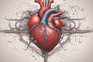
Anatomy of the Heart
- The heart consists of two pumps: the right heart and the left heart.
- The right heart receives deoxygenated blood from peripheral tissues and pumps it to the lungs for oxygenation.
- The left heart receives oxygenated blood from the lungs and pumps it back to peripheral tissues.
- Each pump consists of an atrium and a ventricle; atria fill the ventricles, while ventricles contract to generate high pressure for blood circulation.
Physiology of Cardiac Muscle
- Cardiac muscle includes three types: atrial muscle, ventricular muscle, and specialized excitatory/conductive fibers.
- Cardiac muscle is striated and contains interdigiting actin and myosin filaments that slide past each other during contraction.
- Intercalated discs have low electrical resistance, enabling rapid action potential propagation between cardiac muscle cells.
A-V Junction and Impulse Conduction
- The atrioventricular (A-V) junction conducts impulses from atria to ventricles; this pathway is insulated by fibrous tissue under normal conditions.
- Atrial syncytium and ventricular syncytium are electrically isolated, ensuring organized conduction.
Action Potentials in Cardiac Muscle
- Resting membrane potential of cardiac muscle ranges from −85 to −95 millivolts, with action potentials reaching 105 millivolts.
- Action potentials feature a plateau phase: 0.2 seconds in atrial muscle and 0.3 seconds in ventricular muscle.
- Plateau is sustained by slow entry of sodium and calcium ions, preventing immediate repolarization.
Sodium and Calcium Ion Dynamics
- Fast sodium channels initiate action potentials, rapidly opening and closing.
- Slow calcium-sodium channels contribute to the plateau by allowing calcium and sodium influx after the initial spike.
- Calcium influx promotes cardiac muscle contraction and sustains the plateau phase.
Potassium Ion Dynamics
- Decreased potassium permeability during the plateau phase prevents membrane repolarization.
- Following the plateau, increased potassium permeability allows potassium ions to exit, restoring resting membrane potential.
Calcium Role in Muscle Contraction
- Action potentials induce calcium diffusion into myofibrils, promoting muscle contraction.
- Transverse (T) tubules in cardiac muscle are larger than in skeletal muscle, containing significant calcium for contraction.
- T tubules connect directly with extracellular fluid, affecting calcium concentration and availability during contraction.
Conclusion of Action Potential
- After the plateau phase, calcium influx ceases, and calcium is reabsorbed into the sarcoplasmic reticulum and T tubules.
- The cessation of calcium entry leads to muscle relaxation, completing the cycle of contraction and initiating the next heartbeat.
Studying That Suits You
Use AI to generate personalized quizzes and flashcards to suit your learning preferences.