Podcast
Questions and Answers
What is the primary function of the myocardium?
What is the primary function of the myocardium?
- To provide structure to the heart
- To form the innermost layer of the heart wall
- To regulate body temperature
- To pump blood throughout the body (correct)
What is a characteristic of cardiac muscle fibers?
What is a characteristic of cardiac muscle fibers?
- Non-striated
- Voluntary in nature
- Involuntary in nature (correct)
- Multinucleated
How many types of muscle fibers are found in the myocardium?
How many types of muscle fibers are found in the myocardium?
- Two
- Three (correct)
- Four
- Five
What is the innermost layer of the heart wall?
What is the innermost layer of the heart wall?
What is a unique property of cardiac muscle?
What is a unique property of cardiac muscle?
What is the main function of capillaries?
What is the main function of capillaries?
What are the three layers of the aorta and artery walls?
What are the three layers of the aorta and artery walls?
What is the result of stimulation of one cardiac muscle cell?
What is the result of stimulation of one cardiac muscle cell?
What is the characteristic of arterioles?
What is the characteristic of arterioles?
What is the main source of energy for the cardiac muscle?
What is the main source of energy for the cardiac muscle?
What is the name of the vessels that offer resistance to blood flow?
What is the name of the vessels that offer resistance to blood flow?
What is the term for the functional unity of cardiac muscle cells?
What is the term for the functional unity of cardiac muscle cells?
What is the diameter of venules?
What is the diameter of venules?
What is the characteristic of venules?
What is the characteristic of venules?
What is the name of the vessels that can hold a large quantity of blood?
What is the name of the vessels that can hold a large quantity of blood?
What is the diameter of superior and inferior venae cavae?
What is the diameter of superior and inferior venae cavae?
What is the primary function of the sinoatrial node?
What is the primary function of the sinoatrial node?
What separates the two left chambers from the two right chambers in the heart?
What separates the two left chambers from the two right chambers in the heart?
Which of the following veins returns venous blood from the lower parts of the body?
Which of the following veins returns venous blood from the lower parts of the body?
What is the main difference between the musculature of the atria and ventricles?
What is the main difference between the musculature of the atria and ventricles?
What is the function of the atrioventricular node?
What is the function of the atrioventricular node?
Which side of the heart receives oxygenated blood from the lungs?
Which side of the heart receives oxygenated blood from the lungs?
What is the purpose of the valves in the heart?
What is the purpose of the valves in the heart?
Which valve separates the left atrium and left ventricle?
Which valve separates the left atrium and left ventricle?
What percentage of caloric needs are provided by free fatty acids under basal conditions?
What percentage of caloric needs are provided by free fatty acids under basal conditions?
What is the main source of energy for skeletal muscles, especially during exercise?
What is the main source of energy for skeletal muscles, especially during exercise?
During which phase of the cardiac cycle does the left ventricle receive its blood supply?
During which phase of the cardiac cycle does the left ventricle receive its blood supply?
Why does the left ventricle receive its blood supply only during diastole?
Why does the left ventricle receive its blood supply only during diastole?
What percentage of oxygen delivered by each unit of blood is consumed by the cardiac muscle?
What percentage of oxygen delivered by each unit of blood is consumed by the cardiac muscle?
What is the primary mechanism of energy generation in the cardiac muscle?
What is the primary mechanism of energy generation in the cardiac muscle?
What happens to the cardiac muscle when oxygen delivery is obstructed?
What happens to the cardiac muscle when oxygen delivery is obstructed?
What percentage of total energy is generated by anaerobic metabolism in the cardiac muscle?
What percentage of total energy is generated by anaerobic metabolism in the cardiac muscle?
What is the main component of the wall of capillaries?
What is the main component of the wall of capillaries?
What is characteristic of sinusoids?
What is characteristic of sinusoids?
What is the functional difference between systemic and pulmonary circulation?
What is the functional difference between systemic and pulmonary circulation?
What is true about the ventricles of the heart?
What is true about the ventricles of the heart?
What is the significance of the basal lamina in capillaries?
What is the significance of the basal lamina in capillaries?
What is the characteristic of the walls of veins and venae cavae?
What is the characteristic of the walls of veins and venae cavae?
What is the result of any discrepancy in the time or quantitative relations of blood flow in the heart?
What is the result of any discrepancy in the time or quantitative relations of blood flow in the heart?
What is the division of the circulatory system that passes through the lungs?
What is the division of the circulatory system that passes through the lungs?
Flashcards are hidden until you start studying
Study Notes
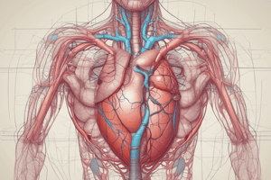
Heart Structure and Function
- The heart is a hollow muscular organ located between the lungs in the mediastinum.
- It consists of 4 chambers: 2 atria and 2 ventricles, separated by a continuous partition (interatrial septum and interventricular septum).
- The musculature is thicker in the ventricles than in the atria.
- The force of contraction of the heart depends on the muscle.
Right Side of the Heart
- The right side of the heart has two chambers: right atrium and right ventricle.
- The right atrium is a thin-walled, low-pressure chamber that receives venous (deoxygenated) blood from the head, neck, and upper limbs through the superior vena cava, and from the lower parts of the body through the inferior vena cava.
- The right atrium has the pacemaker (sinoatrial node) and the atrioventricular node that conducts impulses to the ventricles.
Left Side of the Heart
- The left side of the heart has two chambers: left atrium and left ventricle.
- The left atrium is a thin-walled, low-pressure chamber that receives oxygenated blood from the lungs through the pulmonary veins.
- The left ventricle pumps arterial blood to different parts of the body through the systemic aorta.
Valves of the Heart
- There are no admixtures between arterial and venous blood.
- The myocardium forms the bulk of the heart and is responsible for the pumping action.
- Myocardium has three types of muscle fibers: contractile, pacemaker, and conductive system.
Endocardium
- Endocardium is the innermost layer of the heart wall, a thin, smooth, and glistening membrane.
- It is formed by a single layer of endothelial cells lining the inner surface of the heart.
Properties of Cardiac Muscle
- The cardiac muscle has two types of muscle fibers: cardiac muscle proper and conductive system.
- It has special properties that characterize it from other types of muscles, including:
- Histology of the cardiac muscle: unlike skeletal muscle, with anatomical connections between myocardial fibers (intercalated discs and gap junctions).
- Functional syncytium: stimulation of one cardiac muscle cell results in stimulation of all cells, allowing the heart to contract as one unit.
- Main source of energy: the cardiac muscle consumes fat as the main source of energy.
- Blood flow (supply): the cardiac muscle receives its blood supply mainly during diastole.
- Oxygen extraction: the cardiac muscle extracts a higher amount of oxygen than skeletal muscle.
Blood Vessels
- The circulatory system consists of:
- Aorta
- Arteries
- Arterioles
- Capillaries
- Venules
- Veins
- Venae cavae
Arterial System
- The arterial system comprises the aorta, arteries, and arterioles.
- Walls of the aorta and arteries are formed by three layers: tunica adventitia, tunica media, and tunica intima.
- Aorta and arteries have two laminae of elastic tissues.
- Arterial branches become narrower and their walls become thinner while reaching the periphery.
Venous System
- The venous system starts from the capillaries and includes venules, veins, and venae cavae.
- Capillaries end in venules, which are smaller vessels with thin muscular walls.
- Venules are continued as veins, which have a larger diameter.
- Veins form superior and inferior venae cavae, which have a larger diameter.
Capillaries
- Capillaries make a connective link between the arterioles and venules.
- They are linked by a single layer of flat endothelial cells.
- The capillary endothelium does not directly touch the elements of other tissues and is always separated from a supporting bed of connective tissue by an intervening layer (basal lamina).
Divisions of Circulation
- Blood flows through two divisions of the circulatory system:
- Systemic circulation (greater circulation with high resistance circuit)
- Pulmonary circulation (lesser circulation with low resistance circuit)
- The two systems meet in the heart.
Studying That Suits You
Use AI to generate personalized quizzes and flashcards to suit your learning preferences.