Podcast
Questions and Answers
What type of dentition is characterized by the development of two sets of teeth in mammals, including humans?
What type of dentition is characterized by the development of two sets of teeth in mammals, including humans?
- Monophyodont dentition
- Oligodont dentition
- Diphyodont dentition (correct)
- Polyphyodont dentition
Which part of the tooth is primarily responsible for forming the hard material called dentine?
Which part of the tooth is primarily responsible for forming the hard material called dentine?
- Cementum
- Enamel
- Odontoblasts (correct)
- Pulp chamber
The anterior bony hard palate is lined by which structure?
The anterior bony hard palate is lined by which structure?
- Teeth
- Uvula
- Soft palate
- Palatine rugae (correct)
Which type of teeth in humans are specifically adapted for grinding food?
Which type of teeth in humans are specifically adapted for grinding food?
What is the dental formula for an adult human's permanent teeth?
What is the dental formula for an adult human's permanent teeth?
Which of the following statements accurately describes canines?
Which of the following statements accurately describes canines?
What is the primary function of the uvula in the digestive system?
What is the primary function of the uvula in the digestive system?
Wisdom teeth in humans typically emerge at what age?
Wisdom teeth in humans typically emerge at what age?
What substance covers the dentine of the crown of a tooth?
What substance covers the dentine of the crown of a tooth?
Which type of papillae on the tongue is primarily responsible for taste sensation and located at the anterior margin?
Which type of papillae on the tongue is primarily responsible for taste sensation and located at the anterior margin?
Which part of the pharynx serves as a common passage for both food and air?
Which part of the pharynx serves as a common passage for both food and air?
Which muscle layer may be present in some regions of the alimentary canal, particularly the stomach?
Which muscle layer may be present in some regions of the alimentary canal, particularly the stomach?
What is the primary function of the vermiform appendix associated with the caecum?
What is the primary function of the vermiform appendix associated with the caecum?
Which section of the small intestine receives the bile and pancreatic secretions via the hepatopancreatic duct?
Which section of the small intestine receives the bile and pancreatic secretions via the hepatopancreatic duct?
What is the role of Brunner's glands in the submucosa of the duodenum?
What is the role of Brunner's glands in the submucosa of the duodenum?
What prevents food from entering the glottis during swallowing?
What prevents food from entering the glottis during swallowing?
Which part of the alimentary canal is primarily responsible for the absorption of water and certain drugs?
Which part of the alimentary canal is primarily responsible for the absorption of water and certain drugs?
The internal anal sphincter is primarily composed of which type of muscle?
The internal anal sphincter is primarily composed of which type of muscle?
Which digestive structure is primarily distensible and J-shaped?
Which digestive structure is primarily distensible and J-shaped?
In which layer of the alimentary canal do you find the gastro-oesophageal sphincter?
In which layer of the alimentary canal do you find the gastro-oesophageal sphincter?
What is the role of microvilli in the small intestine?
What is the role of microvilli in the small intestine?
Which part of the colon is primarily responsible for water absorption?
Which part of the colon is primarily responsible for water absorption?
Which statement accurately describes the types of teeth in human dentition?
Which statement accurately describes the types of teeth in human dentition?
Which part of the alimentary canal is responsible for the initial processing of food?
Which part of the alimentary canal is responsible for the initial processing of food?
What is the function of palatine rugae in the buccal cavity?
What is the function of palatine rugae in the buccal cavity?
What distinguishes diphyodont dentition from other dentitions?
What distinguishes diphyodont dentition from other dentitions?
Which part of the tooth is responsible for anchoring it into the jawbone?
Which part of the tooth is responsible for anchoring it into the jawbone?
The dental formula for an adult human reflects which aspect of their dentition?
The dental formula for an adult human reflects which aspect of their dentition?
Which type of epithelium primarily lines the buccal cavity?
Which type of epithelium primarily lines the buccal cavity?
Why do third molars often cause problems when they emerge?
Why do third molars often cause problems when they emerge?
The three main parts of a tooth include the crown, neck, and root. What primarily fills the crown?
The three main parts of a tooth include the crown, neck, and root. What primarily fills the crown?
How do teeth develop in relation to a person's age?
How do teeth develop in relation to a person's age?
What is the primary function of the periodontal membrane in the tooth structure?
What is the primary function of the periodontal membrane in the tooth structure?
Which of the following statements accurately describes the structure of the tongue?
Which of the following statements accurately describes the structure of the tongue?
What is the role of the epiglottis in the pharynx?
What is the role of the epiglottis in the pharynx?
Which layer of the alimentary canal is responsible for the absorption of nutrients?
Which layer of the alimentary canal is responsible for the absorption of nutrients?
The vermiform appendix is considered a vestigial organ because it is believed to have what function?
The vermiform appendix is considered a vestigial organ because it is believed to have what function?
Which feature distinguishes the small intestine from the large intestine?
Which feature distinguishes the small intestine from the large intestine?
What is the primary role of the gastro-oesophageal sphincter?
What is the primary role of the gastro-oesophageal sphincter?
How does the structure of the colon differ from that of the small intestine?
How does the structure of the colon differ from that of the small intestine?
Which type of muscle is primarily involved in the voluntary control of swallowing?
Which type of muscle is primarily involved in the voluntary control of swallowing?
What unique structural feature is formed by the mucosa of the small intestine that aids in digestion?
What unique structural feature is formed by the mucosa of the small intestine that aids in digestion?
Flashcards are hidden until you start studying
Study Notes
Alimentary Canal: The Digestive Journey
- The alimentary canal, or digestive tract, begins at the mouth and ends at the anus.
- It is composed of the buccal cavity, pharynx, esophagus, stomach, small intestine, and large intestine.
Mouth and Buccal Cavity
- Lips: Muscles that border the mouth, allowing for movement.
- Palate: Separates the mouth from the nasal cavity, enabling simultaneous chewing and breathing.
- Hard Palate: Anterior portion, bony, lined with palatine rugae (ridges).
- Soft Palate: Posterior portion, hangs into the pharynx, with a hanging structure called the uvula.
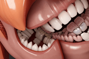
- Teeth: Embedded in sockets of jaw bones (thecodont), diphyodont (two sets of teeth).
- Deciduous Teeth: Temporary/milk teeth.
- Permanent Teeth: Replace deciduous teeth; humans have 32 permanent teeth.
- Heterodont Dentition: Different types of teeth (incisors, canines, premolars, molars).
- Dental Formula: Representation of tooth arrangement (I 2/2; C 1/1; PM 2/2; M 3/3 = 32).
- Wisdom Teeth: Third molars, appear late in life (around 21 years old).
- Incisors: Chisel-shaped for cutting.
- Canines: Dagger-like for tearing.
- Premolars and Molars: Grinding teeth.
- Tooth Structure:
- Crown: Exposed part.
- Neck: Middle part.
- Root: Innermost part, embedded in the jawbone.
- Dentine: Hard material forming the bulk of the tooth, secreted by odontoblasts.
- Enamel: Hardest substance in the body, covers crown, secreted by ameloblasts.
- Pulp Cavity: Internal cavity filled with pulp (odontoblasts, nerves, blood vessels).
- Cementum: Covers the root, attaches tooth to the socket.
- Periodontal Membrane: Anchors the tooth in the socket.
- Gums (Gingiva): Cover the neck and roots of teeth.
- Tongue:
- Freely movable muscular sense organ attached to the floor of the mouth by the frenulum.
- Papillae: Small projections on the tongue's surface, some bearing taste buds.
- Types of Papillae:
- Fungiform: Anterior margin and tip of the tongue.
- Filiform: Surface of the tongue.
- Circumvallate: Posterior surface/base of the tongue.
- Foliate: Lateral sides of the posterior 1/3rd of the tongue, rudimentary in adults.
- Functions of the Tongue:
- Mixing saliva with food.
- Taste detection.
- Deglutition (swallowing).
- Speech.
Pharynx
- Passageway for Food and Air: Short, muscular tube connecting the buccal cavity to the esophagus.
- Regions:
- Nasopharynx: Above the soft palate.
- Oropharynx: Middle part.
- Laryngopharynx: Lower part.
- Glottis: The opening into the larynx.
- Epiglottis: Cartilaginous flap that prevents food from entering the larynx during swallowing.
- Tonsils: Lymphoid tissues in the pharynx:
- Pharyngeal Tonsils (Adenoids)
- Palatine Tonsils (pair)
- Lingual Tonsils (pair)
- Eustachian Tubes: Connect the middle ear cavities to the nasopharynx.
Esophagus
- Muscular Tube: Thin and long, extends from the pharynx through the neck, thorax, and diaphragm.
- Sphincters:
- Gastro-oesophageal/Cardiac Sphincter: Regulates the opening between the esophagus and the stomach.
- Upper Oesophageal Sphincter: Located at the beginning of the esophagus.
Stomach
- Muscular Sac: Wide, J-shaped, located in the upper left portion of the abdominal cavity.
- Regions:
- Cardiac Region: Anterior portion, where the esophagus enters.
- Fundic Region: Middle region, main body of the stomach.
- Pyloric Region: Posterior portion, connects to the small intestine.
- Pyloric Sphincter: Muscular valve that controls the opening between the stomach and the small intestine.
Small Intestine
- Longest Part of the Alimentary Canal: Divided into three regions:
- Duodenum: Proximal region, receives secretions from the liver and pancreas.
- Jejunum: Middle, long, coiled region.
- Ileum: Distal, highly coiled region.
- Ileo-caecal Valve: Valve between the ileum and large intestine, prevents backflow of waste.
Large Intestine
- Regions:
- Caecum: Small blind sac at the beginning of the large intestine, hosts symbiotic microorganisms.
- Vermiform Appendix: Narrow, finger-like projection from the caecum, considered a vestigial organ.
- Colon:
- Ascending Colon: Ascends on the right side of the abdomen.
- Transverse Colon: Crosses the abdomen.
- Descending Colon: Descends on the left side of the abdomen.
- Sigmoid Colon: S-shaped portion leading to the rectum.
- Rectum: Dilated sac at the end of the colon, stores feces.
- Anal Canal: Short, terminal portion of the large intestine, opens to the anus.
- Sphincters: Control the expulsion of feces.
- Internal Anal Sphincter: Involuntary, smooth muscle.
- External Anal Sphincter: Voluntary, striped muscle.
- Function: Absorption of water, minerals, and certain drugs.
- Mucus Secretion: Lubricates the passage of feces.
Histology of the Alimentary Canal
- Layers from Esophagus to Rectum:
- Serosa: Outermost layer, composed of mesothelium and connective tissue.
- Muscularis Externa: Smooth muscle layer, arranged in longitudinal (outer) and circular (inner) layers.
- Submucosa: Connective tissue layer containing nerves, blood vessels, and lymph vessels.
- Brunner's Glands (Duodenum): Secrete mucus.
- Mucosa: Innermost layer, lines the lumen.
- Gastric Rugae (Stomach): Irregular folds to increase surface area.
- Villi (Small Intestine): Small folds to increase surface area.
- Microvilli: Microscopic projections on the surface of epithelial cells in villi, further increasing surface area.
- Brush Border Enzymes: Enzymes on microvilli membranes, such as disaccharidases.
- Lacteals: Lymphatic capillaries within villi.
- Goblet Cells: Secrete mucus for protection and lubrication.
- Crypts of Lieberkuhn: Invaginations in the mucosa between villi.
Alimentary Canal
- Starts at the mouth and ends at the anus
- Consists of the buccal cavity, pharynx, oesophagus, stomach, small intestine, large intestine
- Functions in the digestion and absorption of food
Mouth and Buccal Cavity
- The mouth is the opening of the alimentary canal
- Lined by the upper and lower lips (labia)
- The palate separates the buccal cavity from the nasal chamber, allowing for simultaneous chewing and breathing
- The anterior palate is bony and lined by palatine rugae
- The posterior soft palate hangs down into the pharynx as the uvula
- The jawbones contain teeth and the tongue is located at the base of the buccal cavity
Teeth
- Ecto-mesodermal in origin
- Thecodont: embedded in sockets of the jaw bones
- Diphyodont: two sets of teeth during lifetime - deciduous (milk) and permanent (adult)
- Heterodont: different types of teeth
- Incisors: chisel-shaped for cutting
- Canines: dagger-like for tearing
- Premolars and molars: grinding food
- Adult dental formula: I 2/2 C 1/1 PM 2/2 M 3/3 = 32 teeth
- Deciduous dental formula: I 2/2 C 1/1 M 2/2 = 20 teeth
- Wisdom teeth: third molars, emerge around 21 years of age
Tooth Structure
- Crown: exposed part
- Neck: middle part
- Root: embedded in the jawbone socket
- Dentine: hard material forming the bulk of the tooth, secreted by odontoblasts
- Enamel: hardest substance in the body, covers the crown, secreted by ameloblasts
- Pulp cavity: contains pulp (odontoblasts, nerves, blood vessels), lined by odontoblasts
- Cementum: covers dentine of the root
- Periodontal membrane: fixes the root in the jawbone socket (alveolus)
- Gums (gingiva): cover the neck and root of the tooth
Tongue
- Freely movable, muscular sense organ
- Attached to the floor of the oral cavity by the frenulum
- Has papillae on its upper surface, some with taste buds
- 4 types of papillae:
- Fungiform: anterior margin and tip
- Filiform: surface of the tongue
- Circumvallate: posterior surface/base
- Foliate: lateral sides of posterior 1/3, rudimentary in adults
- Functions in mixing saliva with food, taste detection, swallowing, and speaking
Pharynx
- Short, common passage for food and air
- Divided into:
- Nasopharynx: above the soft palate
- Oropharynx: middle part
- Laryngopharynx: lower part
- Trachea opens into the larynx, which connects to the laryngopharynx through the glottis
- Epiglottis: cartilaginous flap preventing food entry into the glottis during swallowing
- Contains voluntary muscles for swallowing
- Lymphoid tissues (tonsils)
- Pharyngeal tonsils (adenoids)
- Palatine tonsils (pair)
- Lingual tonsils (pair)
- Eustachian tubes from the middle ear cavities open into the nasopharynx
Oesophagus
- Thin, long tube extending posteriorly through the neck, thorax, and diaphragm
- Leads into the stomach
- Gastro-oesophageal (cardiac) sphincter: regulates opening between the oesophagus and stomach
- Upper oesophageal sphincter: at the beginning of the oesophagus
Stomach
- Wide, J-shaped, distensible muscular bag
- Located in the upper left portion of the abdominal cavity, below the diaphragm
- 3 major parts:
- Cardiac: anterior, receives the oesophagus
- Fundic: middle, main body
- Pyloric: posterior, opens into the small intestine through the pyloric aperture
- Pyloric sphincter: guards the pyloric aperture
Small Intestine
- Longest part of the alimentary canal
- Divided into 3 regions:
- Duodenum: proximal, receives the hepato-pancreatic duct
- Jejunum: middle, long and coiled
- Ileum: distal, highly coiled
- Opens into the large intestine through the ilio-caecal valve
- Ilio-caecal valve: prevents backflow of faecal matter from the large intestine into the ileum
Large Intestine
- Consists of caecum, colon, and rectum
- Caecum: small blind sac, hosts symbiotic microorganisms
- Vermiform appendix (abdominal tonsil): narrow, finger-like projection, vestigial organ, arises from the caecum
- Colon: divided into ascending, transverse, descending, and sigmoid colon
- Sigmoid colon: continues behind into the rectum
- Haustra: external bulged out pouches in the colon
- Taenia coli: three longitudinal smooth muscle folds
- Rectum: small, dilated sac leading into the anal canal
- Anal canal: opens through the anus
- Internal anal sphincter: smooth muscle
- External anal sphincter: voluntary, striped muscle
- No significant digestive activity in the large intestine
- Absorbs water, minerals, and certain drugs
- Secretes mucus for lubricating the passage of undigested particles
Histology of the Alimentary Canal
- Oesophagus to rectum: wall has 4 layers
- Serosa: outermost, thin mesothelium with connective tissue
- Muscularis externa: smooth muscles, outer longitudinal and inner circular layers (oblique layer present in some regions like the stomach)
- Submucosa: loose connective tissue with nerves, blood vessels, and lymph vessels; Brunner's glands in the duodenum secrete mucus
- Mucosa: innermost, lines the lumen, forms gastric rugae in the stomach, villi in the small intestine
- Villi: small folds in the small intestine mucosa, increase surface area for absorption
- Microvilli: microscopic projections on the columnar epithelial cells lining the villi, create a brush border appearance, further increase absorption
- Brush border enzymes: disaccharidases located on the plasma membrane of microvilli
- Each villus has a network of capillaries and a lacteal (large lymph capillary)
- Goblet cells: secrete mucus, protect the wall from enzyme activity and lubricate food passage
- Crypts of Lieberkuhn: mucosal invaginations between the bases of villi
Studying That Suits You
Use AI to generate personalized quizzes and flashcards to suit your learning preferences.


