Podcast
Questions and Answers
What is the primary function of hematoxylin in the H&E staining process?
What is the primary function of hematoxylin in the H&E staining process?
- To stain basic components blue (correct)
- To bind to acidic components red
- To provide a background color for microscopy
- To enhance the visibility of collagen fibers
Which of the following best describes the process of sectioning tissue samples using the freezing technique?
Which of the following best describes the process of sectioning tissue samples using the freezing technique?
- Tissue samples are cut at room temperature without freezing
- Tissue samples are fixed and embedded in paraffin
- Tissue samples are cut immediately after surgical removal without processing
- Tissue samples are rapidly frozen in liquid nitrogen for easy handling (correct)
What is the primary difference between light microscopy and transmission electron microscopy (TEM)?
What is the primary difference between light microscopy and transmission electron microscopy (TEM)?
- Light microscopy relies on light interaction, whereas TEM is based on electron interaction (correct)
- Light microscopy uses electrons for imaging, while TEM uses light
- Light microscopy has a lower magnification power than TEM
- Light microscopy is used for non-biological samples, while TEM is only for biological samples
Which of the following components does eosin primarily bind to during the staining process?
Which of the following components does eosin primarily bind to during the staining process?
What is the maximum magnification power achieved by light microscopy as mentioned?
What is the maximum magnification power achieved by light microscopy as mentioned?
What is the primary purpose of tissue fixation during the paraffin technique?
What is the primary purpose of tissue fixation during the paraffin technique?
Which of the following best describes the process of dehydration in the paraffin technique?
Which of the following best describes the process of dehydration in the paraffin technique?
What role does the clearing step play in the paraffin embedding process?
What role does the clearing step play in the paraffin embedding process?
In which temperature range should the tissue be impregnated with melted soft paraffin?
In which temperature range should the tissue be impregnated with melted soft paraffin?
What is the maximum suggested size for tissue samples before fixation in the paraffin technique?
What is the maximum suggested size for tissue samples before fixation in the paraffin technique?
What is the purpose of sectioning in the parraffin embedding process?
What is the purpose of sectioning in the parraffin embedding process?
Which component is NOT involved in the dehydration step of the paraffin technique?
Which component is NOT involved in the dehydration step of the paraffin technique?
What is the significance of using a microtome in the paraffin technique?
What is the significance of using a microtome in the paraffin technique?
What is the magnifying power range of a light microscope (LM)?
What is the magnifying power range of a light microscope (LM)?
Which characteristic differentiates Transmission Electron Microscopes (TEM) from Scanning Electron Microscopes (SEM)?
Which characteristic differentiates Transmission Electron Microscopes (TEM) from Scanning Electron Microscopes (SEM)?
What does resolution in microscopy refer to?
What does resolution in microscopy refer to?
Why is fixation of tissue essential before microscopy?
Why is fixation of tissue essential before microscopy?
What is the resolving power of an electron microscope?
What is the resolving power of an electron microscope?
What does a phase contrast microscope primarily allow for?
What does a phase contrast microscope primarily allow for?
In histology, what is the common thickness for sections prepared for light microscopy using a microtome?
In histology, what is the common thickness for sections prepared for light microscopy using a microtome?
Which statements are true regarding acidophilia and basophilia in H&E stained sections?
Which statements are true regarding acidophilia and basophilia in H&E stained sections?
Flashcards
Micro-technique
Micro-technique
The process of preparing tissues for microscopic examination, involving a series of steps to preserve, harden, and section the tissue.
Fixation
Fixation
A method of preserving tissue structure by preventing decomposition and autolysis.
Dehydration
Dehydration
A process of removing water from tissue using a series of increasing alcohol concentrations.
Clearing
Clearing
Signup and view all the flashcards
Impregnation
Impregnation
Signup and view all the flashcards
Sectioning
Sectioning
Signup and view all the flashcards
Paraffin Technique
Paraffin Technique
Signup and view all the flashcards
Light Microscope
Light Microscope
Signup and view all the flashcards
H&E Staining
H&E Staining
Signup and view all the flashcards
Electron microscopy
Electron microscopy
Signup and view all the flashcards
Freezing technique
Freezing technique
Signup and view all the flashcards
Transmission electron microscopy (TEM)
Transmission electron microscopy (TEM)
Signup and view all the flashcards
Scanning electron microscopy (SEM)
Scanning electron microscopy (SEM)
Signup and view all the flashcards
Resolution Power
Resolution Power
Signup and view all the flashcards
Microtome
Microtome
Signup and view all the flashcards
Magnification
Magnification
Signup and view all the flashcards
Study Notes
Histology Lab 1: Micro-techniques
- Course: BMS111
- Lab: Histology, Micro-technique
- Instructor: Dr. Manal Shaaban Hafez
- Institution: Galala University, Faculty of Medicine
- Semester: Fall 2024-2025
Learning Objectives (ILOs)
- Identify different micro-techniques used in tissue preparation
- Demonstrate the paraffin technique steps and their significance
- Recognize light microscope components
- Use a microscope to examine histological sections
Histology
- The study of cellular organization in tissues and organs
Light Microscopy
- Used in histological and pathological examinations
Micro-techniques
- Microscopic study of cells & tissues:
- In tissue culture and stem cells
- In histological sections
- Paraffin technique
- Freezing technique
- Celloidin technique
Paraffin Technique
- Steps:
- Obtain specimen
- Fixation
- Dehydration (ascending grades of alcohol)
- Clearing in xylol
- Impregnation in paraffin
- Embedding in paraffin
- Cut sections
- Pick sections on glass slides
1-Tissue Sampling
- Small pieces (1 cm³)
- Cut with sharp instruments
- Methods:
- Immediately: biopsy, after death, after operation, from experimental animals
2- Fixation (10% Formalin Saline)
- Aim:
- Prevent putrefaction
- Prevent autolysis
- Preserve tissue structure
- Harden tissue
- Coagulate proteins
3- Dehydration
- Gradual removal of water from tissues using ascending alcohol concentrations (50%, 70%, 90%, then 100%)
4- Clearing
- Replacing alcohol with a clearing fluid (e.g., benzene, xylol)
- Tissue becomes transparent
5- Impregnation (Infiltration)
- Soak tissue in melted paraffin in an electric oven (55-60°C)
- Allows paraffin to infiltrate cellular spaces, for easier cutting
6- Embedding
- Embedding tissue in melted paraffin to form a paraffin block
7- Sectioning
- Cut tissues into thin sections (5-8 µm) using a microtome
8- Mounting
- Attaching the sections to glass slides for easy handling
- Staining for light microscopy
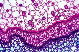
9- Staining of Paraffin Sections (H&E)
- Haematoxylin and Eosin (H&E)
- used for staining the paraffin sections.
Hematoxylin & Eosin
- Haematoxylin:
- Basophilic stain
- Stains nuclei blue/purple
- Eosin:
- Acidophilic stain
- Stains cytoplasm pink/red
Freezing Technique
- Tissue samples removed during surgery or operations
- Biopsies rapidly frozen in liquid nitrogen
- Cutting sections using a cryostat in a sub-freezing temperature cabinet
Microscopy
- Light microscopy: Based on light interaction with tissue
- Electron microscopy: Transmission and Scanning
- Based on electron interaction with tissue
- Transmission electron microscope (TEM):
- Two-dimensional images
- Beams of electrons transmit through cells and tissues
- Examines internal structure
- Scanning electron microscope (SEM):
- Three-dimensional images
- Beam of electrons scans surface of cells and tissues
- Examines surfaces
Microscope Parts
- Body tube
- Ocular lens
- Revolving nosepiece
- Objective lens
- Stage
- Clips
- Diaphragm
- Light source
- Coarse adjustment knob
- Fine adjustment knob
Light Microscope Beam
- Light beam from a lamp (or sun)
Phase Contrast Microscope
- Used to study growth and proliferation of living cells and tissues without stains
Additional Topics
- Trials of OSPE stations: Course on observed skills performance evaluation
- Station 1 (Fixation issues): Fixation is essential for preserving tissue structure.
- Station 2 (Microtome): Microtome is used to cut tissues into thin sections.
- Station 3 (Microscope components): Microscope parts for identification.
- **Station 4 (H&E staining):**Identifying basophilia and acidophilia
- Station 5 (Phase contrast microscope): Use of Phase contrast microscope for evaluating cells.
Studying That Suits You
Use AI to generate personalized quizzes and flashcards to suit your learning preferences.