Podcast
Questions and Answers
What is the approximate resting membrane potential of the sinoatrial (SA) node?
What is the approximate resting membrane potential of the sinoatrial (SA) node?
- -40 millivolts
- 0 millivolts
- -85 to -90 millivolts
- -55 to -60 millivolts (correct)
What causes the self-excitation property of the sinoatrial (SA) node?
What causes the self-excitation property of the sinoatrial (SA) node?
- Naturally leaky sodium and calcium ion channels (correct)
- A low concentration of calcium ions outside the cell
- Impermeability to sodium and calcium ions
- High concentration of potassium ions inside the cell
What occurs when the resting membrane potential of a cardiac cell reaches approximately -40 millivolts?
What occurs when the resting membrane potential of a cardiac cell reaches approximately -40 millivolts?
- Sodium channels close, causing repolarization
- Calcium channels become activated, initiating an action potential (correct)
- The cell remains in a stable resting state
- Potassium channels open, causing hyperpolarization
What primarily causes the repolarization phase of the action potential in the sinoatrial (SA) node?
What primarily causes the repolarization phase of the action potential in the sinoatrial (SA) node?
What is the effect of the potassium channels remaining open for a short period after repolarization?
What is the effect of the potassium channels remaining open for a short period after repolarization?
What creates a positive drift after potassium channels close in the SA node?
What creates a positive drift after potassium channels close in the SA node?
At what rate does the action potential spread through the atrial muscle from the sinoatrial (SA) node?
At what rate does the action potential spread through the atrial muscle from the sinoatrial (SA) node?
What are the anterior, middle, and posterior pathways between the SA node and AV node referred to as?
What are the anterior, middle, and posterior pathways between the SA node and AV node referred to as?
What is the total delay in seconds for an excitatory signal to travel from the atria to the ventricles?
What is the total delay in seconds for an excitatory signal to travel from the atria to the ventricles?
What is a key feature of the AV bundle?
What is a key feature of the AV bundle?
What prevents cardiac impulses from directly passing between the atria and ventricles?
What prevents cardiac impulses from directly passing between the atria and ventricles?
Approximately how much faster do Purkinje fibers transmit impulses compared to ventricular muscle fibers?
Approximately how much faster do Purkinje fibers transmit impulses compared to ventricular muscle fibers?
What is believed to cause the high velocity of transmission in the Purkinje fibers?
What is believed to cause the high velocity of transmission in the Purkinje fibers?
About how long in seconds does it take for an impulse to spread through the Purkinje fibers once it enters the bundle branches?
About how long in seconds does it take for an impulse to spread through the Purkinje fibers once it enters the bundle branches?
At what rate do Purkinje fibers discharge when not stimulated from an outside source?
At what rate do Purkinje fibers discharge when not stimulated from an outside source?
What happens when an AV block prevents impulses from the SA node from reaching the AV bundle?
What happens when an AV block prevents impulses from the SA node from reaching the AV bundle?
What effect does the release of acetylcholine from vagal nerve endings have on the heart?
What effect does the release of acetylcholine from vagal nerve endings have on the heart?
How does acetylcholine reduce heart rate?
How does acetylcholine reduce heart rate?
Which receptor is stimulated by norepinephrine released from sympathetic nerve endings to increase heart rate?
Which receptor is stimulated by norepinephrine released from sympathetic nerve endings to increase heart rate?
What is the effect of stimulating beta-1 adrenergic receptors in the heart?
What is the effect of stimulating beta-1 adrenergic receptors in the heart?
Flashcards
What controls heart beat rate?
What controls heart beat rate?
The sinus node controls the beat rate of the heart.
SA node resting membrane potential
SA node resting membrane potential
The resting membrane potential is about -55 to -60 millivolts.
Cause of SA node rhythmicity
Cause of SA node rhythmicity
Leaky sodium and calcium channels cause self-excitation and automaticity.
Sodium's role in SA node
Sodium's role in SA node
Signup and view all the flashcards
Role of calcium in action potential
Role of calcium in action potential
Signup and view all the flashcards
Potassium's role in repolarization
Potassium's role in repolarization
Signup and view all the flashcards
Internodal pathways
Internodal pathways
Signup and view all the flashcards
AV node delay duration
AV node delay duration
Signup and view all the flashcards
AV bundle directionality
AV bundle directionality
Signup and view all the flashcards
Fibrous barrier function
Fibrous barrier function
Signup and view all the flashcards
Purkinje fibers
Purkinje fibers
Signup and view all the flashcards
Bundle branches location
Bundle branches location
Signup and view all the flashcards
Why ventricles spiral?
Why ventricles spiral?
Signup and view all the flashcards
Intrinsic rate of AV node
Intrinsic rate of AV node
Signup and view all the flashcards
Intrinsic rate of Purkinje fibers
Intrinsic rate of Purkinje fibers
Signup and view all the flashcards
Ectopic pacemaker
Ectopic pacemaker
Signup and view all the flashcards
AV block
AV block
Signup and view all the flashcards
Parasympathetic stimulation
Parasympathetic stimulation
Signup and view all the flashcards
Effect of acetylcholine
Effect of acetylcholine
Signup and view all the flashcards
Sympathetic stimulation
Sympathetic stimulation
Signup and view all the flashcards
Study Notes
- The sinus node typically governs the heart's beat rate.
- The sinus node's resting membrane potential is -55 to -60 mV.
- Ventricular muscle fibers have a resting membrane potential of -85 to -90 mV.
- Leaky sodium and calcium ion channels in the S.A. node cause self-excitation and rhythmicity.
- A high extracellular sodium ion concentration and leaky sodium channels cause sodium to diffuse into the cell.
- The inward sodium diffusion gradually shifts the resting membrane potential in a positive direction.
- Calcium channels become activated, generating an action potential, once the potential reaches about -40 mV.
- After approximately 100 to 150 milliseconds open calcium channels close.
- Simultaneously, numerous potassium channels open, enabling potassium to diffuse out of the cell, repolarizing the fiber.
- Potassium channels stay open briefly, causing hyperpolarization to around -55 or -60 mV following repolarization
- Potassium channels then close, and leaky sodium channels initiate a positive drift in membrane potential.
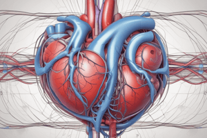
- S.A. node fibers directly connect with surrounding atrial muscle fibers.
- When the S.A. node depolarizes, the impulse spreads through the atrial muscle to the AV node at 0.3 m/s.
- Faster conduction at about 1 m/s occurs through the anterior, middle, and posterior intermodal pathways.
- These pathways transmit impulses from the S.A. node to the A.V. node.
- There is an approximate 0.16-second delay for impulses traveling from the atria to the ventricles.
- The delay allows the atria to empty blood into the ventricles before ventricular contraction.
- The delay is primarily due to a reduced number of gap junctions between cells in the conducting pathways.
- A key feature of the AV bundle is its inability to conduct action potentials backward, except in certain abnormal conditions.
Insulation
- A continuous fibrous barrier separates atrial from ventricular muscle.
- The fibrous barrier insulates and prevents cardiac impulses from passing directly between the atria and ventricles.
- Due to insulation, atrial fibrillation can occur without affecting the ventricles.
- Purkinje fibers are large and conduct impulses at high velocity, roughly six times faster than ventricular muscle.
- This rapid conduction is attributed to a high permeability of gap junctions at the intercalated discs.
- The AV bundle descends within the interventricular septum.
- The AV bundle divides into the left and right bundle branches, which proceed toward the apex of the ventricles.
- Branches subdivide further before connecting with cardiac muscle fibers.
- Impulses spread through the entire ventricular muscle almost immediately, within approximately 0.03 seconds after entering the bundle branches.
- Rapid spread ensures coordinated ventricular muscle contraction for effective pumping.
- Once the impulse enters the ventricular muscle, its velocity slows to about one-sixth the speed of the Purkinje fibers.
- Impulses spiral and take approximately 0.03 seconds to traverse the ventricle, causing a spiral contraction.
Intrinsic Discharge Rates
- Without external stimulation, the S.A. node discharges at 60-100 beats per minute.
- Purkinje fibers discharge at a rate of 15 to 40 beats per minute.
- The S.A. node controls rhythmicity due to its faster discharge rate, a phenomenon called overdriving or over pacing.
- An ectopic pacemaker can develop elsewhere in the heart, leading to disorganized contraction.
- An AV block occurs when impulses from the S.A. node fail to transmit through the AV bundle.
- During an AV block, a new pacemaker may arise in the Purkinje system, driving the heart at 15 to 40 bpm.
- A delay occurs before the Purkinje system takes over, due to the over pacing effect of the S.A. node.
Autonomic Stimulation
- Parasympathetic stimulation releases acetylcholine at vagal nerve endings.
- Acetylcholine reduces the rate and rhythm of the S.A. node.
- Acetylcholine decreases excitability of the AV junctional fibers.
- Mild stimulation can slow the heart, while strong stimulation can halt the S.A. node or block impulse transmission for up to 20 seconds.
- Acetylcholine increases membrane permeability to potassium ions, causing rapid potassium efflux and hyperpolarization.
- Hyperpolarization reduces cell excitability.
- Sympathetic stimulation causes increased S.A. node discharge, enhancing conduction rate, excitability, and contraction force.
- Norepinephrine, released from sympathetic nerve endings, stimulates beta one adrenergic receptors.
- Beta one receptor activation increases membrane permeability to sodium and calcium ions.
- Increased sodium and calcium permeability results in a more positive resting potential, accelerating self-excitation.
- Action potentials conduct more easily through the bundles, reducing conduction time from atria to ventricles.
Studying That Suits You
Use AI to generate personalized quizzes and flashcards to suit your learning preferences.