Podcast
Questions and Answers
Which part of the heart's conduction system is located in the right atrium?
Which part of the heart's conduction system is located in the right atrium?
- Purkinje Fibres
- SA-Node (correct)
- AV-Node
- AV-Bundle
What is the primary function of erythrocytes?
What is the primary function of erythrocytes?
- Circulating immune cells
- Help regulate pulse pressure
- Distribute oxygen and remove CO₂ (correct)
- Detect changes in blood pressure
Which wave on an ECG represents atrial depolarization?
Which wave on an ECG represents atrial depolarization?
- S-T segment
- P wave (correct)
- QRS complex
- T wave
What is the function of baroreceptors?
What is the function of baroreceptors?
Which type of circulation involves the right side of the heart and the lungs?
Which type of circulation involves the right side of the heart and the lungs?
Which hormones can affect heart rate?
Which hormones can affect heart rate?
What is the QRS complex associated with?
What is the QRS complex associated with?
Where are the cardiovascular system's baroreceptors primarily located?
Where are the cardiovascular system's baroreceptors primarily located?
Which of the following is NOT a layer of the heart?
Which of the following is NOT a layer of the heart?
Which structure is located on the left side of the heart, anterior view?
Which structure is located on the left side of the heart, anterior view?
Which type of cell is a major component of blood?
Which type of cell is a major component of blood?
What is characteristic of cardiac muscle histology?
What is characteristic of cardiac muscle histology?
Which artery is part of the right side of the heart, anterior view?
Which artery is part of the right side of the heart, anterior view?
Which layer of the heart is composed of dense irregular connective tissue?
Which layer of the heart is composed of dense irregular connective tissue?
Which vessel returns deoxygenated blood from the lower body to the heart?
Which vessel returns deoxygenated blood from the lower body to the heart?
Which component is primarily involved in generating and conducting electrical impulses in the heart?
Which component is primarily involved in generating and conducting electrical impulses in the heart?
Which division of the aorta directly supplies the thoracic region?
Which division of the aorta directly supplies the thoracic region?
Where is venous blood drained after circulating through the body?
Where is venous blood drained after circulating through the body?
Which of the following describes the tunica media of an artery?
Which of the following describes the tunica media of an artery?
What function do elastic arteries primarily serve?
What function do elastic arteries primarily serve?
Which type of artery is adapted for greater vasoconstriction and vasodilation?
Which type of artery is adapted for greater vasoconstriction and vasodilation?
What is the purpose of precapillary sphincters in arterioles?
What is the purpose of precapillary sphincters in arterioles?
Which of the following is NOT a characteristic of capillaries?
Which of the following is NOT a characteristic of capillaries?
What is one defining feature of venules as they approach the size of veins?
What is one defining feature of venules as they approach the size of veins?
Flashcards are hidden until you start studying
Study Notes
Heart Valves and Circulation of Blood
- Heart valves open and close in response to pressure changes as the heart contracts and relaxes.
- Dense connective tissue rings surround the valves of the heart, providing:
- Support for heart valves
- Insertion point for cardiac muscle bundles
- Electrical insulator between atria and ventricles
Electrical Conductance
- Electrical conduction pathway:
- SA-Node (in right atrium)
- AV-Node
- AV-Bundle (Bundle of His)
- Right & Left Bundle Branches
- Purkinje Fibres
Regulation of Heart Rate
- Nervous control from cardiovascular centre in the medulla:
- Sympathetic impulses increase heart rate and force of contraction
- Parasympathetic impulses decrease heart rate
- Baroreceptors (pressure receptors) detect changes in BP and send info to the cardiovascular center:
- Located in the arch of the aorta and carotid sinuses
- Heart rate is also affected by:
- Hormones (adrenaline, noradrenaline, thyroid hormones)
- Ions (Na+, K+, Ca2+)
- Age, gender, physical fitness, and temperature
Physiological Functions of the Cardiovascular System
Heart
- Pumps over 1 million gallons per year
- Over 60,000 miles of blood vessels
Vessels
- Retain and circulate blood
- Help regulate pulse pressure
Blood
- Erythrocytes:
- Distribute Oxygen from & CO₂ to lungs
- Leukocytes:
- Circulating immune cells to combat infection
Electrocardiogram (ECG or EKG)
- Measures action potentials of all active cells
- Components:
- P wave: atrial depolarization
- P to Q interval: conduction time from atrial to ventricular excitation
- QRS complex: ventricular depolarization
- T wave: ventricular repolarization
- Q-T Interval: time for ventricular depolarization and repolarization to occur
- S-T segment: ventricular fibres are depolarized (plateau phase of AP)
Circulatory Routes
- Systemic circulation: left side heart to body & back to heart
- Hepatic Portal circulation: capillaries of GI tract to capillaries in liver
- Pulmonary circulation: right-side heart to lungs & back to heart
- Foetal circulation: from fetal heart through umbilical cord to placenta & back
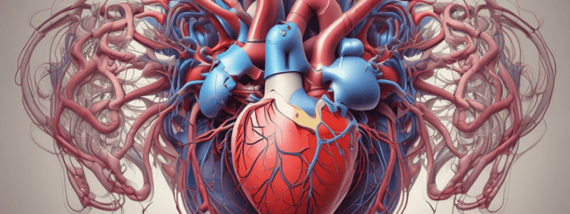
Cardiovascular System
Organs/Structures
- Organs:
- Heart
- Blood vessels
- Blood
- Structures:
- Layers of the heart
- Pericardium
- Epicardium
- Myocardium
- Endocardium
- Valves
- Chambers
- Major vessels
- Major cell types of blood
Gross Anatomy
Right Side of the Heart (Anterior View)
- Brachiocephalic trunk
- Superior vena cava
- Ascending aorta
- Right pulmonary artery
- Parietal pericardium (cut)
- Right Auricle of Right Atrium
- Right coronary artery
- Right Atrium
- Coronary Sulcus
- Right ventricle
- Inferior vena cava
Left Side of the Heart (Anterior View)
- Left common carotid artery
- Left subclavian artery
- Arch of aorta
- Ligamentum arteriosum
- Left pulmonary artery
- Pulmonary trunk
- Left pulmonary veins
- Left Auricle of Left Atrium
- Branch of left coronary artery
- Left ventricle
- Anterior interventricular sulcus
- Descending aorta
Cell Types
Heart
- Pericardium
- Epicardium
- Myocardium (Cardiac myocytes)
- Valves
- Conducting Nerve Bundles (SA/AV Nodes)
- Pericardium
Blood Vessels
- Endothelium
Blood
- Leukocytes: white blood cells
- Erythrocytes: red blood cells
Layers of Heart Wall
- Pericardium:
- Dense irregular connective tissue
- Epicardium:
- Visceral layer of serous pericardium
- Myocardium:
- Cardiac muscle layer
- Endocardium:
- Chamber lining & valves, smooth lubricating layer
Thickness of Cardiac Walls
- Myocardium of left ventricle is thicker than right ventricle
Cardiac Muscle Histology
- Branching, intercalated discs with gap junctions, involuntary, striated, single central nucleus per cell
- Desmosomes between myocytes allow depolarization of adjacent fibers
Arteries and Veins
Arterial Branches of Systemic Circulation
- 4 major divisions of aorta:
- Ascending aorta
- Arch of aorta
- Thoracic aorta
- Abdominal aorta
Veins of the Systemic Circulation
- Drain blood from entire body and return it to the right side of the heart
- Deep veins parallel the arteries in the region
- Superficial veins are found just beneath the skin
- All venous blood drains to either superior or inferior vena cava or coronary sinus
Arteries & Veins
Tunica Interna
- Endothelium
- Basement Membrane
- Internal Elastic Lamina
Tunica Media
- Smooth Muscle
- External Elastic Lamina
Tunica Externa
Elastic Arteries
- Large arteries with more elastic fibers and less smooth muscle
- Conducting arteries because they conduct blood from the heart to medium-sized muscular arteries
Muscular Arteries
- Medium-sized arteries with more muscle than elastic fibers in tunica media
- Capable of greater vasoconstriction and vasodilation to adjust rate of flow
- Walls are relatively thick
- Called distributing arteries because they direct blood flow
Arterioles
- Small arteries delivering blood to capillaries
- Tunica media containing few layers of muscle
- Metarterioles from branches into capillary bed
- To bypass capillary bed, precapillary sphincters close, and blood flows out of bed in the thoroughfare channel
- Vasomotion is intermittent: contraction and relaxation of sphincters that allow filling of capillary beds 5-10 times/minute
Capillaries
- Found near every cell but more extensive in highly active tissue (muscles, liver, kidneys & brain)
- Entire capillary bed fills with blood when tissue is active
- Capillary walls are composed of only a single layer of cells (endothelium) and a basement membrane
- Red blood cells passing through a capillary
Venules
- Small veins collecting blood from capillaries
- Tunica media contains only a few smooth muscle cells and scattered fibroblasts
- Very porous endothelium allows for escape of many phagocytic white blood cells
- Venules that approach size of veins more closely resemble structure of veins
Studying That Suits You
Use AI to generate personalized quizzes and flashcards to suit your learning preferences.


