Podcast
Questions and Answers
What part of the heart receives deoxygenated blood and pumps it to the lungs?
What part of the heart receives deoxygenated blood and pumps it to the lungs?
- Left atrium
- Right ventricle (correct)
- Right atrium
- Left ventricle
The epicardium is the deepest layer of the heart wall.
The epicardium is the deepest layer of the heart wall.
False (B)
What is the function of the myocardium?
What is the function of the myocardium?
It is responsible for the contraction of the heart and pumping blood.
The __________ pericardium is the sac that the heart sits in.
The __________ pericardium is the sac that the heart sits in.
Match the following heart structures with their functions:
Match the following heart structures with their functions:
Which blood vessel returns deoxygenated blood from the head and neck to the heart?
Which blood vessel returns deoxygenated blood from the head and neck to the heart?
The aortic semilunar valve has two cusps.
The aortic semilunar valve has two cusps.
What are the muscular ridges found beneath the auricle of the atrial wall called?
What are the muscular ridges found beneath the auricle of the atrial wall called?
The ___ is the flattened superior portion of the heart where blood vessels emerge.
The ___ is the flattened superior portion of the heart where blood vessels emerge.
Match the following vessels to their function:
Match the following vessels to their function:
What is the function of chordae tendineae?
What is the function of chordae tendineae?
What is the primary function of hemoglobin in red blood cells?
What is the primary function of hemoglobin in red blood cells?
The foramen ovale allows blood to flow from the left atrium to the right atrium.
The foramen ovale allows blood to flow from the left atrium to the right atrium.
Cardiac muscle tissue has a multinucleated structure.
Cardiac muscle tissue has a multinucleated structure.
What do the structures foramen ovale and ductus arteriosus become after birth?
What do the structures foramen ovale and ductus arteriosus become after birth?
What mechanism allows electrical signals to pass easily between cardiac muscle cells?
What mechanism allows electrical signals to pass easily between cardiac muscle cells?
The smallest cell fragment involved in clotting is called a ______.
The smallest cell fragment involved in clotting is called a ______.
Match each type of blood cell with its function:
Match each type of blood cell with its function:
Which component of cardiac muscle tissue assists in holding cells tightly together?
Which component of cardiac muscle tissue assists in holding cells tightly together?
Neutrophils are responsible for producing antibodies.
Neutrophils are responsible for producing antibodies.
What are two major structures located in the dissected heart that are involved in preventing backflow of blood?
What are two major structures located in the dissected heart that are involved in preventing backflow of blood?
Describe the role of the right atrium in the heart's function.
Describe the role of the right atrium in the heart's function.
What are the components of the pericardium and their functions?
What are the components of the pericardium and their functions?
Explain the significance of the interventricular septum.
Explain the significance of the interventricular septum.
Identify and describe the three layers of the heart wall.
Identify and describe the three layers of the heart wall.
What is the role of the auricle in the atria?
What is the role of the auricle in the atria?
Explain the function of the semilunar valves.
Explain the function of the semilunar valves.
Describe the anatomical significance of the anterior interventricular sulcus.
Describe the anatomical significance of the anterior interventricular sulcus.
What prevents the cusps of the atrioventricular valves from being pushed back into the atria?
What prevents the cusps of the atrioventricular valves from being pushed back into the atria?
Where does the superior vena cava return blood from?
Where does the superior vena cava return blood from?
How does the structure of the tricuspid valve differ from the bicuspid valve?
How does the structure of the tricuspid valve differ from the bicuspid valve?
What is the role of intercalated disks in cardiac muscle tissue?
What is the role of intercalated disks in cardiac muscle tissue?
Describe the primary structural features of erythrocytes.
Describe the primary structural features of erythrocytes.
What distinguishes leukocytes from erythrocytes?
What distinguishes leukocytes from erythrocytes?
Explain the process of hemopoiesis.
Explain the process of hemopoiesis.
What is the function of the pulmonary trunk?
What is the function of the pulmonary trunk?
What role do platelets play in the hemostatic process?
What role do platelets play in the hemostatic process?
Identify the primary components of hemoglobin.
Identify the primary components of hemoglobin.
Match the parts of the pericardium with their descriptions:
Match the parts of the pericardium with their descriptions:
Match the layers of the heart wall with their characteristics:
Match the layers of the heart wall with their characteristics:
Match the following components of cardiac muscle tissue with their functions:
Match the following components of cardiac muscle tissue with their functions:
Match the following heart valves with their respective locations:
Match the following heart valves with their respective locations:
Match the following anatomical positions of the heart with their descriptions:
Match the following anatomical positions of the heart with their descriptions:
Match the following structures involved in fetal circulation with their functions:
Match the following structures involved in fetal circulation with their functions:
Flashcards
Heart Chambers
Heart Chambers
Separate compartments within the heart (2 atria & 2 ventricles), responsible for receiving and pumping blood; right receives deoxygenated, pumps to lungs; left receives oxygenated, pumps to body
Pericardium Layers
Pericardium Layers
Protective covering of the heart; composed of fibrous (outer bag) and serous membranes (inner layers); serous with parietal (outer) and visceral (inner, epicardium) layers, with serous fluid preventing friction
Myocardium
Myocardium
Muscle layer of the heart wall, responsible for pumping blood.
Atria
Atria
Signup and view all the flashcards
Ventricle function
Ventricle function
Signup and view all the flashcards
Superior Vena Cava
Superior Vena Cava
Signup and view all the flashcards
Atrioventricular (AV) Valve
Atrioventricular (AV) Valve
Signup and view all the flashcards
Tricuspid Valve
Tricuspid Valve
Signup and view all the flashcards
Semilunar Valves
Semilunar Valves
Signup and view all the flashcards
Foramen Ovale
Foramen Ovale
Signup and view all the flashcards
Chordae Tendineae
Chordae Tendineae
Signup and view all the flashcards
Papillary Muscles
Papillary Muscles
Signup and view all the flashcards
Cardiac Muscle Tissue
Cardiac Muscle Tissue
Signup and view all the flashcards
Intercalated Disks
Intercalated Disks
Signup and view all the flashcards
Erythrocytes
Erythrocytes
Signup and view all the flashcards
Hemoglobin
Hemoglobin
Signup and view all the flashcards
Leukocytes
Leukocytes
Signup and view all the flashcards
Neutrophils
Neutrophils
Signup and view all the flashcards
Platelets
Platelets
Signup and view all the flashcards
Blood (general)
Blood (general)
Signup and view all the flashcards
What are the chambers of the heart?
What are the chambers of the heart?
Signup and view all the flashcards
Right side of the heart
Right side of the heart
Signup and view all the flashcards
Left side of the heart
Left side of the heart
Signup and view all the flashcards
Pericardium
Pericardium
Signup and view all the flashcards
Base of the Heart
Base of the Heart
Signup and view all the flashcards
Apex of the Heart
Apex of the Heart
Signup and view all the flashcards
Atrioventricular Valve (AV Valve)
Atrioventricular Valve (AV Valve)
Signup and view all the flashcards
Bicuspid (Mitral) Valve
Bicuspid (Mitral) Valve
Signup and view all the flashcards
Gap Junctions
Gap Junctions
Signup and view all the flashcards
Desmosomes
Desmosomes
Signup and view all the flashcards
Platelet Plug
Platelet Plug
Signup and view all the flashcards
Pulmonary Arteries
Pulmonary Arteries
Signup and view all the flashcards
Pulmonary Veins
Pulmonary Veins
Signup and view all the flashcards
Pulmonary Semilunar Valve
Pulmonary Semilunar Valve
Signup and view all the flashcards
Heart Location
Heart Location
Signup and view all the flashcards
Right Heart Function
Right Heart Function
Signup and view all the flashcards
Left Heart Function
Left Heart Function
Signup and view all the flashcards
Pericardium Roles
Pericardium Roles
Signup and view all the flashcards
Heart Wall Layers
Heart Wall Layers
Signup and view all the flashcards
Study Notes
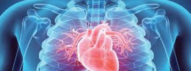
Heart Structure and Function
- The heart is a four-chambered organ in the mediastinum of the thoracic cavity.
- The right side receives deoxygenated blood from the body and pumps it to the lungs.
- The left side receives oxygenated blood from the lungs and pumps it to the body.
- The pericardium surrounds the heart, comprising fibrous and serous layers.
- The fibrous pericardium is a protective sac.
- The serous pericardium has parietal and visceral layers, separated by fluid to reduce friction.
- Atria are the upper chambers receiving blood. The right atrium receives from systemic circulation, and the left atrium from the pulmonary circulation.
- Ventricles are the lower chambers, pumping blood. The right ventricle pumps to the lungs, and the left ventricle pumps to the body.
Heart Wall Layers
- The heart wall has three layers:
- Epicardium (visceral pericardium): the outermost superficial layer.
- Myocardium: the middle muscular layer responsible for contraction.
- Endocardium: the innermost layer lining the heart chambers and blood vessels.
Heart Valves
- Atrioventricular (AV) valves prevent backflow between atria and ventricles.
- Tricuspid valve: between the right atrium and ventricle (three cusps).
- Bicuspid (mitral) valve: between the left atrium and ventricle (two cusps).
- Chordae tendineae and papillary muscles anchor the AV valve cusps.
- Semilunar (SL) valves prevent backflow from arteries into ventricles.
- Pulmonary semilunar valve: between the right ventricle and pulmonary trunk.
- Aortic semilunar valve: between the left ventricle and aorta.
Fetal Circulation
- During fetal development, blood flow is rerouted away from the lungs.
- The foramen ovale allows blood to flow from the right to the left atrium.
- The ductus arteriosus shunts blood from the pulmonary trunk to the aorta.
- These structures close after birth.
Blood Vessels Entering/Exiting the Heart
- Superior vena cava: returns deoxygenated blood from the head and upper body to the right atrium.
- Inferior vena cava: returns deoxygenated blood from the lower body to the right atrium.
- Coronary sinus: returns coronary circulation blood to the right atrium.
- Pulmonary trunk: carries deoxygenated blood from the right ventricle to the lungs.
- Aorta: carries oxygenated blood from the left ventricle to the body.
Additional Heart Structures
- Auricle: external extension of the atria, increases volume.
- Interatrial septum: divides the atria.
- Interventricular septum: divides the ventricles.
- Pectinate muscles: ridges in the atria walls.
- Trabeculae carneae: ridges in the ventricles.
- Interventricular sulci: grooves separating ventricles.
Studying That Suits You
Use AI to generate personalized quizzes and flashcards to suit your learning preferences.


