Podcast
Questions and Answers
What is the primary function of the pulmonary trunk in the heart's circulation system?
What is the primary function of the pulmonary trunk in the heart's circulation system?
The pulmonary trunk transports deoxygenated blood from the right ventricle to the lungs.
Identify the two sets of valves in the heart and their respective locations.
Identify the two sets of valves in the heart and their respective locations.
The two sets are the atrioventricular (AV) valves located between the atria and ventricles, and the semilunar valves located at the boundary between each ventricle and its associated arterial trunk.
Describe the role of the aortic semilunar valve.
Describe the role of the aortic semilunar valve.
The aortic semilunar valve is located between the left ventricle and the aorta, allowing oxygenated blood to flow into the systemic circulation and preventing backflow.
What distinguishes the pulmonary arteries from other arteries in the body?
What distinguishes the pulmonary arteries from other arteries in the body?
Explain the term 'pulmonary circulation' and its significance.
Explain the term 'pulmonary circulation' and its significance.
What is the path of blood flow through the heart starting from the right atrium?
What is the path of blood flow through the heart starting from the right atrium?
Differentiate between systemic circulation and pulmonary circulation.
Differentiate between systemic circulation and pulmonary circulation.
What clinical signs indicate right ventricle impairment due to congestive heart failure?
What clinical signs indicate right ventricle impairment due to congestive heart failure?
Why is the oxygenated blood returned from the lungs to the left atrium important?
Why is the oxygenated blood returned from the lungs to the left atrium important?
What is the effect of left ventricle impairment in congestive heart failure?
What is the effect of left ventricle impairment in congestive heart failure?
What are the three major types of blood vessels in the cardiovascular system?
What are the three major types of blood vessels in the cardiovascular system?
Describe the position of the heart within the thoracic cavity.
Describe the position of the heart within the thoracic cavity.
What is the primary function of pulmonary circulation in the human body?
What is the primary function of pulmonary circulation in the human body?
What is the primary function of the heart in the cardiovascular system?
What is the primary function of the heart in the cardiovascular system?
Describe the flow of oxygenated blood starting from the left atrium.
Describe the flow of oxygenated blood starting from the left atrium.
What structures ensure one-way blood flow within the heart?
What structures ensure one-way blood flow within the heart?
Explain the structural composition of the pericardium and its layers.
Explain the structural composition of the pericardium and its layers.
Explain the role of capillaries in the cardiovascular system.
Explain the role of capillaries in the cardiovascular system.
How does the positioning of the heart within the thoracic cavity differ between the left and right sides?
How does the positioning of the heart within the thoracic cavity differ between the left and right sides?
What role does the pericardial cavity play in heart function?
What role does the pericardial cavity play in heart function?
Identify the components of the heart's conduction system responsible for initiating the action potential.
Identify the components of the heart's conduction system responsible for initiating the action potential.
Identify the route taken by deoxygenated blood from the systemic veins to the right atrium.
Identify the route taken by deoxygenated blood from the systemic veins to the right atrium.
What occurs during the refractory period of the cardiac cycle?
What occurs during the refractory period of the cardiac cycle?
Differentiate between the blood supplied to the right side and the left side of the heart.
Differentiate between the blood supplied to the right side and the left side of the heart.
What is the relationship between the pulmonary arteries and the lungs, specifically regarding blood supply?
What is the relationship between the pulmonary arteries and the lungs, specifically regarding blood supply?
Explain how systemic circulation differs from pulmonary circulation?
Explain how systemic circulation differs from pulmonary circulation?
What occurs during a myocardial infarction?
What occurs during a myocardial infarction?
Describe the protective functions of the fibrous pericardium.
Describe the protective functions of the fibrous pericardium.
How do arteries and veins differ in their structural features?
How do arteries and veins differ in their structural features?
How does the heart's shape and size influence its function?
How does the heart's shape and size influence its function?
What is cardiac output, and why is it important?
What is cardiac output, and why is it important?
What are the main consequences of cardiovascular system failure to maintain adequate perfusion?
What are the main consequences of cardiovascular system failure to maintain adequate perfusion?
Name the two major veins that return deoxygenated blood to the right atrium.
Name the two major veins that return deoxygenated blood to the right atrium.
How does the structure of the heart support its function as a pump?
How does the structure of the heart support its function as a pump?
What are the main symptoms associated with Hypertrophic Cardiomyopathy?
What are the main symptoms associated with Hypertrophic Cardiomyopathy?
How does Hypertrophic Cardiomyopathy differ from Cardiomegaly in terms of structural changes in the heart?
How does Hypertrophic Cardiomyopathy differ from Cardiomegaly in terms of structural changes in the heart?
What role do heart sounds play in diagnosing heart conditions?
What role do heart sounds play in diagnosing heart conditions?
Explain the significance of valvular insufficiency related to heart murmurs.
Explain the significance of valvular insufficiency related to heart murmurs.
What diagnostic method is primarily used to detect Hypertrophic Cardiomyopathy?
What diagnostic method is primarily used to detect Hypertrophic Cardiomyopathy?
What structure covers the heart and consists of three layers?
What structure covers the heart and consists of three layers?
Describe the location and function of the pericardial cavity.
Describe the location and function of the pericardial cavity.
Identify the main vessels connected to the left atrium from an anterior view.
Identify the main vessels connected to the left atrium from an anterior view.
What are the effects of excess fluid accumulation in the pericardial cavity?
What are the effects of excess fluid accumulation in the pericardial cavity?
What is the function of the myocardial layer of the heart wall?
What is the function of the myocardial layer of the heart wall?
Name the two sulci that separate the ventricles from each other.
Name the two sulci that separate the ventricles from each other.
What is pericarditis and its primary symptom?
What is pericarditis and its primary symptom?
Which layer of the heart wall is also known as the visceral pericardium?
Which layer of the heart wall is also known as the visceral pericardium?
List the main vessels seen in the anterior view of the heart.
List the main vessels seen in the anterior view of the heart.
What is the significance of the coronary sulcus?
What is the significance of the coronary sulcus?
What is the main function of the myocardium layer in the heart?
What is the main function of the myocardium layer in the heart?
How does the thickness of the left ventricular wall compare to that of the right ventricular wall?
How does the thickness of the left ventricular wall compare to that of the right ventricular wall?
What are the fossa ovalis and its significance in the heart?
What are the fossa ovalis and its significance in the heart?
Where do deoxygenated blood and oxygenated blood enter the heart?
Where do deoxygenated blood and oxygenated blood enter the heart?
What role do papillary muscles play in heart function?
What role do papillary muscles play in heart function?
Which valves are involved in preventing backflow during ventricular contraction?
Which valves are involved in preventing backflow during ventricular contraction?
What condition is characterized by an enlargement of the heart?
What condition is characterized by an enlargement of the heart?
Describe the structure of semilunar valves and their function.
Describe the structure of semilunar valves and their function.
What separates the left and right atria in the heart?
What separates the left and right atria in the heart?
What happens to the flexibility and elasticity of heart valves with age or disease?
What happens to the flexibility and elasticity of heart valves with age or disease?
What are trabeculae carneae and where are they found?
What are trabeculae carneae and where are they found?
Name the two types of heart valves and their locations.
Name the two types of heart valves and their locations.
What condition is characterized by thickening of the heart muscle?
What condition is characterized by thickening of the heart muscle?
What structures prevent the AV valves from being forced back into the atria?
What structures prevent the AV valves from being forced back into the atria?
What is the significance of the resting membrane potential (RMP) of -60 mV in SA nodal cells?
What is the significance of the resting membrane potential (RMP) of -60 mV in SA nodal cells?
Explain the role of voltage-gated Ca2+ channels during depolarization in SA nodal cells.
Explain the role of voltage-gated Ca2+ channels during depolarization in SA nodal cells.
How does parasympathetic stimulation affect the heart rate regulated by the SA node?
How does parasympathetic stimulation affect the heart rate regulated by the SA node?
Describe the process of repolarization in SA nodal cells.
Describe the process of repolarization in SA nodal cells.
What is autorhythmicity, and why is it vital for SA nodal cells?
What is autorhythmicity, and why is it vital for SA nodal cells?
What distinguishes SA nodal cells from neurons in terms of action potential generation?
What distinguishes SA nodal cells from neurons in terms of action potential generation?
How does the fibrous skeleton of the heart affect the conduction rate of action potentials?
How does the fibrous skeleton of the heart affect the conduction rate of action potentials?
What are ectopic pacemakers, and how do they differ from the SA node?
What are ectopic pacemakers, and how do they differ from the SA node?
Explain how pacemaker potentials facilitate the SA node's ability to set the heart's rhythm.
Explain how pacemaker potentials facilitate the SA node's ability to set the heart's rhythm.
What is the typical intrinsic heart rate set by SA nodal cells, and how does it get modified?
What is the typical intrinsic heart rate set by SA nodal cells, and how does it get modified?
What role does the SA node play in the cardiac conduction system?
What role does the SA node play in the cardiac conduction system?
What happens if the SA node fails to function properly?
What happens if the SA node fails to function properly?
How does an action potential propagate through the cardiac conduction system?
How does an action potential propagate through the cardiac conduction system?
What is the significance of the delay in conduction at the AV node?
What is the significance of the delay in conduction at the AV node?
What is the membrane potential of cardiac muscle cells at rest?
What is the membrane potential of cardiac muscle cells at rest?
What triggers the depolarization in cardiac muscle cells?
What triggers the depolarization in cardiac muscle cells?
Define the plateau phase in cardiac muscle electrical events.
Define the plateau phase in cardiac muscle electrical events.
What is the role of calcium ions in cardiac muscle contraction?
What is the role of calcium ions in cardiac muscle contraction?
What are the three key electrical events during an action potential in cardiac muscle cells?
What are the three key electrical events during an action potential in cardiac muscle cells?
What is the refractory period in cardiac cells?
What is the refractory period in cardiac cells?
Explain the role of mechanical pacemakers in cardiac function.
Explain the role of mechanical pacemakers in cardiac function.
How do cardiac muscle cells at rest maintain a depolarized state?
How do cardiac muscle cells at rest maintain a depolarized state?
Describe the mechanical events that occur during cardiac muscle contraction.
Describe the mechanical events that occur during cardiac muscle contraction.
What occurs during repolarization of cardiac muscle cells?
What occurs during repolarization of cardiac muscle cells?
Why is the plateau phase essential for cardiac muscle function?
Why is the plateau phase essential for cardiac muscle function?
What is the primary structural component of the fibrous skeleton of the heart?
What is the primary structural component of the fibrous skeleton of the heart?
How do coronary arteries differ from coronary veins in their function?
How do coronary arteries differ from coronary veins in their function?
What complication can arise from a blockage in the anterior interventricular artery?
What complication can arise from a blockage in the anterior interventricular artery?
What are the main veins that drain deoxygenated blood from the heart?
What are the main veins that drain deoxygenated blood from the heart?
Describe the role of the coronary sinus in cardiac circulation.
Describe the role of the coronary sinus in cardiac circulation.
What characterizes the coronary arteries as functional end arteries?
What characterizes the coronary arteries as functional end arteries?
What is valvular stenosis and what can cause it?
What is valvular stenosis and what can cause it?
How does a myocardial infarction typically occur?
How does a myocardial infarction typically occur?
Identify an unusual symptom of myocardial infarction that may be experienced by women.
Identify an unusual symptom of myocardial infarction that may be experienced by women.
What effect does the compression of coronary vessels during heart contraction have?
What effect does the compression of coronary vessels during heart contraction have?
What function do intercalated discs serve in cardiac muscle cells?
What function do intercalated discs serve in cardiac muscle cells?
Describe the significance of the sinoatrial (SA) node in the heart.
Describe the significance of the sinoatrial (SA) node in the heart.
What is the role of myoglobin in cardiac muscle cells?
What is the role of myoglobin in cardiac muscle cells?
Explain how cardiac muscle metabolism differs from that of skeletal muscle.
Explain how cardiac muscle metabolism differs from that of skeletal muscle.
What role do the SA node and AV node play in heart function?
What role do the SA node and AV node play in heart function?
How do parasympathetic and sympathetic innervations differ in their effects on heart rate?
How do parasympathetic and sympathetic innervations differ in their effects on heart rate?
Explain the significance of baroreceptors in the cardiovascular system.
Explain the significance of baroreceptors in the cardiovascular system.
Describe the three primary phases of action potential in SA nodal cells.
Describe the three primary phases of action potential in SA nodal cells.
What determines the unstable resting membrane potential of SA nodal cells?
What determines the unstable resting membrane potential of SA nodal cells?
How does the cardiac center in the medulla oblongata influence heart activity?
How does the cardiac center in the medulla oblongata influence heart activity?
What happens during the depolarization phase in SA nodal cells?
What happens during the depolarization phase in SA nodal cells?
Discuss the consequences of ischemic conditions on cardiac muscle.
Discuss the consequences of ischemic conditions on cardiac muscle.
What is the role of Purkinje fibers in the conduction system of the heart?
What is the role of Purkinje fibers in the conduction system of the heart?
How do sympathetic and parasympathetic innervations affect coronary artery function?
How do sympathetic and parasympathetic innervations affect coronary artery function?
Explain the relationship between cardiac muscle cells and aerobic metabolism.
Explain the relationship between cardiac muscle cells and aerobic metabolism.
Describe the function of voltage-gated channels in SA nodal cells.
Describe the function of voltage-gated channels in SA nodal cells.
What does repolarization involve in SA nodal cells, and why is it important?
What does repolarization involve in SA nodal cells, and why is it important?
What impact does the cardiac center have on the overall cardiovascular system during stress?
What impact does the cardiac center have on the overall cardiovascular system during stress?
What are the five phases of the cardiac cycle?
What are the five phases of the cardiac cycle?
How does pressure change in the heart contribute to the opening and closing of valves?
How does pressure change in the heart contribute to the opening and closing of valves?
What is the significance of end-diastolic volume (EDV) in the cardiac cycle?
What is the significance of end-diastolic volume (EDV) in the cardiac cycle?
Define isovolumic contraction and its importance in the cardiac cycle.
Define isovolumic contraction and its importance in the cardiac cycle.
What role does atrial systole play in the cardiac cycle?
What role does atrial systole play in the cardiac cycle?
Explain the phenomenon of isovolumic relaxation.
Explain the phenomenon of isovolumic relaxation.
What happens during ventricular ejection and its significance?
What happens during ventricular ejection and its significance?
How does the contraction of the atria prevent backflow of blood?
How does the contraction of the atria prevent backflow of blood?
Describe the relationship between stroke volume (SV) and end-systolic volume (ESV).
Describe the relationship between stroke volume (SV) and end-systolic volume (ESV).
What is the role of the semilunar valves during the cardiac cycle?
What is the role of the semilunar valves during the cardiac cycle?
How do semilunar valves function during ventricular contraction?
How do semilunar valves function during ventricular contraction?
What condition occurs if there is sustained unequal blood pumping from the left and right ventricles?
What condition occurs if there is sustained unequal blood pumping from the left and right ventricles?
What components are involved in the calculation of cardiac output?
What components are involved in the calculation of cardiac output?
Explain the significance of the dicrotic notch in the aortic pressure curve.
Explain the significance of the dicrotic notch in the aortic pressure curve.
What distinguishes the left ventricle from the right ventricle concerning its structure and function?
What distinguishes the left ventricle from the right ventricle concerning its structure and function?
Describe the phases of the cardiac cycle.
Describe the phases of the cardiac cycle.
What physiological changes occur in the heart during isovolumic relaxation?
What physiological changes occur in the heart during isovolumic relaxation?
How can cardiac reserve be assessed?
How can cardiac reserve be assessed?
What role does the P wave play in an ECG reading?
What role does the P wave play in an ECG reading?
Identify the primary causes behind the opening and closing of the AV valves.
Identify the primary causes behind the opening and closing of the AV valves.
What is the consequence of a higher heart rate in individuals with smaller hearts?
What is the consequence of a higher heart rate in individuals with smaller hearts?
What does stroke volume signify in terms of cardiac function?
What does stroke volume signify in terms of cardiac function?
Explain the relationship between cardiac output and daily blood volume circulation.
Explain the relationship between cardiac output and daily blood volume circulation.
What distinguishes the systemic circulation from the pulmonary circulation in the heart's function?
What distinguishes the systemic circulation from the pulmonary circulation in the heart's function?
What role does ATP play in the release and reset of the myosin head during muscle contraction?
What role does ATP play in the release and reset of the myosin head during muscle contraction?
Why is the refractory period in cardiac muscle cells longer than in skeletal muscle fibers?
Why is the refractory period in cardiac muscle cells longer than in skeletal muscle fibers?
What are the three principal deflections observed in a typical ECG tracing?
What are the three principal deflections observed in a typical ECG tracing?
How do Ca2+ channel blockers affect the heart's contraction strength and rate?
How do Ca2+ channel blockers affect the heart's contraction strength and rate?
Define the P-R interval in relation to cardiac conduction.
Define the P-R interval in relation to cardiac conduction.
Describe the consequences of a third-degree AV block.
Describe the consequences of a third-degree AV block.
What is the significance of the T wave on an ECG?
What is the significance of the T wave on an ECG?
Explain ventricular fibrillation and its potential consequences.
Explain ventricular fibrillation and its potential consequences.
How does the cardiac cycle ensure that the heart chambers contract and relax efficiently?
How does the cardiac cycle ensure that the heart chambers contract and relax efficiently?
Describe how high-frequency stimulation affects skeletal muscle contraction.
Describe how high-frequency stimulation affects skeletal muscle contraction.
What is the role of the AV node in cardiac conduction?
What is the role of the AV node in cardiac conduction?
What is the Q-T interval and its clinical significance?
What is the Q-T interval and its clinical significance?
Discuss how abnormal waves on an ECG can indicate potential heart problems.
Discuss how abnormal waves on an ECG can indicate potential heart problems.
Why is the presence of an AED important in public spaces?
Why is the presence of an AED important in public spaces?
What are the potential consequences if a patent foramen ovale remains open in a newborn?
What are the potential consequences if a patent foramen ovale remains open in a newborn?
How do calcium channel blockers affect heart function?
How do calcium channel blockers affect heart function?
Describe the role of intercalated discs in cardiac muscle tissue.
Describe the role of intercalated discs in cardiac muscle tissue.
What structural differences exist between the walls of the atria and ventricles?
What structural differences exist between the walls of the atria and ventricles?
Explain the significance of the coronary vessels in heart health.
Explain the significance of the coronary vessels in heart health.
What is the impact of cutting the right vagus nerve on heart rate?
What is the impact of cutting the right vagus nerve on heart rate?
How does the right ventricle's wall thickness compare to the left ventricle, and why?
How does the right ventricle's wall thickness compare to the left ventricle, and why?
What symptoms may indicate the presence of angina?
What symptoms may indicate the presence of angina?
How does bradycardia affect cardiac output?
How does bradycardia affect cardiac output?
What is the embryonic origin of the heart's four chambers?
What is the embryonic origin of the heart's four chambers?
Describe the role of the pericardial fluid.
Describe the role of the pericardial fluid.
What is a common result of an incomplete partition in the interatrial septum?
What is a common result of an incomplete partition in the interatrial septum?
How does the position of the heart within the thoracic cavity affect its visibility?
How does the position of the heart within the thoracic cavity affect its visibility?
What structural feature separates the atria from the ventricles?
What structural feature separates the atria from the ventricles?
What are intercalated discs, and why are they important in cardiac muscle?
What are intercalated discs, and why are they important in cardiac muscle?
How does the heart's electrical conduction system initiate a heartbeat?
How does the heart's electrical conduction system initiate a heartbeat?
What happens to blood flow if the foramen ovale remains open after birth?
What happens to blood flow if the foramen ovale remains open after birth?
What is the function of the coronary arteries?
What is the function of the coronary arteries?
Identify a potential consequence of inadequate oxygen supply to cardiac muscle cells.
Identify a potential consequence of inadequate oxygen supply to cardiac muscle cells.
Describe the role of desmosomes in cardiac muscle.
Describe the role of desmosomes in cardiac muscle.
What structural alteration characterizes tetralogy of Fallot?
What structural alteration characterizes tetralogy of Fallot?
How does sympathetic stimulation affect the heart rate?
How does sympathetic stimulation affect the heart rate?
What are chronotropic agents, and how do they influence heart function?
What are chronotropic agents, and how do they influence heart function?
Explain the Frank-Starling law of the heart.
Explain the Frank-Starling law of the heart.
Identify one factor that influences stroke volume.
Identify one factor that influences stroke volume.
What role do inotropic agents play in cardiac function?
What role do inotropic agents play in cardiac function?
Discuss the relationship between afterload and stroke volume.
Discuss the relationship between afterload and stroke volume.
How do thyroid hormones affect heart rate?
How do thyroid hormones affect heart rate?
What physiological change occurs in the heart due to sympathetic nerve stimulation?
What physiological change occurs in the heart due to sympathetic nerve stimulation?
Describe how venous return impacts preload.
Describe how venous return impacts preload.
What happens to stroke volume if afterload increases?
What happens to stroke volume if afterload increases?
What distinguishes positive chronotropic agents from negative ones?
What distinguishes positive chronotropic agents from negative ones?
Why might individuals with a weakened heart have reduced cardiac reserve?
Why might individuals with a weakened heart have reduced cardiac reserve?
How does an increase in preload affect myocardial contraction?
How does an increase in preload affect myocardial contraction?
What is the influence of exercise training on adrenal medulla secretion?
What is the influence of exercise training on adrenal medulla secretion?
Summarize how both heart rate and stroke volume determine cardiac output.
Summarize how both heart rate and stroke volume determine cardiac output.
Identify a consequence of decreased stroke volume on the cardiovascular system.
Identify a consequence of decreased stroke volume on the cardiovascular system.
What triggers the SA nodal cells to reach their threshold during depolarization?
What triggers the SA nodal cells to reach their threshold during depolarization?
Describe the sequence of the conduction system that the action potential follows.
Describe the sequence of the conduction system that the action potential follows.
Explain the relationship between preload and stroke volume.
Explain the relationship between preload and stroke volume.
What effect do positive inotropic agents have on cardiac function?
What effect do positive inotropic agents have on cardiac function?
How does the refractory period in cardiac muscle cells compare to that in skeletal muscle fibers?
How does the refractory period in cardiac muscle cells compare to that in skeletal muscle fibers?
What physiological change occurs due to the activation of positive chronotropic agents?
What physiological change occurs due to the activation of positive chronotropic agents?
What is the significance of the foramen ovale in embryonic heart development?
What is the significance of the foramen ovale in embryonic heart development?
Identify the phases of the cardiac cycle involved in the contraction of the heart.
Identify the phases of the cardiac cycle involved in the contraction of the heart.
What happens during the atrial reflex in the heart?
What happens during the atrial reflex in the heart?
Discuss how chronotropic agents influence heart rate.
Discuss how chronotropic agents influence heart rate.
What characterizes the mechanical events of cardiac muscle contraction?
What characterizes the mechanical events of cardiac muscle contraction?
Explain the role of calcium channels in SA nodal cells.
Explain the role of calcium channels in SA nodal cells.
How does increased venous return affect stroke volume?
How does increased venous return affect stroke volume?
What technique is primarily utilized for diagnosing abnormal heart function?
What technique is primarily utilized for diagnosing abnormal heart function?
What is the relationship between venous return and cardiac output?
What is the relationship between venous return and cardiac output?
How do positive inotropic agents affect cardiac output?
How do positive inotropic agents affect cardiac output?
What is the impact of afterload on stroke volume?
What is the impact of afterload on stroke volume?
During exercise, how does venous return change compared to rest?
During exercise, how does venous return change compared to rest?
What occurs in the body to increase venous return?
What occurs in the body to increase venous return?
Explain how electrolyte imbalances influence cardiac output.
Explain how electrolyte imbalances influence cardiac output.
What effects does atherosclerosis have on cardiac function?
What effects does atherosclerosis have on cardiac function?
Describe the role of chronotropic agents on heart rate.
Describe the role of chronotropic agents on heart rate.
How do Ca2+ levels in the sarcoplasm affect myocardial contractility?
How do Ca2+ levels in the sarcoplasm affect myocardial contractility?
What defines bradycardia and its potential impacts on cardiac output?
What defines bradycardia and its potential impacts on cardiac output?
What major developmental event occurs by day 21 in embryonic heart development?
What major developmental event occurs by day 21 in embryonic heart development?
Why is it difficult to predict cardiac output when heart rate and stroke volume change oppositely?
Why is it difficult to predict cardiac output when heart rate and stroke volume change oppositely?
What factors influence stroke volume?
What factors influence stroke volume?
In what ways do positive inotropic agents and negative inotropic agents differ?
In what ways do positive inotropic agents and negative inotropic agents differ?
Flashcards are hidden until you start studying
Study Notes
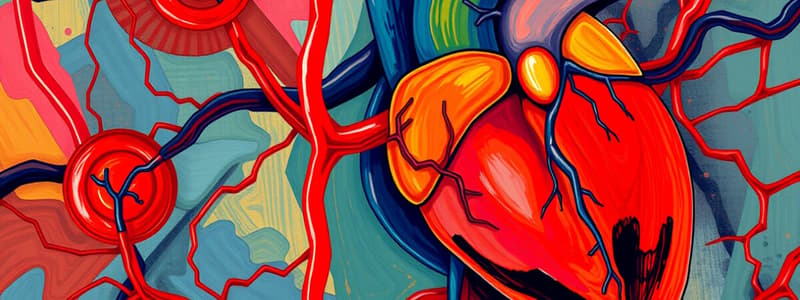
Introduction to the Cardiovascular System
- Responsible for transporting blood throughout the body
- Composed of the heart and blood vessels: arteries, capillaries, and veins
The Heart
- Located in the thoracic cavity, posterior to the sternum and between the lungs
- Comprised of four chambers: left atrium, left ventricle, right atrium, right ventricle
- Functions as a pump to deliver oxygen and nutrients, remove carbon dioxide and waste
Blood Vessels
- Arteries transport blood away from the heart, while veins carry it toward the heart
- Capillaries facilitate exchange between blood and body cells
- Arteries and veins have thicker walls than capillaries to withstand higher blood pressures
Cardiac Cycle
- Involves electrical and mechanical events regulated by the heart's conduction system
- SA node initiates action potentials, leading to synchronized heart contractions
- Cycle includes phases of systole (contraction) and diastole (relaxation)
Cardiac Output
- Influenced by heart rate (number of beats per minute) and stroke volume (amount of blood pumped per beat)
- Variables affecting heart rate include autonomic nervous system activity, hormones, and fitness level
- Stroke volume variable influenced by cardiac muscle contractility, blood volume, and vascular resistance
Heart's Conduction System
- SA node acts as the primary pacemaker; AV node and bundle of His facilitate conduction
- Rapid spread of action potentials through Purkinje fibers ensures organized contractions
Great Vessels
- Include superior and inferior vena cavae (deoxygenated blood to right atrium) and the aorta (oxygenated blood to the body)
- Pulmonary arteries transport deoxygenated blood from right ventricle to lungs; pulmonary veins return oxygenated blood to left atrium
Cardiac Valves
- Atrioventricular (AV) valves prevent backflow between atria and ventricles
- Semilunar valves prevent backflow from arteries into the ventricles
- Right AV valve (tricuspid) and left AV valve (bicuspid or mitral) are critical for one-way blood flow
Pulmonary and Systemic Circulation
- Pulmonary circulation involves blood flow from the heart to the lungs for gas exchange
- Systemic circulation delivers oxygenated blood from the heart to body tissues
- Blood flow sequence: right atrium → right ventricle → pulmonary artery → lungs → pulmonary veins → left atrium → left ventricle → aorta → systemic cells
Anatomy of the Heart and Pericardium
- Heart enclosed in three layers: fibrous pericardium, parietal layer, visceral layer (epicardium)
- Pericardial cavity contains serous fluid, reducing friction during heartbeats
- Heart positioned slightly rotated within the thoracic cavity
Clinical Considerations
- Congestive Heart Failure: Impaired heart pumping ability leading to fluid accumulation and edema
- Pericarditis: Inflammation of pericardial layers; excessive fluid can cause cardiac tamponade
- Common Conditions: Myocardial infarction, heart murmurs, and atherosclerosis impact cardiovascular health
Layers of the Heart Wall
- Composed of epicardium (outer layer), myocardium (middle, muscular layer), and endocardium (inner lining of heart chambers)
Sulci of the Heart
- Coronary sulcus: Separates atria from ventricles
- Interventricular sulcus: Separates left and right ventricles, contains coronary vessels
Summary of Blood Flow
- Deoxygenated blood enters right atrium → right ventricle → lungs for oxygenation → returns to left atrium → left ventricle → distributes oxygen-rich blood via aorta to the body.### Heart Structure and Layers
- The heart consists of three layers: epicardium (outermost), myocardium (thickest, responsible for pumping), and endocardium (innermost).
- The myocardium generates the force required to pump blood; it is thicker in ventricles compared to atria.
- The left ventricular wall is approximately three times thicker than the right ventricular wall.
Heart Chambers and Blood Flow
- The heart has four chambers: left atrium, right atrium, left ventricle, and right ventricle.
- The right atrium receives deoxygenated blood from the body through the superior and inferior venae cava.
- The right atrioventricular opening, which contains the right AV valve, allows blood flow from the right atrium to the right ventricle.
- The left atrium receives oxygenated blood from the lungs and pumps it into the left ventricle.
Valvular Structure and Function
- The heart's four valves include right and left atrioventricular valves and aortic and pulmonary semilunar valves.
- Atrioventricular valves prevent backflow during ventricular contraction; they are supported by papillary muscles and tendinous cords.
- Semilunar valves prevent backflow into the ventricles when the heart relaxes, opening during ventricular contraction.
Congenital and Acquired Heart Conditions
- Fossa ovalis: A remnant of the foramen ovale, located in the interatrial septum.
- Cardiomegaly: Enlargement of the heart due to increased chamber or wall size; can lead to heart failure.
- Hypertrophic cardiomyopathy: A genetic condition causing thickening of the heart muscle, can impede blood flow.
Coronary Circulation
- The coronary arteries supply oxygenated blood and branch from the ascending aorta; major branches include the right coronary artery and left coronary artery (including the anterior interventricular and circumflex arteries).
- Coronary arteries function as end arteries with no anastomoses, making blockages critical.
- Deoxygenated blood is returned via coronary veins, including the great cardiac vein and coronary sinus.
Cardiac Muscle Structure
- Cardiac muscle is striated and composed of short, branched cells interconnected by intercalated discs containing desmosomes and gap junctions for effective contraction and communication.
- T-tubules and less extensive sarcoplasmic reticulum (SR) play a role in muscle contraction.
Heart Conduction System
- The heart's conduction system comprises the SA node (pacemaker), AV node, bundle of His, and Purkinje fibers, which facilitate synchronized contraction.
- Electrical impulses initiate in the SA node and propagate to ensure atria contract before ventricles.
Heart Sounds and Murmurs
- Heart sounds (S1 and S2) are associated with valve closure; murmurs indicate potential valve dysfunction (stenosis or insufficiency).
- Valvular stenosis narrows openings; valvular insufficiency causes leaks leading to backflows.
Clinical Considerations
- Angina pectoris: Chest pain from inadequate blood supply; can present differently in women.
- Myocardial infarction (MI): Heart attack due to arterial blockage; characterized by severe chest pain and other symptoms like fatigue and shortness of breath.
Membrane and Blood Flow Dynamics
- Coronary sinus collects deoxygenated blood from myocardium before draining into the right atrium.
- Blood flow through coronary vessels is influenced by heart contraction and relaxation cycles.
Summary of Key Arteries and Conditions
- Major arteries: Right coronary artery supplies right ventricle; left coronary artery branches into anterior interventricular and circumflex arteries.
- Critical artery: Anterior interventricular artery, known as "widowmaker" if occluded due to high risk of heart attack.### Anatomy of the Heart's Conduction System
- The SA node serves as the primary pacemaker, initiating heartbeats.
- Key components of the conduction system include the AV node, AV bundle, bundle branches, and Purkinje fibers, facilitating electrical activity for heart contractions.
Regulation of Heart Rate and Contraction
- Parasympathetic innervation via the vagus nerve leads to decreased heart rate.
- Sympathetic innervation increases both heart rate and force of contraction.
Characteristics of Cardiac Muscle
- Cardiac muscle is highly vulnerable to ischemic conditions, which can lead to failure.
- Limited ability to utilize glycolysis or accumulate oxygen debt compared to skeletal muscle.
- Primarily relies on aerobic cellular respiration for energy.
Innervation and Physiological Responses
- Parasympathetic innervation originates from the cardioinhibitory center, reducing conduction at the SA and AV nodes.
- Sympathetic innervation increases both heart rate and contraction force, emanating from the cardioacceleratory center in the brainstem.
Receptors and Sensors
- Baroreceptors in the right atrium monitor blood pressure changes.
- Chemoreceptors detect variations in oxygen, carbon dioxide, and hydrogen ion levels in the blood.
SA Nodal Cells
- SA nodal cells spontaneously depolarize, generating action potentials.
- The resting membrane potential (RMP) of SA nodal cells is -60 mV, influenced by various ion channels.
Action Potential Propagation
- Conducted through the heart's conduction system, the action potential triggers contractile events in cardiac muscle cells.
- An effective heart rhythm relies on coordinated contraction of atria and ventricles.
Ectopic Pacemakers
- Ectopic pacemakers can arise, taking over pacing when the SA node fails, but typically have slower rates.
Mechanical Pacemaker
- A mechanical pacemaker is surgically implanted to regulate heartbeat in patients with conduction system deficiencies.
Electrical Events in Cardiac Muscle Cells
- Cardiac muscle cells undergo distinct phases: depolarization, plateau, and repolarization, maintaining proper cycling for contraction.
- Fast voltage-gated Na+ channels trigger rapid depolarization, while slow Ca2+ channels maintain the plateau phase.
Calcium and Contraction Mechanism
- Calcium ions play a crucial role in excitation-contraction coupling, promoting muscle contraction by binding to troponin.
- Crossbridge cycling involves myosin heads attaching to actin, facilitating muscle contraction.
Refractory Period
- The refractory period prevents premature stimulation of cardiac muscle cells and ensures coordinated contractions.
- The plateau phase extends contraction duration, enhancing cardiac output.
Effects of Calcium Channel Blockers
- Calcium channel blockers may be prescribed to reduce cardiac contractility and heart rate by inhibiting calcium influx into cardiac cells.
Studying That Suits You
Use AI to generate personalized quizzes and flashcards to suit your learning preferences.


