Podcast
Questions and Answers
What anatomical direction describes the orientation of the base of the heart in a canine?
What anatomical direction describes the orientation of the base of the heart in a canine?
- Laterally
- Ventrocaudally
- Ventro laterally
- Dorsocranially (correct)
Which term describes the period of heart muscle contraction?
Which term describes the period of heart muscle contraction?
- Asystole
- Diastole
- Systole (correct)
- Arrhythmia
What is the primary anatomical landmark used to differentiate the left side of the heart from the right side externally?
What is the primary anatomical landmark used to differentiate the left side of the heart from the right side externally?
- Ventricles
- Interventricular Grooves (correct)
- Coronary Groove
- Atria
Which of the following best describes the location of the Left AV valve?
Which of the following best describes the location of the Left AV valve?
What is the function of the chordae tendineae?
What is the function of the chordae tendineae?
During which phase of the cardiac cycle does the pulmonary semilunar valve prevent the backflow of blood into the right ventricle?
During which phase of the cardiac cycle does the pulmonary semilunar valve prevent the backflow of blood into the right ventricle?
What vessels carry oxygenated blood from the lungs to the left atrium?
What vessels carry oxygenated blood from the lungs to the left atrium?
Which valve prevents backflow of blood from the aorta into the left ventricle?
Which valve prevents backflow of blood from the aorta into the left ventricle?
Where is the 'T' point of maximal intensity (PMI) typically auscultated?
Where is the 'T' point of maximal intensity (PMI) typically auscultated?
What causes the first normal heart sound (S1)?
What causes the first normal heart sound (S1)?
In a normal, healthy animal, are heart sounds associated with blood flow through open valves usually audible?
In a normal, healthy animal, are heart sounds associated with blood flow through open valves usually audible?
Which of the following best describes the location of the heart within the canine thorax?
Which of the following best describes the location of the heart within the canine thorax?
What surface feature of the heart is characterized by its separation of the atria and ventricles and often contains fat?
What surface feature of the heart is characterized by its separation of the atria and ventricles and often contains fat?
If a veterinarian auscultates a 'lub-dub' sound followed by a murmur during a cardiac examination, what is the most likely cause of the murmur?
If a veterinarian auscultates a 'lub-dub' sound followed by a murmur during a cardiac examination, what is the most likely cause of the murmur?
Which of the following vessels does NOT directly supply blood to the heart itself?
Which of the following vessels does NOT directly supply blood to the heart itself?
Which of the following statements accurately describes the orientation of the apex of the heart?
Which of the following statements accurately describes the orientation of the apex of the heart?
In the context of heart anatomy, what is the significance of the term 'auricular surface'?
In the context of heart anatomy, what is the significance of the term 'auricular surface'?
Which structure is responsible for preventing the atrioventricular valves from inverting into the atria during ventricular contraction?
Which structure is responsible for preventing the atrioventricular valves from inverting into the atria during ventricular contraction?
When auscultating the heart, at which intercostal space is the aortic valve sound typically best heard?
When auscultating the heart, at which intercostal space is the aortic valve sound typically best heard?
Which of the following describes the path of deoxygenated blood in pulmonary circulation?
Which of the following describes the path of deoxygenated blood in pulmonary circulation?
In the cardiac cycle, what event causes the 'dub' or S2 heart sound?
In the cardiac cycle, what event causes the 'dub' or S2 heart sound?
During which phase of the cardiac cycle does blood flow through normal open valves?
During which phase of the cardiac cycle does blood flow through normal open valves?
Which of the following is the correct sequence of blood flow through the heart's valves?
Which of the following is the correct sequence of blood flow through the heart's valves?
What distinguishes the 'atrial surface' of the heart from other views?
What distinguishes the 'atrial surface' of the heart from other views?
What could the presence of S3 or S4 heart sounds indicate?
What could the presence of S3 or S4 heart sounds indicate?
Which statement accurately describes the anatomical relationship between the aorta and the left ventricle?
Which statement accurately describes the anatomical relationship between the aorta and the left ventricle?
An incompetent mitral valve would directly cause backflow of blood into which chamber?
An incompetent mitral valve would directly cause backflow of blood into which chamber?
Which circuit receives deoxygenated blood from systemic circulation and sends blood into pulmonary circulation?
Which circuit receives deoxygenated blood from systemic circulation and sends blood into pulmonary circulation?
Which of the following statements correctly describes the relative locations of the ventricles when viewing a transverse section of the heart?
Which of the following statements correctly describes the relative locations of the ventricles when viewing a transverse section of the heart?
A veterinarian detects a heart murmur that is loudest over the left apex of the heart. Which valve is most likely affected?
A veterinarian detects a heart murmur that is loudest over the left apex of the heart. Which valve is most likely affected?
If the subsinuosal interventricular groove is occluded, which chamber's blood supply would be most affected?
If the subsinuosal interventricular groove is occluded, which chamber's blood supply would be most affected?
What happens when the left AV valve stops backflow of blood from the left ventricle to the left atrium during systole?
What happens when the left AV valve stops backflow of blood from the left ventricle to the left atrium during systole?
Identify the vessel that carries deoxygenated blood from the right ventricle to the lungs.
Identify the vessel that carries deoxygenated blood from the right ventricle to the lungs.
Which of the following chambers of the heart has the thickest myocardium (muscle layer)?
Which of the following chambers of the heart has the thickest myocardium (muscle layer)?
Which structure ensures one-way blood flow from the right atrium to the right ventricle?
Which structure ensures one-way blood flow from the right atrium to the right ventricle?
Which structure prevents backflow of blood from the right ventricle into right atrium?
Which structure prevents backflow of blood from the right ventricle into right atrium?
Which heart valve sound is best auscultated at the 5th intercostal space?
Which heart valve sound is best auscultated at the 5th intercostal space?
What can the S3 heart sound indicate?
What can the S3 heart sound indicate?
What vessels carries oxygenated blood to the heart?
What vessels carries oxygenated blood to the heart?
What is the difference between the Parietal cusp and Septal cusp?
What is the difference between the Parietal cusp and Septal cusp?
Flashcards
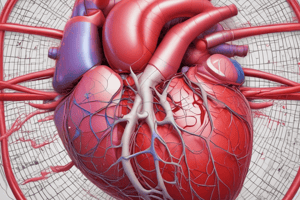
Heart Base and Apex Orientation
Heart Base and Apex Orientation
The base is oriented dorsocranially and the apex points ventrocaudally and to the left.
Coronary Groove
Coronary Groove
Separates the atria from the ventricles; contains coronary vessels and fat; encircles the base of the heart.
Auricular Surface
Auricular Surface
The left view of the heart, where both auricles are prominently observed.
Interventricular Grooves
Interventricular Grooves
Signup and view all the flashcards
Atrial Surface
Atrial Surface
Signup and view all the flashcards
AV Valve Cusps
AV Valve Cusps
Signup and view all the flashcards
Right Side Heart Flow
Right Side Heart Flow
Signup and view all the flashcards
Left Side Heart Flow
Left Side Heart Flow
Signup and view all the flashcards
Pulmonary Semilunar Valve
Pulmonary Semilunar Valve
Signup and view all the flashcards
Aortic Semilunar Valve
Aortic Semilunar Valve
Signup and view all the flashcards
First Tissue Supplied
First Tissue Supplied
Signup and view all the flashcards
Points of Maximum Intensity (PMI)
Points of Maximum Intensity (PMI)
Signup and view all the flashcards
S1 Heart Sound
S1 Heart Sound
Signup and view all the flashcards
S2 Heart Sound
S2 Heart Sound
Signup and view all the flashcards
Left AV Valve
Left AV Valve
Signup and view all the flashcards
Right AV Valve
Right AV Valve
Signup and view all the flashcards
Pulmonary semilunar valve
Pulmonary semilunar valve
Signup and view all the flashcards
Aortic semilunar valve
Aortic semilunar valve
Signup and view all the flashcards
Systole
Systole
Signup and view all the flashcards
Diastole
Diastole
Signup and view all the flashcards
Study Notes
- Heart anatomy objectives include:
- Position/orientation of the heart
- Surface and internal features
- Normal blood flow (pulmonary and systemic circulation)
- Blood supply to the heart muscle
- Sounds of cardiac auscultation
Heart Position
- The base is oriented dorsocranially
- Apex points ventrocaudally and to the left
- The right ventricle and septum are cranial to the left ventricle
Heart Views
- Left view (auricular surface) shows both auricles
- Right view (atrial surface) shows the right atrium prominently
Surface Topography
- The coronary groove separates the atria and ventricles, contains coronary vessels, often contains fat and encircles the base
- Interventricular grooves marks separation of the ventricles
- Paraconal interventricular groove
- Subsinuosal interventricular groove
Internal Features
- Marginal (parietal) cusps originate from the outer ventricular wall
- Septal cusp originates from the interventricular septal wall
- Cusps are anchored to the inner walls of the ventricles by chordae tendineae
- Papillary muscles secure the chordae tendineae to the inner walls of the ventricles
- AV valves close during systole to prevent backflow of blood into the atria
Blood Circulation
- Systemic and pulmonary circuits are separated
- Gas exchange in the lungs occurs via the pulmonary circuit
- Gas exchange to all other body cells occurs via the systemic circuit
- The right side receives deoxygenated blood from systemic circulation and sends blood into pulmonary circulation
- The left side receives oxygenated blood from pulmonary circulation and sends blood into systemic circulation
Blood Flow
- Right atrium receives blood from the cranial and caudal vena cava
- The right atrium pumps blood into the right ventricle through the right atrioventricular valve (tricuspid)
- The right atrioventricular valve stops backflow of blood from the right ventricle into the right atrium during systole
- The right ventricle contracts to send blood to the lungs through the pulmonary trunk and pulmonary arteries
- The pulmonary semilunar valve prevents backflow of blood from the pulmonary trunk into the right ventricle during diastole
- Oxygenated blood flows back to the heart through the pulmonary veins into the left atrium
- The left atrium pumps blood into the left ventricle through the left atrioventricular valve (Mitral)
- The left atrioventricular valve stops backflow from the left ventricle to the left atrium during systole
- The left ventricle contracts to send blood through the aortic semilunar valve and into the aorta
- Supplies the heart muscle
- The aortic semilunar valve prevents backflow of blood from the aorta into the left ventricle during diastole
- Pulmonary semilunar valve is located between the right ventricle and the pulmonary arteries (trunk)
- Left AV valve is located between the left atria and left ventricle
- Aortic semilunar valve is located between the left ventricle and the aorta
- Right AV valve is located between the right atria and right ventricle
- Systole is heart contraction
- Diastole is heart rest
- The first tissue supplied by the heart is the heart itself
Heart Auscultation
- Listening for points of maximum intensity (PMI) of each valve
- Left side:
- P: pulmonary valve sound (3rd intercostal space)
- A: aortic valve sound (4th intercostal space)
- M: mitral/left AV sound (5th intercostal space)
- Right side:
- T: tricuspid/right AV sound (4th intercostal space)
Heart Sounds
- Two normal sounds of the heart
- S1 and S2 are caused by blood stopping at closed valves
- S1: Closure of Left and Right Atrioventricular Valves
- S2: Closure of Aortic and Pulmonic semilunar Valves
- Blood flow through normal open valves is silent to the human ear in normal dogs and cats
Abnormal Heart Sounds
- S3 and S4 are not normally heard in small animals
- S3: Left and right ventricular filling during diastole can be audible with congestive disease and abnormal ventricular dilation
- S4: Left and right atrial contraction immediately before ventricular contraction is audible with non-compliant ventricular conditions e.g. Hypertrophic Cardiomyopathy
Studying That Suits You
Use AI to generate personalized quizzes and flashcards to suit your learning preferences.