Podcast
Questions and Answers
If a patient has a congenital defect resulting in a hole between the heart's pumping chambers, which structure is most likely affected?
If a patient has a congenital defect resulting in a hole between the heart's pumping chambers, which structure is most likely affected?
- Semilunar valve
- Interatrial septum
- Atrioventricular valve
- Interventricular septum (correct)
Damage to the papillary muscles following a myocardial infarction is most likely to result in:
Damage to the papillary muscles following a myocardial infarction is most likely to result in:
- Increased blood flow through the valve
- Stenosis of the valve
- Increased valve strength
- Valve regurgitation (correct)
Which vessel directly delivers oxygenated blood to the left atrium?
Which vessel directly delivers oxygenated blood to the left atrium?
- Inferior vena cava
- Pulmonary vein (correct)
- Superior vena cava
- Pulmonary artery
A surgeon needs access to the heart's myocardium. Which layer must they cut through, after the pericardium, to reach it?
A surgeon needs access to the heart's myocardium. Which layer must they cut through, after the pericardium, to reach it?
What is the primary function of the fibrous pericardium?
What is the primary function of the fibrous pericardium?
Which valve prevents the backflow of blood from the right ventricle into the right atrium?
Which valve prevents the backflow of blood from the right ventricle into the right atrium?
If a doctor is examining an X-ray and notes that the apex of the heart is displaced further to the left than normal, towards which anatomical landmark would it be pointing?
If a doctor is examining an X-ray and notes that the apex of the heart is displaced further to the left than normal, towards which anatomical landmark would it be pointing?
Which heart chamber would be directly affected by a blockage in the coronary sinus?
Which heart chamber would be directly affected by a blockage in the coronary sinus?
Which layer of the heart is in direct contact with the blood within the cardiac chambers?
Which layer of the heart is in direct contact with the blood within the cardiac chambers?
After passing through the aortic semilunar valve, blood enters which structure?
After passing through the aortic semilunar valve, blood enters which structure?
Which valve is located between the left atrium and the left ventricle?
Which valve is located between the left atrium and the left ventricle?
What is the remnant of the fetal foramen ovale found in the adult heart?
What is the remnant of the fetal foramen ovale found in the adult heart?
Which of the following vessels does not directly empty into the right atrium?
Which of the following vessels does not directly empty into the right atrium?
Which of the following arteries does not branch directly off the aortic arch?
Which of the following arteries does not branch directly off the aortic arch?
What is the function of the chordae tendineae?
What is the function of the chordae tendineae?
Which layer of the heart wall is responsible for the heart's pumping action?
Which layer of the heart wall is responsible for the heart's pumping action?
A doctor auscultates a heart murmur indicative of valve regurgitation between the left atrium and left ventricle. Which valve is most likely malfunctioning?
A doctor auscultates a heart murmur indicative of valve regurgitation between the left atrium and left ventricle. Which valve is most likely malfunctioning?
If a patient experiences a blockage in the left subclavian artery, which region of the body is most likely to be affected?
If a patient experiences a blockage in the left subclavian artery, which region of the body is most likely to be affected?
Which of the following is a function of the pericardial fluid?
Which of the following is a function of the pericardial fluid?
Which heart chamber has the thickest myocardium?
Which heart chamber has the thickest myocardium?
Flashcards
Heart Location
Heart Location
Located within the thoracic cavity in the mediastinum, shifted about 2/3 to the left of the midsternal line.
Atria
Atria
Upper chambers of the heart that receive blood.
Right Atrium Receivers
Right Atrium Receivers
Inferior vena cava (IVC), Superior vena cava, and Coronary sinus.
Fossa Ovalis
Fossa Ovalis
Signup and view all the flashcards
Left Atrium Function
Left Atrium Function
Signup and view all the flashcards
Heart Valves Function
Heart Valves Function
Signup and view all the flashcards
Tricuspid Valve
Tricuspid Valve
Signup and view all the flashcards
Bicuspid Valve
Bicuspid Valve
Signup and view all the flashcards
Chordae Tendineae
Chordae Tendineae
Signup and view all the flashcards
Ventricles
Ventricles
Signup and view all the flashcards
Pulmonary Semilunar Valve
Pulmonary Semilunar Valve
Signup and view all the flashcards
Aortic Semilunar Valve
Aortic Semilunar Valve
Signup and view all the flashcards
Aortic Arch Branches
Aortic Arch Branches
Signup and view all the flashcards
Endocardium
Endocardium
Signup and view all the flashcards
Myocardium
Myocardium
Signup and view all the flashcards
Epicardium
Epicardium
Signup and view all the flashcards
Pericardial Cavity
Pericardial Cavity
Signup and view all the flashcards
Fibrous Pericardium
Fibrous Pericardium
Signup and view all the flashcards
Study Notes
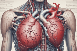
Heart Anatomy Overview
- The heart is located within the thoracic cavity, specifically in the mediastinum.
- The heart is shifted about 2/3 to the left of the midsternal line.
- The apex of the heart points towards the left hip.
- The base of the heart points towards the right shoulder.
- The heart weighs approximately 200-300 grams and is about the size of a fist.
Heart Chambers
- The two upper chambers of the heart are called the atria (right and left).
- The right atrium receives blood from three major vessels:
- Inferior vena cava (IVC) brings blood from below the diaphragm.
- Superior vena cava brings blood from the head, neck, and upper limbs.
- Coronary sinus receives blood from the myocardium.
- The fossa ovalis is a scar tissue in the right atrium, a remnant of the foramen ovale in the fetus.
- The left atrium receives oxygenated blood from the pulmonary veins.
- The left pulmonary veins bring blood from the left lung.
- The right pulmonary veins bring blood from the right lung.
Heart Valves
- Valves prevent backflow and ensure one-way blood flow.
- The tricuspid valve (or right atrioventricular valve) separates the right atrium from the right ventricle.
- The bicuspid valve (mitral valve or left atrioventricular valve) separates the left atrium from the left ventricle.
- Valves consist of layers called the Zona fibrosa, Zona spongiosa, and Zona ventricularis, with an endothelial lining.
- Chordae tendineae are collagen cords attaching to the valve cusps, anchoring the valves.
- Papillary muscles attach to the chordae tendineae and anchor them, preventing valve prolapse.
- Ischemia or damage to the papillary muscles (e.g., during myocardial infarction) can cause valve regurgitation.
Ventricles and Septa
- The right ventricle is the pumping chamber on the right side of the heart.
- The left ventricle is the pumping chamber on the left side of the heart.
- The interventricular septum separates the right ventricle from the left ventricle.
- Ventricular septal defect, a congenital disorder, involves a hole in this septum.
- The interatrial septum separates the left atrium and the right atrium
Pulmonary and Aortic Valves and Vessels
- The pulmonary semilunar valve separates the right ventricle from the pulmonary trunk.
- The aortic semilunar valve separates the left ventricle from the aorta.
- The pulmonary trunk splits into the left and right pulmonary arteries leading to the lungs.
- Blood flows through the ascending aorta, then the aortic arch.
- Three important vessels branch off the aortic arch:
- Brachiocephalic trunk (which splits into the right common carotid artery and the right subclavian artery).
- Left common carotid artery.
- Left subclavian artery.
Heart Layers
- The layers of the heart, from innermost to outermost, are:
- Endocardium: Innermost layer made of endothelial tissue.
- Myocardium: Middle muscular layer made of cardiac muscle tissue; thicker in the left ventricle.
- Epicardium (or visceral layer of the serous pericardium): Outer layer.
- The pericardial cavity contains pericardial fluid, which reduces friction during heartbeats.
- The parietal layer of the serous pericardium is continuous with the visceral layer (epicardium).
- The fibrous pericardium is the outermost layer, made of dense irregular connective tissue.
- Anchors the heart to surrounding structures.
- Protects the heart.
- Prevents the heart from overfilling with blood.
Studying That Suits You
Use AI to generate personalized quizzes and flashcards to suit your learning preferences.