Podcast
Questions and Answers
Where is Point 1 located in the surface anatomy of the heart?
Where is Point 1 located in the surface anatomy of the heart?
- Lower border of the 3rd Lt costal cartilage one & half inches from the median plane.
- Lower border of the 2nd Lt costal cartilage one & half inches from the median plane. (correct)
- Upper border of the 3rd Rt costal cartilage one inch from the median plane.
- Upper border of the 6th Rt costal cartilage one inch from its junction with the sternum.
If a patient has a mitral valve disorder, where would you primarily auscultate to hear the murmur?
If a patient has a mitral valve disorder, where would you primarily auscultate to hear the murmur?
- Left 2nd intercostal space
- Left 4th intercostal space at the parasternal line
- Right 2nd intercostal space
- Left 5th intercostal space at the midclavicular line (correct)
Which layer of the heart wall is the most superficial?
Which layer of the heart wall is the most superficial?
- Pericardium
- Epicardium (correct)
- Endocardium
- Myocardium
During a cardiac examination, where would you palpate to assess for aortic valve issues?
During a cardiac examination, where would you palpate to assess for aortic valve issues?
What causes cyanosis to decrease when a patient with tetralogy of Fallot squats?
What causes cyanosis to decrease when a patient with tetralogy of Fallot squats?
What is a key hemodynamic effect of sustained squatting in patients with Tetralogy of Fallot?
What is a key hemodynamic effect of sustained squatting in patients with Tetralogy of Fallot?
What condition is suggested by a patient presenting with a lean body build, thin face, and long, spidery fingers?
What condition is suggested by a patient presenting with a lean body build, thin face, and long, spidery fingers?
A patient has a mauve discoloration in the butterfly distribution of the nose and cheeks. Which condition is most likely indicated?
A patient has a mauve discoloration in the butterfly distribution of the nose and cheeks. Which condition is most likely indicated?
What is the cause of differential cyanosis, where the lower extremity is bluish but the upper extremity and head are not?
What is the cause of differential cyanosis, where the lower extremity is bluish but the upper extremity and head are not?
Which of the following is characteristic of venous pulsation?
Which of the following is characteristic of venous pulsation?
What does blue clubbing, associated with cyanotic congenital heart disease, indicate?
What does blue clubbing, associated with cyanotic congenital heart disease, indicate?
What is suggested by observing pulsations in the epigastric area of a patient?
What is suggested by observing pulsations in the epigastric area of a patient?
What is the most common cause of an invisible and impalpable apex beat?
What is the most common cause of an invisible and impalpable apex beat?
In cases of cardiac enlargement, which direction does the apex shift with left ventricular hypertrophy (LVH)?
In cases of cardiac enlargement, which direction does the apex shift with left ventricular hypertrophy (LVH)?
You are palpating the suprasternal area. What are you assessing?
You are palpating the suprasternal area. What are you assessing?
If there is a pulsation to the left and up in the epigastric area, what potential condition is indicated?
If there is a pulsation to the left and up in the epigastric area, what potential condition is indicated?
Regarding thrills, what is a key characteristic?
Regarding thrills, what is a key characteristic?
When assessing Jugular Venous Pressure (JVP), what angle should the head of the bed be at?
When assessing Jugular Venous Pressure (JVP), what angle should the head of the bed be at?
A patient has an increased JVP. What could this indicate?
A patient has an increased JVP. What could this indicate?
In a Hepatojugular Reflux test, what does a sustained rise of equal to or more than 4cm indicate?
In a Hepatojugular Reflux test, what does a sustained rise of equal to or more than 4cm indicate?
What sound is confirmed in percussion with the normal heart, liver, and stomach?
What sound is confirmed in percussion with the normal heart, liver, and stomach?
During percussion, if there is resonance instead of the expected dullness of the bare area of the heart, what could this indicate?
During percussion, if there is resonance instead of the expected dullness of the bare area of the heart, what could this indicate?
Which of the following can cause a 'dull' sound upon percussion of the lungs?
Which of the following can cause a 'dull' sound upon percussion of the lungs?
What is the 1st heart sound (S1) caused by?
What is the 1st heart sound (S1) caused by?
When is S3, the Ventricular gallop, heart sound directly heard?
When is S3, the Ventricular gallop, heart sound directly heard?
Describe what happens to the aortic valve in order for splitting to occur.
Describe what happens to the aortic valve in order for splitting to occur.
Abnormal musical sound produced due to turbulence of the blood flow results in:
Abnormal musical sound produced due to turbulence of the blood flow results in:
What is the term for a backward displacement of the valve?
What is the term for a backward displacement of the valve?
Flashcards
Point 1: Heart Surface Anatomy
Point 1: Heart Surface Anatomy
Lower border of 2nd left costal cartilage, 1.5 inches from the median plane.
Point 2: Heart Surface Anatomy
Point 2: Heart Surface Anatomy
Upper border of the right 3rd costal cartilage, 1 inch from the median plane.
Point 3: Heart Surface Anatomy
Point 3: Heart Surface Anatomy
Upper border of the right 6th costal cartilage, 1 inch from its junction with the sternum.
Point 4: Heart Surface Anatomy
Point 4: Heart Surface Anatomy
Signup and view all the flashcards
Epicardium
Epicardium
Signup and view all the flashcards
Myocardium
Myocardium
Signup and view all the flashcards
Endocardium
Endocardium
Signup and view all the flashcards
Squatting position with Tetralogy of Fallot
Squatting position with Tetralogy of Fallot
Signup and view all the flashcards
Cyanosis decreases with Squatting
Cyanosis decreases with Squatting
Signup and view all the flashcards
Long sitting position
Long sitting position
Signup and view all the flashcards
Prayers position
Prayers position
Signup and view all the flashcards
Cyanosis
Cyanosis
Signup and view all the flashcards
Differential cyanosis
Differential cyanosis
Signup and view all the flashcards
Angle disappears
Angle disappears
Signup and view all the flashcards
Local Examination
Local Examination
Signup and view all the flashcards
APEX beat
APEX beat
Signup and view all the flashcards
Causes from heart
Causes from heart
Signup and view all the flashcards
Suprasternal area
Suprasternal area
Signup and view all the flashcards
Hepatojagular reflex (HJR)
Hepatojagular reflex (HJR)
Signup and view all the flashcards
Percussion
Percussion
Signup and view all the flashcards
CVP
CVP
Signup and view all the flashcards
Thrills
Thrills
Signup and view all the flashcards
Resonance
Resonance
Signup and view all the flashcards
Dullness
Dullness
Signup and view all the flashcards
Suprasternal area
Suprasternal area
Signup and view all the flashcards
Study Notes
- The provided document contains study notes on the anatomy of the heart, cardiac examination, and related topics.
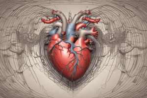
Surface Anatomy of The Heart
- Point 1: The lower border of the left 2nd costal cartilage is located one and a half inches from the median plane.
- Point 2: The upper border of the right 3rd costal cartilage is one inch from the median plane.
- Point 3: The upper border of the right 6th costal cartilage is one inch from its junction with the sternum.
- Point 4: The apex is located in the left 5th intercostal space, 3 1/2 inches from the median plane.
Heart Chambers, Valves and Circulation
- The heart has four chambers: the right atrium, right ventricle, left atrium, and left ventricle.
- The valves include the tricuspid valve (between the right atrium and ventricle), the pulmonary valve (leading to the pulmonary artery), the mitral valve (between the left atrium and ventricle), and the aortic valve (leading to the aorta).
- Blood flows from the superior and inferior vena cavae into the right atrium, through the tricuspid valve to the right ventricle, through the pulmonary valve to the pulmonary artery, to the lungs and back through pulmonary veins to the left atrium, through the mitral valve to the left ventricle, and finally through the aortic valve to the aorta.
Layers of Heart Wall
- The heart wall consists of three layers: the epicardium, myocardium, and endocardium.
Surfaces of Heart
- Key anatomical features on the surfaces of the heart include the right atrium, right atrial appendage, left atrial appendage, atrioventricular groove, right ventricle, left ventricle, apex of the heart, and anterior interventricular groove.
Borders of the Heart
- Borders of the heart include the superior vena cava, arch of the aorta, right auricle, left auricle, right atrium, right ventricle, left ventricle, inferior vena cava and the apex.
Conduction System of the Heart
- The heart's conduction system includes the interatrial pathway, sinoatrial (SA) node, atrioventricular (AV) node, right atrium, left atrium, internodal pathway, right ventricle, left branch of bundle of His, right branch of bundle of His, left ventricle, and Purkinje fibers.
Coronary Arteries
- The main coronary arteries are the left coronary artery, circumflex artery, right coronary artery, and the left anterior descending artery.
Cardiac Examination - History
- Key aspects of a cardiac history include personal factors (occupation, habits), past medical history (surgeries, diseases, medications), present symptoms (onset, sudden MI/angina), and family history of IHD/HF.
Cardiac Examination - Assessment
- General Examination: Includes assessing decubitus position, body build, color, mental state, and other problems.
- Decubitus Position: Squatting position may indicate tetralogy of Fallot.
- Tetralogy of Fallot: It is a cyanotic congenital heart disease involving pulmonary stenosis, right ventricular hypertrophy, overriding aorta, and ventricular septal defect (VSD).
- Squatting helps relieve dyspnea by increasing pulmonary blood flow and arterial oxygen saturation. Squatting does this by kinking the femoral arteries, compressing splanchnic vessels, and trapping oxygenated venous blood.
- Hemodynamics of Squatting: has two phases, immediate drop in venous return followed by a sustained increase in venous return and systemic vascular resistance.
- Long Sitting Position: Patients with orthopnea, induced by supine lying position, are often more comfortable in a long sitting position, with the head of the bed elevated.
- Prayers Position: Leaning forward helps to decrease pain sensation in pericarditis by increasing intra-abdominal pressure and decreasing venous return to the heart.
Body Build
- Thin (Cachetic): May indicate advanced left side heart failure.
- Obese: May indicate right side heart failure or coronary artery disease.
- Inverted Pyramid: May indicate coarctation of the aorta.
- Marfan's Syndrome: Patient with lean built, thin face, long spidery fingers, and atrial septal defect.
Color
- Malar Flushes: Mauve discoloration of nose and cheeks, indicates tight mitral stenosis.
- Pale Color: Face indicates rheumatic fever or aortic valve disease.
- Jaundice: Yellowish discoloration of skin and mucus membrane, >3mg bilirubin, indicates sclera of eye.
- Cyanosis: Bluish discoloration of skin and mucous membrane, more than 5gm reduced hemoglobin, indicates poor oxygen delivery.
- Differential Cyanosis: Bluish coloration of lower extremity without upper extremity and head indicates patent ductus arteriosus (PDA).
Central vs. Peripheral Cyanosis
- Central cyanosis: Gas exchange problem in patients with fallot of tetralogy, coronary artery disease, and advanced heart failure, increase with exercise.
- Peripheral cyanosis: Low cardiac output in patients with atherosclerosis causes cyanosis in the lips, nose, hands etc.
- Exercise reduces this type of cyanosis
Mental Status
- Mood: Anxiety or depression following myocardial infarction.
- Level of Coordination: Cooperative or uncooperative.
- Level of Consciousness: Includes alert, confused , automatic state, stupor, delirious, semi-comatose, and comatose.
Other Problems During Physical Examination
- Nodules: Osler nodules (endocarditis), Subcutaneous nodules (rheumatic fever).
- Puffy Eyelids: Indicates heart failure.
- Rheumatic Chorea: Involuntary jerky movements.
- Venous Pulsation: Evaluate Jugular Venous Pressure (JVP).
- Arterial vs Venous Pulsation: Carotid pulse vs jugular pulse.
Additional Findings
- Clubbing Fingers: Hypertrophy of connective tissue of nail-bed, indicative of poor oxygen perfusion of extremities, and indicates heart disease.
- Lower Extremities: Check for cardiac edema, cardiac edema in right heart failure. 6- lower extremities ( cardiac edema in right heart failure). Test for pitting and non pitting edema.
- Neck: Assess vigorous neck pulsations for sign of aortic regurgitation.
- Fever: May indicate rheumatic fever or infective endocarditis.
- Vital Signs: Check pulse, rhythm, volume and pressure.
- Palpate radial, carotid and peripheral pulses: Evaluate rate, rhythm, and radial-femoral delay.
- Blood Pressure: Measure blood pressure in both arms.
- Orthostatic Symptoms: Assess after standing for 5 minutes.
Local Examination: Inspection, Palpation, Percussion
- I- Inspection:
- Previous operation
- Skeletal deformities
- Suprasternal pulsation
- Pericardial area
- Parasternal area
- Apex beat
- Dilated veins of chest wall
- Epigastric area
Previous Operation
- Scars: From median sternotomy, lateral thoracotomy, supraclavicular (pacemaker), or midaxillary line (pacer-cardioverter-defibrillator).
Skeletal Deformities
- Include scoliosis, kyphosis, kyphoscoliosis, barrel chest, and pectus excavatum.
Suprasternal Pulsation
- Related to tension, Carotid pulsation due to Aortic Rergurge, venous pulsation due to CHF
Pericardial Area
- Note that a Bulge indicates large heart due to infantile disease
Parasternal Area
- Pulsations indicate pulmonary hypertension, enlargement of atrium, or other heart conditions
Apex Beat:
- Sometimes visible.
Dilated Veins
- Indicates a Superior Vena Cava obstruction
Palpation
- APEX beat, Suprasternal area, Epigastric pulsation, THRILLS, Jugular Venous Pressure (JVP), HEPATOJAGULAR REFLEX (HJR), and Tracheal position
Apex Beat
- Lower most point of Contraction of left ventricle during systole. * sometimes visible and usually palpable in 75% of subjects.
- Located normally at the 5th left intercostal space along the midclavicular line
- Should be dime sized
- Difficult palpation with obesity thick chest, etc.
- Apex beat shifts toward other areas with chest and abdominal diesease
Size
- Normally around 3cm
Intensity
- Normally you wont feel it
Force
- Left ventricle enlargement
Causes of shifting of apex
- Outside Heart Causes: Include chest and abdomen diseases, fibrosis and collapse, pleural effusion, deformity of chest and abdominal distension.
- Heart Causes: Include cardiac enlargement (LVH/RVH) and dextrocardia
Percussion
- Confirms the position of the heart, liver, and stomach.
- Aids in assessing heart size.
- Dullness = Blood/Fluid.
Percussion Areas
- Pulmonary area
- Aortic area
- Bare area of heart
- Lower third of sternum
- Liver
- Spleen.
Auscultation
- Important for listening to murmurs, heart sounds, and pericardial rub
Heart Sounds
- 1ST, 2nd, 3rd and 4th heart sounds
- Spliting, Gallop
- Surface anatomy of valves
Stethoscope
- Diaphragmatic high-pitched murmur
- Bell :low pitched murmur
Studying That Suits You
Use AI to generate personalized quizzes and flashcards to suit your learning preferences.