Podcast
Questions and Answers
What is the central ray for the PA Hand projection?
What is the central ray for the PA Hand projection?
- 3rd metacarpophalangeal joint (MCP) (correct)
- Distal wrist
- Midcarpals
- Metacarpophalangeal joint (MCP)
What is the positioning for a PA Oblique Hand projection?
What is the positioning for a PA Oblique Hand projection?
Place the patient 90 degrees to the table, rotate hand 45 degrees lateral, place all fingers on IR.
Which structure is seen in the Lateral Hand projection?
Which structure is seen in the Lateral Hand projection?
- PA oblique projection of the hand
- Superimposed carpals (correct)
- PA projection of 2nd through 5th digit
- PA projection of carpal bones
The central ray for the PA Wrist is located at the ______.
The central ray for the PA Wrist is located at the ______.
What anatomy is demonstrated in the PA Oblique Wrist?
What anatomy is demonstrated in the PA Oblique Wrist?
What is the breathing instruction for the projections mentioned?
What is the breathing instruction for the projections mentioned?
What type of shielding is recommended for the projections?
What type of shielding is recommended for the projections?
What structures are seen in the PA projection of the wrist?
What structures are seen in the PA projection of the wrist?
Flashcards are hidden until you start studying
Study Notes
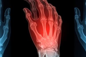
PA Hand
- Central Ray (CR) is at the 3rd metacarpophalangeal joint (MCP).
- Positioning involves placing the patient at a 90-degree angle to the table, with the hand pronated and fingers extended on the image receptor (IR).
- Collimation should include the distal wrist area.
- Breathing should be at normal respiration.
- Shielding is recommended for patient safety.
- Structures visible include PA projections of the 2nd through 5th digits and an oblique view of the 1st digit.
PA Oblique Hand
- CR is directed to the 3rd MCP joint.
- Position the patient at a 90-degree angle and rotate the hand laterally by 45 degrees, ensuring all fingers are on the IR.
- Collimation should extend to the distal wrist.
- Normal respiration is required for breathing instruction.
- Recommended shielding for patient safety.
- Recognized structures include the PA oblique projection of the hand.
Lateral Hand
- CR is at the metacarpophalangeal joint (MCP).
- Place patient at a 90-degree angle with the hand in a lateral position, fingers either extended or in a fan position.
- Collimation to include the distal wrist is essential.
- Breathing is performed at normal respiration.
- Shielding is recommended for patient safety.
- Key structures seen include superimposed carpals, metacarpals, and phalanges; phalanges are shown without superimposition when in a fanned position.
PA Wrist
- The central ray is directed at the midcarpals.
- Patient positioning is at a 90-degree angle with the hand pronated on the IR, fingers rolled in.
- Collimation should reach 2.5 inches beyond the wrist.
- Breathing instructions specify normal respiration.
- Recommended shielding for protection.
- Structures visualized include PA projection of carpal bones, proximal metacarpals, and distal ulna and radius.
PA Oblique Wrist
- Positioning is similar to previous wrist projections with slight overlap of the distal radius and ulna.
- Well-demonstrated structures include the scaphoid and trapezium.
- Anatomy visualized includes distal radius and ulna, carpals, and proximal half of metacarpals.
Gaynor Hart Method
- Information not provided.
AP Forearm
- Information not provided.
AP Elbow
- Information not provided.
Lateral Elbow
- Information not provided.
Jones Method Elbow #1
- Information not provided.
Jones Method Elbow #2
- Information not provided.
AP Shoulder (External Rotation)
- Information not provided.
AP Shoulder (Internal Rotation)
- Information not provided.
Scapular Y
- Information not provided.
Inferosuperior Axial Shoulder (Lawrence Method)
- Information not provided.
RAO Sternum
- Information not provided.
PA Chest
- Information not provided.
Choleangiogram
- Information not provided.
Abdomen (KUB)
- Information not provided.
KUB w/ Contrast
- Information not provided.
UGI w/ Contrast
- Information not provided.
Left Lateral Decubitus (Pneumothorax)
- Information not provided.
AP Pelvis
- Information not provided.
AP Hip
- Information not provided.
Lateral Hip
- Information not provided.
Danelius Miller (X-Table Hip)
- Information not provided.
Lateral Knee
- Information not provided.
Settegast (Sunrise) Knee
- Information not provided.
Intercondylar Fossa (Holmblad or Camp Conventry)
- Information not provided.
Ankle (Mortise)
- Information not provided.
AP Foot
- Information not provided.
Lateral Calcaneus
- Information not provided.
Axial Calcaneus
- Information not provided.
Lateral C-Spine
- Information not provided.
AP Open Mouth
- Information not provided.
Lateral Skull
- Information not provided.
Lateral T-Spine
- Information not provided.
AP T-Spine
- Information not provided.
Swimmer's (Pawlow or Twining Method) T-Spine
- Information not provided.
Oblique L-Spine (w/ Scotty Dog)
- Information not provided.
Oblique C-Spine
- Information not provided.
PA Skull (Caldwell)
- Information not provided.
SMV (Schuller) Skull
- Information not provided.
Lateral Facial Bones
- Information not provided.
AP Axial Skull (Towne)
- Information not provided.
Waters
- Information not provided.
AP Mandible
- Information not provided.
Axiolateral Mandible (Body)
- Information not provided.
Rhese Method
- Information not provided.
Sphenoid
- Information not provided.
TMJ (Open vs Closed)
- Information not provided.
Lateral Oblique TMJ
- Information not provided.
Sacrum & Coccyx
- Information not provided.
Intravenous Urogram
- Information not provided.
Studying That Suits You
Use AI to generate personalized quizzes and flashcards to suit your learning preferences.