Podcast
Questions and Answers
Which structure does NOT contribute to the boundaries of the cubital fossa?
Which structure does NOT contribute to the boundaries of the cubital fossa?
- Lateral margin of pronator teres (correct)
- Epicondylar line (correct)
- Medial border of brachioradialis (correct)
- Tendon of biceps brachii (correct)
In relation to the anterior aspect of the upper forearm, where is the cubital fossa located?
In relation to the anterior aspect of the upper forearm, where is the cubital fossa located?
- Medial
- Posterior
- Lateral
- Anterior (correct)
What structures form the roof of the cubital fossa?
What structures form the roof of the cubital fossa?
- Median nerve and brachial artery
- Supinator and brachialis muscles
- Skin, superficial fascia, and deep fascia (correct)
- Radial and ulnar arteries
Which set of veins lie within the superficial fascia of the cubital fossa, proceeding from medial to lateral?
Which set of veins lie within the superficial fascia of the cubital fossa, proceeding from medial to lateral?
What two muscles constitute the floor of the cubital fossa?
What two muscles constitute the floor of the cubital fossa?
From medial to lateral, which sequence of structures is found within the cubital fossa?
From medial to lateral, which sequence of structures is found within the cubital fossa?
What anatomical division does the palmar aponeurosis create within the hand?
What anatomical division does the palmar aponeurosis create within the hand?
Where is the thenar space located relative to the tendons in the hand?
Where is the thenar space located relative to the tendons in the hand?
What is the clinical significance of the lumbrical canals in the hand?
What is the clinical significance of the lumbrical canals in the hand?
A patient presents with acute occlusion of the axillary artery. Why might potential vascular anastomoses around the shoulder girdle be unreliable?
A patient presents with acute occlusion of the axillary artery. Why might potential vascular anastomoses around the shoulder girdle be unreliable?
Flashcards
Palmar Aponeurosis
Palmar Aponeurosis
Divides the hand into thenar, hypothenar, and central compartments.
Midpalmar Space
Midpalmar Space
Located deep to the long flexor tendons of the medial 3 fingers.
Thenar Space
Thenar Space
Deep to the long flexor tendons of the first 2 fingers and superficial to adductor pollicis.
Cubital Fossa
Cubital Fossa
Signup and view all the flashcards
Roof of Cubital Fossa
Roof of Cubital Fossa
Signup and view all the flashcards
Epicondylar Line
Epicondylar Line
Signup and view all the flashcards
Veins in Superficial Cubital Fossa
Veins in Superficial Cubital Fossa
Signup and view all the flashcards
Floor of Cubital Fossa
Floor of Cubital Fossa
Signup and view all the flashcards
Medial contents of Cubital Fossa
Medial contents of Cubital Fossa
Signup and view all the flashcards
Acute Axillary Artery Occlusion
Acute Axillary Artery Occlusion
Signup and view all the flashcards
Study Notes
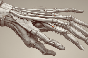
- Two septa deepen from the medial and lateral borders of the palmar aponeurosis and blend with the 5th and 3rd metacarpals respectively.
- The hand is divided into thenar, hypothenar, and central compartments.
- The midpalmar space is deep to the long flexor tendons of the medial 3 fingers.
- The thenar space is deep to the long flexor tendons of the first 2 fingers and superficial to adductor pollicis.
- Spaces in the hand can be drained along the lumbrical canals.
Cubital Fossa Definition and Boundaries
- The cubital fossa is a triangular depression located on the anterior aspect of the upper forearm.
- Proximally, the cubital fossa is bordered by the epicondylar line.
- Medially, the cubital fossa is bordered by the lateral margin of the pronator teres muscle.
- Laterally, the cubital fossa is bordered by the medial face of the brachioradialis muscle.
- The cubital fossa is bisected by the tendon of the biceps brachii muscle.
Roof of Cubital Fossa
- The roof of the cubital fossa is formed by the skin, superficial fascia, and deep fascia.
- The basilic, median cubital, and cephalic veins lie within the superficial fascia from medial to lateral.
- Branches of the medial cutaneous nerve of the forearm are closely related to the basilic vein.
- The lateral cutaneous nerve of the forearm runs alongside the lateral edge of the tendon of biceps brachii and then surfaces.
- The deep fascia of the forearm forms the roof and is continuous with the deep fascia of the anterior arm.
- The deep fascia is attached to the medial and lateral humeral epicondyles
- The deep fascia is augmented by the bicipital aponeurosis, which sweeps across the medial and inferior part of the fossa to the fascia over the superficial flexor muscles.
Floor and Contents of Cubital Fossa
- The floor of the cubital fossa is formed by the supinator and brachialis muscles.
- From medial to lateral, the median nerve and brachial artery descend, inclining from the medial side of the arm.
- The radial and ulnar arteries, along with their accompanying veins, originate in the lower part of the fossa.
- The radial nerve enters the fossa laterally in the groove between the brachioradialis and brachialis muscles.
- The radial nerve divides into the superficial radial and posterior interosseous nerves, leveling with the lateral epicondyle's tip.
- The superficial radial nerve continues distally down the forearm under cover of the brachioradialis muscle.
Scapular Anastomosis
- Dorsal scapular artery is a part of the scapular anastomosis
- Suprascapular artery is a part of the scapular anastomosis
- Subscapular artery is a part of the scapular anastomosis
- Circumflex scapular artery is a part of the scapular anastomosis
- Thoracodorsal artery, is a part of the scapular anastomosis
- In acute occlusion of the axillary artery, the potential vascular anastomoses around the shoulder girdle are not reliable.
- The axillary artery is most 'at risk' of occlusion due to intimal separation and dissection in its second part, behind the pectoralis minor.
- Magnetic resonance imaging with angiography is recommended for the diagnosis of the poorly perfused limb after distraction trauma.
- The site of intimal rupture is consistent enough to make urgent surgical exploration a reasonable clinical decision without imaging.
Skin Creases
- Three anterior transverse lines cross the wrist.
- The proximal line marks the proximal limit of the flexor synovial sheaths.
- The intermediate line (proximal wrist crease) overlies the radiocarpal joint.
- The distal line (distal wrist crease) marks the proximal border of the flexor retinaculum.
- A curved radial longitudinal line (radial longitudinal crease) bounds the thenar eminence, ending close to the distal wrist crease.
- A proximal transverse line (proximal transverse crease) begins at the distal end of the thenar crease and runs obliquely to the middle of the hypothenar eminence across the shafts of the metacarpals.
- A distal transverse line (distal transverse crease) begins at or near the cleft between the index and middle finger, and curves across the palm over the third to fifth metacarpal heads, near the proximal ends of the fibrous flexor sheaths.
- A proximal transverse, often double, line (proximal digital crease), lies approximately 2 cm distal to the metacarpophalangeal joints in the second to fifth digits.
- A middle transverse crease (middle digital crease) is usually double; the proximal line lies directly over the proximal interphalangeal joint.
- A distal transverse line (distal digital crease) is usually single and lies proximal to the distal interphalangeal joints.
- The metacarpophalangeal joint of the thumb is crossed by a crease that starts on the radial side and ends in the web space level with the base of the proximal phalanx.
- In the thumb, there is a second, shorter crease, usually 1 cm distal to the first line.
- Two creases cross the interphalangeal joint of the thumb.
Studying That Suits You
Use AI to generate personalized quizzes and flashcards to suit your learning preferences.


