Podcast
Questions and Answers
What is the primary function of the gastrointestinal tract?
What is the primary function of the gastrointestinal tract?
- Breaking down food into smaller molecules
- Absorbing nutrients into the bloodstream
- Eliminating waste products from the body
- Transporting food and liquids through the body (correct)
Which of the following is NOT a component of the upper GIT?
Which of the following is NOT a component of the upper GIT?
- Oesophagus
- Pharynx (throat)
- Mouth
- Rectum (correct)
What is the purpose of a Barium swallow?
What is the purpose of a Barium swallow?
- To visualize the chest cavity
- To visualize the abdominal cavity
- To visualize the upper GIT (correct)
- To visualize the lower GIT
What type of imaging is used to rule out intraperitoneal free air?
What type of imaging is used to rule out intraperitoneal free air?
What is the purpose of a contrast study?
What is the purpose of a contrast study?
What is the term for the appearance of the inner surface of the bowel?
What is the term for the appearance of the inner surface of the bowel?
What is an intraluminal filling defect characterized by?
What is an intraluminal filling defect characterized by?
What is the significance of shouldering in a radiological diagnosis?
What is the significance of shouldering in a radiological diagnosis?
What is the characteristic of a false diverticulum in the oesophagus?
What is the characteristic of a false diverticulum in the oesophagus?
What is the term for circumferential or annular narrowing of the bowel?
What is the term for circumferential or annular narrowing of the bowel?
What is the term for a visible crater filled with barium?
What is the term for a visible crater filled with barium?
What is the characteristic of an extramural filling defect?
What is the characteristic of an extramural filling defect?
Flashcards are hidden until you start studying
Study Notes
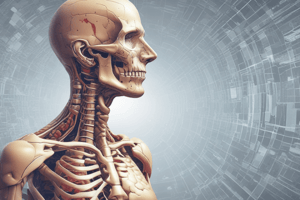
Gastrointestinal Tract (GIT)
- Part of the digestive system
- Organs that food and liquids travel through from mouth to anus
- Includes: mouth, pharynx, oesophagus, stomach, small intestine, large intestine, rectum, and anus
GIT Imaging Techniques
- Radiography: Chest X-ray, Abdominal X-ray (erect and supine)
- Contrast Study: Fluoroscopy (Barium or water-soluble contrast)
- Upper Gastrointestinal Series: Barium swallow, Barium Meal and follow-through, Enteroclysis (small bowel enema)
- Lower Gastrointestinal Series: Barium Enema
- Other Special Fluoroscopy Examinations: Fistulography
- Ultrasound
- Computed Tomography: CT enterography, CT colonoscopy
- Magnetic Resonance Imaging: MR enterography
- Nuclear Medicine: Chest Radiograph (for ruling out chest infection, pre-op prep, post-op evaluation, and intraperitoneal free air)
Contrast Studies
- Make the GIT more visible on imaging
- Coat inner surfaces of the GIT
- Radio-opaque: Barium sulphate, Water soluble
- Radiolucent: Gas
- Administered orally or rectally, or via Enteroclysis (small bowel enema)
Mucosal Pattern and Filling Defects
- Mucosal pattern: appearance of the inner surface of bowel
- Abnormalities: smoothing, irregularity
- Filling defects: any process occupying space within the bowel, resulting in an area of total or relative radiolucency within the barium column
- Types of filling defects:
- Intraluminal: barium all around it, may be freely mobile (e.g. food, faeces)
- Intramural: causes indentation from one side only, forming a sharp angle with the bowel wall, not completely surrounded by barium (e.g. tumour)
- Extramural: causes indentation from one side only, forming a shallow angle with the bowel wall, mucosa is preserved but stretched over the filling defect (e.g. enlarged adjacent organ)
Other Radiological Signs
- Stricture: circumferential or annular narrowing (differentiated from peristaltic waves), may have tapered ends, abrupt end (or shouldering)
- Ulceration: visible when the crater is filled with barium, appears as an outward projection from the lumen
- Haematemesis: possible lesion of the oesophagus
Normal and Abnormal Lesions of the Oesophagus
- Normal: develops in the hypopharynx, typically between the cricopharyngeus muscle and the inferior pharyngeal constrictor muscle, only involves the mucosa and submucosal layers, does not involve the muscular layer
- Abnormal: causes malignant strictures
Studying That Suits You
Use AI to generate personalized quizzes and flashcards to suit your learning preferences.