Podcast
Questions and Answers
What is the function of transseptal fibers in the periodontal ligament?
What is the function of transseptal fibers in the periodontal ligament?
- Prevent tooth extrusion
- Bear vertical masticatory stresses
- Keep teeth aligned (correct)
- Guide tissue regeneration
What is the direction of extension of horizontal fibers in the periodontal ligament?
What is the direction of extension of horizontal fibers in the periodontal ligament?
- Coronal direction
- Right angles to the long axis of the tooth (correct)
- Apical direction
- Oblique direction
What is the largest group of periodontal ligament fibers?
What is the largest group of periodontal ligament fibers?
- Alveolar crest fibers
- Apical fibers
- Oblique fibers (correct)
- Horizontal fibers
What is the function of alveolar crest fibers in the periodontal ligament?
What is the function of alveolar crest fibers in the periodontal ligament?
What is the location of the apical termination of the junctional epithelium in healthy tissue?
What is the location of the apical termination of the junctional epithelium in healthy tissue?
What is the function of the internal basal lamina in the junctional epithelium?
What is the function of the internal basal lamina in the junctional epithelium?
What is the term for the process of guiding the growth of wanted tissues to fill in a defect?
What is the term for the process of guiding the growth of wanted tissues to fill in a defect?
What is the characteristic of the mesial surface of the maxillary central?
What is the characteristic of the mesial surface of the maxillary central?
What is the condition for a new attachment to form after periodontal treatment?
What is the condition for a new attachment to form after periodontal treatment?
What is the term for the junctional epithelium in disease?
What is the term for the junctional epithelium in disease?
What is the direction of the cementoenamel junction curvature on the facial and lingual surfaces of a tooth?
What is the direction of the cementoenamel junction curvature on the facial and lingual surfaces of a tooth?
What determines the width of the interdental alveolar bone?
What determines the width of the interdental alveolar bone?
What is the width of the attached gingiva calculated by?
What is the width of the attached gingiva calculated by?
Which of the following is NOT a characteristic of masticatory mucosa?
Which of the following is NOT a characteristic of masticatory mucosa?
What is the term for the oral mucosa that covers the gingiva and hard palate?
What is the term for the oral mucosa that covers the gingiva and hard palate?
What is the definition of a 'functionally adequate' zone of gingiva?
What is the definition of a 'functionally adequate' zone of gingiva?
Which of the following is a characteristic of lining mucosa?
Which of the following is a characteristic of lining mucosa?
What is the function of the submucosa?
What is the function of the submucosa?
Which of the following cells is NOT typically found in the epithelium of the lining mucosa?
Which of the following cells is NOT typically found in the epithelium of the lining mucosa?
What is the term for the movement of the free gingival margin on stretching the lip or cheek?
What is the term for the movement of the free gingival margin on stretching the lip or cheek?
What is the dominant cellular element in the gingival connective tissue?
What is the dominant cellular element in the gingival connective tissue?
What is the origin of fibroblasts in the gingival connective tissue?
What is the origin of fibroblasts in the gingival connective tissue?
What is the role of fibroblasts in the gingival connective tissue?
What is the role of fibroblasts in the gingival connective tissue?
Where are mast cells typically found in the oral mucosa?
Where are mast cells typically found in the oral mucosa?
What is the primary source of blood supply to the gingiva?
What is the primary source of blood supply to the gingiva?
What is the route of lymphatic drainage of the gingiva?
What is the route of lymphatic drainage of the gingiva?
What is the characteristic of nerve fibers in the gingival connective tissue?
What is the characteristic of nerve fibers in the gingival connective tissue?
What is the composition of principal fibers in the periodontal ligament?
What is the composition of principal fibers in the periodontal ligament?
What is the outcome of phosphatase liberating from dental plaque, desquamated epithelial cells, or bacteria?
What is the outcome of phosphatase liberating from dental plaque, desquamated epithelial cells, or bacteria?
What is the term referred to as the concept of small foci of calcification that enlarge and coalesce to form a calcified mass?
What is the term referred to as the concept of small foci of calcification that enlarge and coalesce to form a calcified mass?
What is suspected to play an active role as the seeding agent in calcification?
What is suspected to play an active role as the seeding agent in calcification?
What is the term for the process of plaque formation?
What is the term for the process of plaque formation?
What is the function of the acquired pellicle?
What is the function of the acquired pellicle?
What is the term for the newly formed bacterial clusters that remain attached to the surface?
What is the term for the newly formed bacterial clusters that remain attached to the surface?
What is the primary mechanism of coaggregation between oral bacteria?
What is the primary mechanism of coaggregation between oral bacteria?
What is the characteristic of the early primary colonizers?
What is the characteristic of the early primary colonizers?
What is the term for the concentration of microorganisms, desquamated epithelial cells, leukocytes, and a mixture of salivary proteins and lipids?
What is the term for the concentration of microorganisms, desquamated epithelial cells, leukocytes, and a mixture of salivary proteins and lipids?
What is the ability of Bacterionema and Veillonella species?
What is the ability of Bacterionema and Veillonella species?
What is the primary source of inorganic constituents of supragingival plaque?
What is the primary source of inorganic constituents of supragingival plaque?
What type of bone loss is characterized by a uniform reduction in bone height, with the margins of the bone remaining perpendicular to the tooth surface?
What type of bone loss is characterized by a uniform reduction in bone height, with the margins of the bone remaining perpendicular to the tooth surface?
What is the term for a bone defect that creates a hollowed-out trough alongside the root of the tooth?
What is the term for a bone defect that creates a hollowed-out trough alongside the root of the tooth?
What is the term for a concavity contained within the facial and lingual walls in the crest of interdental bone?
What is the term for a concavity contained within the facial and lingual walls in the crest of interdental bone?
What is the number of osseous walls left surrounding the tooth that determines the classification of an angular defect?
What is the number of osseous walls left surrounding the tooth that determines the classification of an angular defect?
What is the term for a defect that has a greater number of osseous walls in the apical portion than in the occlusal portion?
What is the term for a defect that has a greater number of osseous walls in the apical portion than in the occlusal portion?
What is the distance between the base of the pocket and a fixed point on the crown, such as the CEJ?
What is the distance between the base of the pocket and a fixed point on the crown, such as the CEJ?
What is the term for a loss of the buccal or lingual bone overlaying the root portion of a tooth, leaving the area covered by soft tissue only?
What is the term for a loss of the buccal or lingual bone overlaying the root portion of a tooth, leaving the area covered by soft tissue only?
What is the term for pockets associated with horizontal bone loss?
What is the term for pockets associated with horizontal bone loss?
What is the critical parameter for the prognosis of a periodontally involved tooth?
What is the critical parameter for the prognosis of a periodontally involved tooth?
What is the characteristic of the epithelium on the dorsal surface of the tongue?
What is the characteristic of the epithelium on the dorsal surface of the tongue?
Which type of cells are the most common in the periodontal ligament?
Which type of cells are the most common in the periodontal ligament?
What is the function of the periodontal ligament in regards to orthodontic treatment?
What is the function of the periodontal ligament in regards to orthodontic treatment?
What is the main component of the ground substance in the periodontal ligament?
What is the main component of the ground substance in the periodontal ligament?
What is the average width of the periodontal ligament space?
What is the average width of the periodontal ligament space?
Which type of cells are responsible for the resorption of cementum in the periodontal ligament?
Which type of cells are responsible for the resorption of cementum in the periodontal ligament?
What is the primary function of the supraperiosteal arterioles in the gingiva?
What is the primary function of the supraperiosteal arterioles in the gingiva?
What is the term for the calcified masses that can be found in the periodontal ligament?
What is the term for the calcified masses that can be found in the periodontal ligament?
What is the type of epithelium found in the oral mucosa?
What is the type of epithelium found in the oral mucosa?
Which of the following is a function of the periodontal ligament?
Which of the following is a function of the periodontal ligament?
What is the primary characteristic of a periodontal pocket?
What is the primary characteristic of a periodontal pocket?
Which of the following is NOT a sign or symptom of periodontal pocket formation?
Which of the following is NOT a sign or symptom of periodontal pocket formation?
What is the most reliable method to determine the location and extent of periodontal pockets?
What is the most reliable method to determine the location and extent of periodontal pockets?
What is the term for the invasion of the bifurcation and trifurcation of multi-rooted teeth by periodontal disease?
What is the term for the invasion of the bifurcation and trifurcation of multi-rooted teeth by periodontal disease?
What is the characteristic of a suprabony pocket?
What is the characteristic of a suprabony pocket?
What is the primary difference between a gingival pocket and a periodontal pocket?
What is the primary difference between a gingival pocket and a periodontal pocket?
What can radiographs show in relation to periodontal pockets?
What can radiographs show in relation to periodontal pockets?
What is the characteristic of an infrabony pocket?
What is the characteristic of an infrabony pocket?
What is the term for the classification of furcation involvement?
What is the term for the classification of furcation involvement?
What is the significance of probing along the gingival margin in periodontal diagnosis?
What is the significance of probing along the gingival margin in periodontal diagnosis?
What is the main function of the periodontal ligament?
What is the main function of the periodontal ligament?
What type of epithelium lines the gingival sulcus?
What type of epithelium lines the gingival sulcus?
What is the main difference between the sulcular epithelium and the junctional epithelium?
What is the main difference between the sulcular epithelium and the junctional epithelium?
What is the function of the nerve fibers in the periodontal ligament?
What is the function of the nerve fibers in the periodontal ligament?
Which type of neural termination is responsible for carrying pain sensation?
Which type of neural termination is responsible for carrying pain sensation?
Which complex is associated with bleeding on probing, an important clinical parameter of active inflammation?
Which complex is associated with bleeding on probing, an important clinical parameter of active inflammation?
What is the histological characteristic that distinguishes the free gingiva from the epithelial attachment?
What is the histological characteristic that distinguishes the free gingiva from the epithelial attachment?
What is the primary function of the water channels in the plaque biofilm?
What is the primary function of the water channels in the plaque biofilm?
How does the loss of carbon dioxide and the formation of ammonia by dental plaque bacteria contribute to mineral precipitation?
How does the loss of carbon dioxide and the formation of ammonia by dental plaque bacteria contribute to mineral precipitation?
What is the main function of the outer oral epithelium?
What is the main function of the outer oral epithelium?
What is the primary etiologic factor for the initiation of periodontal disease?
What is the primary etiologic factor for the initiation of periodontal disease?
During the early stages of plaque formation, which type of bacteria predominates?
During the early stages of plaque formation, which type of bacteria predominates?
What is the consequence of stagnation of saliva on the supersaturated state of calcium phosphate salts?
What is the consequence of stagnation of saliva on the supersaturated state of calcium phosphate salts?
As plaque ages, what is the shift in the type of bacteria present?
As plaque ages, what is the shift in the type of bacteria present?
What is the consequence of orthodontic treatment on the periodontal ligament?
What is the consequence of orthodontic treatment on the periodontal ligament?
What is the mechanism by which bacteria in a biofilm communicate with each other?
What is the mechanism by which bacteria in a biofilm communicate with each other?
What is the significance of the redox potential in the plaque biofilm?
What is the significance of the redox potential in the plaque biofilm?
What is the result of the exchange of genetic information among bacterial cells in a biofilm?
What is the result of the exchange of genetic information among bacterial cells in a biofilm?
What is the role of the intercellular matrix in the plaque biofilm?
What is the role of the intercellular matrix in the plaque biofilm?
What is the significance of the water channels in the plaque biofilm in relation to nutrient availability?
What is the significance of the water channels in the plaque biofilm in relation to nutrient availability?
What is the purpose of adding the recession measurement to the probe depth at a particular site?
What is the purpose of adding the recession measurement to the probe depth at a particular site?
What is the correct way to place the probe for measuring probing depth?
What is the correct way to place the probe for measuring probing depth?
What is the consequence of using a hard-bristle toothbrush?
What is the consequence of using a hard-bristle toothbrush?
What is the primary symptom of root hypersensitivity?
What is the primary symptom of root hypersensitivity?
Why is it important to keep roots free of plaque after removal of a periodontal dressing?
Why is it important to keep roots free of plaque after removal of a periodontal dressing?
What is the term for the narrow grooves that extend from the crest of the gingiva to the attached gingiva?
What is the term for the narrow grooves that extend from the crest of the gingiva to the attached gingiva?
What is the term for trauma from toothbrushing that results in recession of the marginal gingiva?
What is the term for trauma from toothbrushing that results in recession of the marginal gingiva?
What is the term for the process by which the pain of root sensitivity results from indirect innervation caused by dentinal fluid movement in the tubules?
What is the term for the process by which the pain of root sensitivity results from indirect innervation caused by dentinal fluid movement in the tubules?
Why is it important to give antibiotic prophylaxis to patients at risk for bacterial endocarditis prior to performing periodontal probing?
Why is it important to give antibiotic prophylaxis to patients at risk for bacterial endocarditis prior to performing periodontal probing?
What is the correct way to measure the width of attached gingiva?
What is the correct way to measure the width of attached gingiva?
What is the characteristic of Grade III furcation involvement?
What is the characteristic of Grade III furcation involvement?
What is the primary method of diagnosing furcation involvement?
What is the primary method of diagnosing furcation involvement?
What is a complication of furcation involvement?
What is a complication of furcation involvement?
What is the characteristic of drug-induced gingival enlargement?
What is the characteristic of drug-induced gingival enlargement?
What is the characteristic of the gingival growth in drug-induced gingival enlargement?
What is the characteristic of the gingival growth in drug-induced gingival enlargement?
What is the treatment for drug-induced gingival enlargement?
What is the treatment for drug-induced gingival enlargement?
What is the characteristic of the histopathology of drug-induced gingival enlargement?
What is the characteristic of the histopathology of drug-induced gingival enlargement?
What is the function of a periodontal probe?
What is the function of a periodontal probe?
What is the amount of pressure typically applied when using a periodontal probe?
What is the amount of pressure typically applied when using a periodontal probe?
What is the characteristic of the lesion that forms in drug-induced gingival enlargement?
What is the characteristic of the lesion that forms in drug-induced gingival enlargement?
What is the most accurate way to assess periodontal pocket depth?
What is the most accurate way to assess periodontal pocket depth?
Why is the periodontal probe angled approximately 10° on each interproximal surface?
Why is the periodontal probe angled approximately 10° on each interproximal surface?
What is the purpose of using a calibrated periodontal probe?
What is the purpose of using a calibrated periodontal probe?
What is the correct way to perform probing?
What is the correct way to perform probing?
What is the purpose of measuring recession?
What is the purpose of measuring recession?
What is the importance of 'walking' the probe along the junctional epithelium?
What is the importance of 'walking' the probe along the junctional epithelium?
What is the significance of the distance from the base of the pocket to the margin of the free gingiva?
What is the significance of the distance from the base of the pocket to the margin of the free gingiva?
What is the purpose of taking measurements before and after scaling and root planing?
What is the purpose of taking measurements before and after scaling and root planing?
What is the characteristic of the correct probe force?
What is the characteristic of the correct probe force?
What is the purpose of the curved #2 Nabers probe?
What is the purpose of the curved #2 Nabers probe?
What is the primary mechanism of action of desensitizing agents in toothpastes?
What is the primary mechanism of action of desensitizing agents in toothpastes?
What is a common cause of gingival recession?
What is a common cause of gingival recession?
What is the importance of evaluating brushing technique and monitoring hard and soft tissue conditions at each recall visit?
What is the importance of evaluating brushing technique and monitoring hard and soft tissue conditions at each recall visit?
Why is it important to wait at least 4 weeks post-operatively before conducting a clinical evaluation of the soft tissue response to scaling and root planing?
Why is it important to wait at least 4 weeks post-operatively before conducting a clinical evaluation of the soft tissue response to scaling and root planing?
What is the purpose of burnishing of dentin in desensitization procedures?
What is the purpose of burnishing of dentin in desensitization procedures?
What is the role of the dental professional in treating patients with hypersensitivity?
What is the role of the dental professional in treating patients with hypersensitivity?
What is the purpose of iontophoresis in desensitization procedures?
What is the purpose of iontophoresis in desensitization procedures?
What is the importance of keeping the roots consistently free of plaque in treating patients with hypersensitivity?
What is the importance of keeping the roots consistently free of plaque in treating patients with hypersensitivity?
What is the purpose of restorative agents in desensitization procedures?
What is the purpose of restorative agents in desensitization procedures?
What is the outcome of acids and toxins produced by plaque on the pulp?
What is the outcome of acids and toxins produced by plaque on the pulp?
Study Notes
Cellular Elements of Gingival Connective Tissue
- Fibroblasts: dominant cellular element, found between fiber bundles, of mesenchymal origin, and play a major role in development, maintenance, and repair of gingival connective tissue.
- Mast cells: numerous in the connective tissue of the oral mucosa and gingiva.
- Fixed macrophages and histiocytes: present in the gingival connective tissue as components of the mononuclear phagocyte system, derived from blood monocytes.
- Adipose cells and eosinophils: scarce in the lamina propria.
- Plasma cells and lymphocytes: found in small foci in the connective tissue near the base of the sulcus in clinically normal gingiva.
Blood Supply to the Gingiva
- Three sources:
- Supraperiosteal arterioles: along the facial and lingual surfaces of the alveolar bone, extending capillaries to the sulcular epithelium and between the rete pegs of the external gingival surface.
- Vessels of the PDL: extending into the gingiva and anastomosing with capillaries in the sulcus area.
- Arterioles emerging from the crest of the interdental septa: extending parallel to the alveolar bone crest and anastomosing with vessels of the PDL.
Nerve Innervation of the Gingiva
- Most nerve fibers are myelinated and closely associated with blood vessels.
- Gingival innervation is derived from fibers arising from nerves in the PDL and from the labial, buccal, and palatal nerves.
Periodontal Ligament
- Principal fibers: collagenous, arranged in bundles, and follow a wavy course when seen on a longitudinal section.
- Six groups of principal fibers:
- Transseptal fibers: extending interproximally over the crest of the alveolar bone, embedded in the adjacent teeth's cementum.
- Alveolar crest fibers: extending in an oblique direction from the cementum to the alveolar crest, preventing tooth extrusion and providing resistance to lateral movement.
- Horizontal fibers: extending from the cementum to the alveolar bone at right angles, perpendicular to the long axis of the tooth.
- Oblique fibers: extending in a coronal direction from the cementum to the alveolar bone, forming the largest group of periodontal ligament fibers.
- Apical fibers: extending from the cementum to the alveolar bone at the apical region of the tooth socket, radiating in an irregular manner.
- Interradicular fibers: extending from the cementum to the alveolar bone in the furcation areas of multi-rooted teeth.
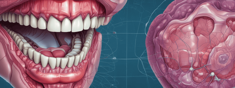
Junctional Epithelium
- A collar-like band of stratified squamous non-keratinizing epithelium, surrounding the tooth and located immediately apical to the sulcular epithelium.
- Length: ranges from 0.25 to 1.35 mm.
- Width: 10-29 cells wide at the coronal end, tapering apically to 1-2 cells wide.
- Apical termination: located at the cementoenamel junction in healthy tissue.
- Consists of two basal laminae: external basal lamina attaching the junctional epithelium to the gingival connective tissue, and internal basal lamina attaching the junctional epithelium to the tooth.
Attached Gingiva
- Width: Total gingival width (gingival margin to the mucogingival line) - sulcus or pocket depth.
- Width differs in different areas of the mouth, generally greatest in the incisors region (3.5-4.5 mm in the maxilla, 3.3-3.9 mm in the mandible), and narrower in the posterior segments (1.9 mm in the maxillary and 1.8 mm in the mandibular first premolars).
Functional Types of Oral Mucosa
- Masticatory mucosa: covers the gingiva and hard palate, with a keratinized or parakeratinized stratified squamous epithelium, and a lamina propria with two layers (papillary and reticular).
- Lining mucosa: covers all soft tissue of the oral cavity except the gingiva, hard palate, and dorsal surface of the tongue, with a non-keratinized epithelium (except on the vermillion border of the lip, where it is keratinized).
- Specialized mucosa: restricted to the dorsal surface of the tongue, with a keratinized epithelium, and characterized by the presence of surface papillae and taste buds.
Periodontal Ligament Functions
- Physical: attachment of the tooth to the bone via principal fibers, and absorption of occlusal forces.
- Formative: formation of connective tissue components by activities of connective tissue cells.
- Remodeling: by activities of connective tissue cells that are able to form as well as resorb cementum, PDL, and alveolar bone.
- Nutritive: through blood vessels that maintain the vitality of its various cells.
- Sensory: proprioceptive and tactile sensitivity is imparted through the PDL, sensed by the trigeminal nerve.
Gingival Epithelium
- Consists of stratified squamous epithelium with three distinct areas: the outer oral epithelium, the sulcular epithelium, and the junctional epithelium.
- Sulcular epithelium: a semipermeable membrane allowing seepage of tissue fluid from the gingiva into the gingival sulcus and entry of injurious bacterial products into the gingiva.
Mechanisms of Plaque Mineralization
- Two main categories:
- Mineral precipitation: results from a local rise in the degree of saturation of calcium and phosphate ions.
- Seeding agents: induce small foci of calcification that enlarge and coalesce to form a calcified mass.### Colonization and Plaque Maturation
- Microorganisms attach to tooth surfaces, forming microcolonies or biofilms, which lead to the development of periodontal disease
- Interbacterial connections occur through specific stereochemical interactions of protein and carbohydrate molecules on bacterial cell surfaces
- At least 18 genera of oral bacteria have shown coaggregation, including Fusobacterium nucleatum, which possesses surface molecules that foster cell-to-cell interactions
Plaque Formation and Maturation
- Plaque formation begins immediately after tooth surface cleaning, with rate affected by diet, age, salivary flow, oral hygiene, tooth alignment, systemic disease, and host factors
- Composition of plaque changes over time, with:
- Days 1-2: Young plaque consists of cocci (e.g. Streptococcus mutans and sanguis)
- Days 2-4: Coccistill dominate, with increasing numbers of filamentous forms and slender rods
- Days 4-7: Filaments increase, with a more mixed flora appearing
- Days 7-14: Vibrios and spirochetes appear, with increased gram-negative and anaerobic organisms
- Days 14-21: Vibrios and spirochetes prevail, with gingivitis evident clinically
Plaque as a Biofilm
- Dental plaque biofilm has a similar structure to all biofilms, with microcolonies encased in a polysaccharide matrix
- Heterogenous structure with open fluid-filled channels, allowing for nutrient passage and communication between bacteria
- Intercellular matrix composed of organic and inorganic materials from saliva, gingival crevicular fluid, and bacterial products
- Biofilm allows for quorum sensing, regulating gene expression through signaling compounds
Periodontal Disease and Bone Loss
- Bacterial plaque is the primary etiologic factor for periodontal disease
- Bone loss occurs through:
- Horizontal bone loss: uniform reduction in bone height, with margins remaining perpendicular to the tooth surface
- Vertical or angular defect: bone loss occurs in an oblique direction, with the base of the defect resting apical to the surrounding bone
- Osseous crater: concavity in the interdental bone, more common in posterior segments
Periodontal Pockets
- Periodontal pocket: pathological extension of the gingival sulcus, leading to destruction of supporting periodontal tissues
- Suprabony pocket: base of the pocket is coronal to the alveolar bone
- Intrabony pocket: base of the pocket is apical to the alveolar bone level
- Signs and symptoms: bleeding, suppuration, tooth mobility, diastema formation, pain, and foul taste
Furcation Involvement
- Invasion of the bifurcation and trifurcation of multi-rooted teeth by periodontal disease
- Classified as grades I-IV, with grade I being incipient bone loss and grade IV being total bone loss with through-and-through opening
- Treatment involves eliminating the involvement, with guided tissue regeneration and bone grafting having better results
Drug-Induced Gingival Enlargement
- Side effect of certain anticonvulsants, immunosuppressants, and calcium channel blockers
- Characteristics: painless, mulberry-shaped, pale pink, and resilient, with a lobulated surface
- Histopathology shows pronounced connective tissue and epithelial hyperplasia
Periodontal Probing
- Used to assess periodontal health, locate, measure, and determine the configuration of periodontal pockets
- Typical probe: thin, rodlike, tapered instrument with a blunt, rounded tip
- Inserts into the pocket with firm, gentle pressure, and walked around each surface of the tooth
- Provides the most accurate assessment of periodontal pocket depth and loss of attachment### Periodontal Probing
- The correct probe force is approximately 10-20 grams, which depresses the thumb pad by 1-2 mm.
- When probing, a hard ledge may block the passage of the probe, which is usually calculus; gently lift the probe away from the tooth and attempt to proceed apically again.
- The probe should be inserted parallel to the vertical axis of the tooth and "walked" circumferentially around each surface of each tooth to detect areas of deepest penetration.
Measuring Recession and Attached Gingiva
- Recession is the measurement of the migration of the free gingival margin apical to the CEJ of the tooth, measured as a positive value.
- The recession measurement added to the probe depth at a particular site indicates the amount of periodontal attachment that has been lost at that site.
- To measure the amount of attached gingiva:
- Place the probe on the external surface of the gingiva and measure from the mucogingival junction to the gingival margin to determine the width of the total gingiva.
- Insert the probe and measure the probing depth.
- Subtract the probing depth from the total gingival measurement to get the width of the attached gingiva.
Probing Techniques
- Tilting the probe can affect the accuracy of the measurements; the probe should be flat against the tooth near the gingival margin with the probe approximately parallel to the long axis of the tooth for insertion.
- In the presence of inflammation, the probe may extend apical to the most coronal extent of the junctional epithelium and give a slightly greater depth than is actually present.
Periodontal Inflammation and Bacteremia
- Patients at risk for bacterial endocarditis need to be given antibiotic prophylaxis prior to performing periodontal probing.
- The presence of inflammation leads to a longer duration of bacteremia with resultant risks for patients at risk of acute bacterial endocarditis.
Toothbrush Trauma and Gingival Recession
- Toothbrush trauma can cause gingival recession and abrasion of enamel and/or cementum.
- Hard-bristle toothbrushes should be avoided, and soft-bristle toothbrushes with a dentifrice of optimum abrasiveness should be used.
- Dentin is abraded 25 times faster and cementum 35 times faster than enamel, leading to root surface abrasion and root sensitivity.
Gingival Clefts and Root Sensitivity
- Gingival clefts (aka Stillman's clefts) are narrow grooves that extend from the crest of the gingiva to the attached gingiva.
- Root hypersensitivity is a common problem, often occurring spontaneously when the root becomes exposed due to gingival recession or pocket formation.
- The primary symptom of root hypersensitivity is cold sensitivity.
Desensitizing Agents and Treatment
- Desensitizing agents act through the precipitation of crystalline salts on the dentin surface, which block dentinal tubules.
- Various in-office products and procedures for desensitization of dentin include cavity varnishes, anti-inflammatory agents, and treatments that partially obturate dentinal tubules.
Gingival Recession and Periodontal Therapy
- Gingival recession can occur secondary to periodontal therapy, increasing the risk for cervical abrasion and dentinal sensitivity.
- The patient will often complain of cold sensitivity, and the hypersensitivity will sometimes subside in time with daily plaque removal using a soft brush.
Oral Hygiene and Dental Professional Evaluation
- The dental professional should evaluate brushing technique and monitor hard and soft tissue conditions at each recall visit.
- Faulty placement, overaggressive movement or pressure, or the use of a hard toothbrush can lead to hard and soft tissue damage.
- The most common cause of gingival recession is tooth injury (abrasion).
Studying That Suits You
Use AI to generate personalized quizzes and flashcards to suit your learning preferences.
Description
This quiz covers the different types of cells present in gingival connective tissue, including fibroblasts, mast cells, macrophages, and histiocytes. Learn about their functions and origins.


