Podcast
Questions and Answers
Which of the following is NOT a primary function of the gastrointestinal tract (GIT)?
Which of the following is NOT a primary function of the gastrointestinal tract (GIT)?
- Absorption of digestive products
- Movement of food through the alimentary tract
- Secretion of digestive juices
- Regulation of body temperature (correct)
The arrangement of GI smooth muscle fibers allows electrical signals to:
The arrangement of GI smooth muscle fibers allows electrical signals to:
- Readily travel from one fiber to the next within each bundle. (correct)
- Travel slowly and inefficiently in all directions.
- Be completely isolated within individual muscle fibers.
- Quickly travel sideways between bundles but slowly along the bundle length.
What cellular structure facilitates the movement of ions between the smooth muscle cells of the gastrointestinal tract?
What cellular structure facilitates the movement of ions between the smooth muscle cells of the gastrointestinal tract?
- Desmosomes
- Tight junctions
- Adherens junctions
- Gap junctions (correct)
What is the primary role of interstitial cells of Cajal in the gastrointestinal tract?
What is the primary role of interstitial cells of Cajal in the gastrointestinal tract?
Which of the following best describes slow waves in the gastrointestinal tract?
Which of the following best describes slow waves in the gastrointestinal tract?
Compared to action potentials in nerve fibers, spike potentials in gastrointestinal smooth muscle fibers are characterized by:
Compared to action potentials in nerve fibers, spike potentials in gastrointestinal smooth muscle fibers are characterized by:
Which of the following factors would most likely lead to hyperpolarization and decreased excitability of gastrointestinal smooth muscle?
Which of the following factors would most likely lead to hyperpolarization and decreased excitability of gastrointestinal smooth muscle?
What is the direct role of calcium ions in smooth muscle contraction?
What is the direct role of calcium ions in smooth muscle contraction?
The myenteric plexus is primarily responsible for:
The myenteric plexus is primarily responsible for:
Stimulation of the parasympathetic nervous system generally results in:
Stimulation of the parasympathetic nervous system generally results in:
Which of the following neurotransmitters typically inhibits gastrointestinal activity?
Which of the following neurotransmitters typically inhibits gastrointestinal activity?
Sensory nerve fibers in the gut can be stimulated by which of the following?
Sensory nerve fibers in the gut can be stimulated by which of the following?
Which reflex involves signals traveling from the stomach to cause evacuation of the colon?
Which reflex involves signals traveling from the stomach to cause evacuation of the colon?
Gastrin secretion is stimulated by:
Gastrin secretion is stimulated by:
What is the primary function of motilin?
What is the primary function of motilin?
Flashcards
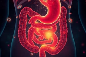
Functions of the GIT
Functions of the GIT
Supplies the body with water, electrolytes, vitamins, and nutrients through movement of food, secretion, absorption, and circulation.
Layers of intestinal wall
Layers of intestinal wall
The intestinal wall consists of the serosa, longitudinal/circular smooth muscle layers, submucosa, and mucosa.
GI muscle as a syncytium
GI muscle as a syncytium
GI smooth muscle fibers act as a syncytium, allowing electrical signals to travel via gap junctions for coordinated contractions.
Slow waves
Slow waves
Signup and view all the flashcards
Interstitial cells of Cajal
Interstitial cells of Cajal
Signup and view all the flashcards
Spike potentials
Spike potentials
Signup and view all the flashcards
Depolarization
Depolarization
Signup and view all the flashcards
Hyperpolarization
Hyperpolarization
Signup and view all the flashcards
Tonic contraction
Tonic contraction
Signup and view all the flashcards
Enteric Nervous System
Enteric Nervous System
Signup and view all the flashcards
Myenteric plexus
Myenteric plexus
Signup and view all the flashcards
Submucosal plexus
Submucosal plexus
Signup and view all the flashcards
Parasympathetic nerves
Parasympathetic nerves
Signup and view all the flashcards
Sympathetic nerves
Sympathetic nerves
Signup and view all the flashcards
Gastrin's function
Gastrin's function
Signup and view all the flashcards
Study Notes
- The GIT continuously supplies the body with water, electrolytes, vitamins, and nutrients. This process requires movement of food, secretion of digestive juices, absorption of water, circulation of blood, and control by local, nervous, and hormonal systems.
General Principles of GI Motility
- The intestinal wall consists of the serosa, longitudinal and circular smooth muscle layers, submucosa, and mucosa, which contains smooth mucosal muscle fibers.
Gastrointestinal Smooth Muscle
- GI smooth muscle fibers are arranged in bundles within the longitudinal and circular muscle layers.
- Muscle fibers are connected by many gap junctions, allowing ion movement and rapid electrical signal transmission within bundles.
- Muscle bundles are partly separated by connective tissue but fuse at points, creating a branching network and causing each muscle layer to function as a syncytium.
- When an action potential is elicited, it generally travels in all directions within the muscle. The distance it travels is dependent on the excitability of the muscle.
Electrical Activity of Gastrointestinal Smooth Muscle
- GI smooth muscle is excited by continuous slow electrical activity along the membranes of the muscle fibers.
- This activity includes slow waves and spikes.
- The resting membrane potential of GI smooth muscle can change, affecting motor activity.
Slow Waves
- Slow waves are undulating changes in resting membrane potential that cause rhythmic GI contractions.
- Intensity varies from 5 to 15 millivolts.
- Frequency varies in the human GI tract: 3/min in the stomach, 12/min in the duodenum, and 8-9/min in the terminal ileum.
- Slow waves are caused by complex interactions between smooth muscle and interstitial cells of Cajal, which act as electrical pacemakers.
- Interstitial cells of Cajal undergo cyclic changes in membrane potential due to unique ion channels.
- Slow waves usually do not cause muscle contraction except in the stomach. Instead, they excite the appearance of intermittent spike potentials, which then excite muscle contraction.
- Rhythmic and tonic GI contractions occur.
Spike Potentials
- These are true action potentials that occur when the resting membrane potential of GI smooth muscle becomes more positive than -40 millivolts.
- The normal resting membrane potential in gut smooth muscle fibers is between -50 and -60 millivolts.
- Spike potentials appear on the peaks of slow waves when they become more positive than -40 millivolts, with higher slow wave potentials leading to greater spike frequency.
Differences Between Action Potentials in Nerve Fibers and Smooth Muscle Fibers
- GI smooth muscle spike potentials last longer than action potentials in large nerve fibers.
- Nerve fiber action potentials are caused by rapid entry of sodium ions through sodium channels.
- GI muscle fiber action potentials are caused by entry of large numbers of calcium ions through calcium-sodium channels.
- Calcium-sodium channels open and close more slowly than sodium channels. The movement of calcium ions into muscle fibers causes contraction, unlike in nerve fibers.
Changes in Voltage of the Resting Membrane Potential
- The baseline voltage (-56 millivolts) of smooth muscle resting membrane potential can change due to multiple factors.
- Depolarization (potential becomes less negative) makes muscle fibers more excitable, while hyperpolarization (potential becomes more negative) makes them less excitable.
- Factors that depolarize the membrane are stretching of the muscle, acetylcholine stimulation from parasympathetic nerves, and stimulation by specific gastrointestinal hormones.
- Factors that hyperpolarize the membrane are norepinephrine or epinephrine and stimulation of sympathetic nerves that secrete norepinephrine.
Calcium Ions and Smooth Muscle Contraction
- Calcium ions use a calmodulin control mechanism to activate myosin filaments, causing muscle contraction.
- Slow waves do not cause calcium ion entry, only sodium ions.
- Significant calcium ion entry occurs during spike potentials, leading to contraction.
Tonic Contraction
- Tonic contraction is continuous and not associated with the electrical rhythm of slow waves.
- This may last for minutes or hours and can increase or decrease in intensity.
- Causes include continuous repetitive spike potentials, hormones, or continuous calcium ion entry.
Neural Control of Gastrointestinal Function
- The enteric nervous system in the wall of the GIT controls GI movements and secretion and has more neurons than the spinal cord.
- There are two plexuses: the myenteric (Auerbach's) plexus between the longitudinal and circular muscle layers, which controls GI movements, and the submucosal (Meissner's) plexus in the submucosa, which controls GI secretions and local blood flow.
- Extrinsic sympathetic and parasympathetic fibers connect to both plexuses.
Differences Between the Myenteric and Submucosal Plexuses
- The myenteric plexus controls muscle activity along the gut's length, increases tonic contraction, rhythmic contraction intensity, contraction rate, and conduction velocity of excitatory waves.
- The myenteric plexus contains inhibitory neurons that use vasoactive intestinal polypeptide (VIP) or other inhibitory peptides which inhibit intestinal sphincter muscles like the pyloric and ileocecal sphincters.
- The submucosal plexus controls function in the inner wall of each intestinal segment, controlling local intestinal secretion, absorption, and submucosal muscle contraction.
Neurotransmitters
- Neurotransmitters secreted by enteric neurons include acetylcholine, norepinephrine/epinephrine, adenosine triphosphate, serotonin, dopamine, cholecystokinin, substance P, vasoactive intestinal polypeptide, somatostatin, leu-enkephalin, met-enkephalin, bombesin, neuropeptide Y, and nitric oxide.
- Acetylcholine excites gastrointestinal activity, while norepinephrine inhibits it.
Autonomic Control of the Gastrointestinal Tract
- Parasympathetic stimulation increases activity via cranial (vagus nerves) and sacral divisions. The cranial fibers innervate the esophagus, stomach, pancreas and first half of the large intestine, while the sacral fibers innervate the distal half of the large intestine to the anus. Postganglionic neurons are located in the myenteric and submucosal plexuses.
- Sympathetic stimulation typically inhibits activity, originating from spinal cord segments T5 to L2.
Sympathetic Innervation
- Preganglionic fibers that innervate the gut enter the sympathetic chains and synapse in ganglia, such as the celiac and mesenteric ganglia, with postganglionic fibers spreading to all parts of the gut.
- Sympathetic innervation is extensive throughout the GI tract.
- Sympathetic nerve endings secrete norepinephrine, while parasympathetic endings secrete acetylcholine.
- Sympathetic stimulation generally inhibits GI tract activity and effects, acting through direct inhibition by norepinephrine or inhibition of enteric nervous system neurons.
- Strong sympathetic stimulation stops food movement.
Afferent Sensory Nerve Fibers
- Afferent sensory nerve fibers in the gut have cell bodies in the enteric nervous system and dorsal root ganglia of the spinal cord.
- These nerves are stimulated by irritation of the gut mucosa, excessive gut distention, or specific chemicals.
- Signals can cause excitation or inhibition of movements/secretion. 80% of vagus nerve fibers are afferent, transmitting sensory signals to the brain medulla to initiate vagal reflexes that control GI functions.
Gastrointestinal Reflexes
- Reflexes integrated within the gut wall (enteric nervous system) control secretion, peristalsis, and mixing contractions.
- Reflexes from the gut to the prevertebral sympathetic ganglia cause long-distance signals.
- Reflexes from the gut to the spinal cord or brain stem control gastric motor and secretory activity (vagus nerves), pain inhibition, and defecation.
Hormonal Control
- Gastrointestinal hormones are released into the portal circulation, acting on target cells with specific receptors. Hormonal effects persist after nervous connections are severed.
Gastrointestinal Hormone Actions
- Gastrin: Stimulated by protein, distention, and nervous signals. Site of secretion is the G cells of the antrum, duodenum, and jejunum. Action is to stimulate gastric acid secretion and mucosal growth.
- Cholecystokinin: Stimulated by protein, fat, and acid. Site of secretion is the I cells in the duodenum, jejunum, and ileum. Actions are to stimulate pancreatic enzyme and bicarbonate secretion, gallbladder contraction, growth of exocrine pancreas, and inhibit gastric emptying.
- Secretin: Stimulated by acid and fat. Site of secretion is the S cells of the duodenum, jejunum, and ileum. Actions are to stimulate pepsin, pancreatic and biliary bicarbonate secretion, growth of exocrine pancreas, and inhibit gastrin release and gastric acid secretion.
- Glucose-dependent insulinotropic peptide: Stimulated by protein, fat, and carbohydrate. Site of secretion is the K cells of the duodenum and jejunum. Actions are to stimulate insulin release and inhibit gastric acid secretion.
- Motilin: Stimulated by fat, acid, and nervous signals. Site of secretion is the M cells of the duodenum and jejunum. Actions are to stimulate gastric and intestinal motility.
Specific Hormones
- Gastrin is secreted by G cells in the stomach antrum and stimulates gastric acid secretion and mucosal growth.
- Cholecystokinin (CCK) is secreted by I cells in the duodenum and jejunum, contracts the gallbladder, and slows stomach contraction.
- Secretin is secreted by S cells in the duodenum and neutralizes acid in the small intestine.
- Glucose-dependent insulinotropic peptide (GIP) is secreted by the mucosa of the upper small intestine, decreases stomach motor activity, and stimulates insulin secretion.
- Motilin is secreted by the stomach and upper duodenum during fasting and increases gastrointestinal motility.
Functional Movements
- Propulsive movements move food forward and mixing movements keep intestinal contents mixed.
Propulsive Movements
- Peristalsis is an inherent property of syncytial smooth muscle tubes, where stimulation causes a contractile ring to appear and spread.
- Peristalsis occurs in the gut organs.
- Distention initiates intestinal peristalsis, and other stimuli include chemical/physical irritation and parasympathetic nervous signals.
- Peristalsis is weak or absent with congenital absence of the myenteric plexus and is depressed by atropine.
Peristaltic Waves
- Peristalsis normally dies out in the orad direction and continues toward the anus because the myenteric plexus is polarized.
- Distention initiates peristalsis, with the contractile ring starting on the orad side.
- At the same time, receptive relaxation occurs, with the gut relaxing downstream toward the anus.
- This complex pattern is called the myenteric or peristaltic reflex.
Mixing Movements
- Mixing movements differ in different alimentary tract parts.
- Peristaltic contractions cause mixing, especially when forward progression is blocked by a sphincter.
- Local intermittent segmentation contractions occur every few centimeters, with new constrictions occurring at other points.
Gastrointestinal Blood Flow
- Splanchnic circulation involves blood flow through the gut, spleen, pancreas, and liver.
- Blood flows from these organs to the liver via the portal vein, then through liver sinusoids, and leaves via the hepatic vein to the vena cava.
- Reticuloendothelial cells in liver sinusoids remove bacteria and particulate matter; non-fat, water-soluble nutrients go to liver sinusoids, while fats are absorbed into intestinal lymphatics and conducted to systemic circulation via the thoracic duct, bypassing the liver.
Studying That Suits You
Use AI to generate personalized quizzes and flashcards to suit your learning preferences.