Podcast
Questions and Answers
In the context of heart evolution, what key adaptation occurred in bony fish that led to the development of the two-sided heart?
In the context of heart evolution, what key adaptation occurred in bony fish that led to the development of the two-sided heart?
- The modification of the swim bladder into lungs, necessitating a more efficient circulatory system. (correct)
- The transformation of the ventral aorta into a pulmonary artery.
- The development of the sinus venosus into a distinct chamber.
- The division of the ventricle by a septum, creating separate pulmonary and systemic circuits.
What is the functional consequence of the inter-venous crest's diversion of blood flow between the cranial and caudal vena cava within the right atrium?
What is the functional consequence of the inter-venous crest's diversion of blood flow between the cranial and caudal vena cava within the right atrium?
- Synchronization of atrial contraction for optimized ventricular filling.
- Facilitation of oxygenation of blood before it enters the ventricle.
- Prevention of turbulence and collision of blood flow from each major vein. (correct)
- Regulation of blood pressure within the atrial chamber.
How does the structure of the auricle contribute to the overall function of the atrium?
How does the structure of the auricle contribute to the overall function of the atrium?
- It contains pectinate muscles that enable forceful atrial contraction.
- It directs blood flow through the atrioventricular valve.
- It increases the volume of the atrium, accommodating a larger blood reservoir. (correct)
- It houses the sinoatrial node, initiating electrical impulses for heart contraction.
What functional significance do the moderator bands (trabecula septomarginalis) have in ventricular contraction, beyond physical support?
What functional significance do the moderator bands (trabecula septomarginalis) have in ventricular contraction, beyond physical support?
In the context of coronary circulation, why are the major coronary vessels positioned outside the myocardium?
In the context of coronary circulation, why are the major coronary vessels positioned outside the myocardium?
How does the presence of ossa cordis in large ungulates influence cardiac function or structure?
How does the presence of ossa cordis in large ungulates influence cardiac function or structure?
What is the primary mechanism by which parasympathetic innervation affects heart rate?
What is the primary mechanism by which parasympathetic innervation affects heart rate?
What is the functional consequence of the ductus arteriosus failing to close after birth?
What is the functional consequence of the ductus arteriosus failing to close after birth?
How does the specialized bundle of His-like bundle contribute to the coordinated contraction of the heart?
How does the specialized bundle of His-like bundle contribute to the coordinated contraction of the heart?
Which structural feature primarily facilitates the directional flow of blood from the right atrium to the right ventricle?
Which structural feature primarily facilitates the directional flow of blood from the right atrium to the right ventricle?
Flashcards
Two-sided Heart Evolution
Two-sided Heart Evolution
Heart evolution from bony fish, septum insertion created a two-sided heart. Swim bladder evolved into lungs, carotid artery divides subsequently.
Right Atrium Vessels
Right Atrium Vessels
Vessels into right atrium: cranial vena cava, caudal vena cava (with fossa ovalis, a remnant of foramen ovale), azygos vein, coronary sinus.
Azygos Vein Drainage
Azygos Vein Drainage
Azygos vein drains dorsal thorax, entering right side in horse, dog, and cat.
Coronary Sinus Function
Coronary Sinus Function
Signup and view all the flashcards
Left Heart Vessels
Left Heart Vessels
Signup and view all the flashcards
Ductus Arteriosus
Ductus Arteriosus
Signup and view all the flashcards
Ossa Cordis
Ossa Cordis
Signup and view all the flashcards
Sinuatrial Node (SAN)
Sinuatrial Node (SAN)
Signup and view all the flashcards
Parasympathetic Heart Nerve
Parasympathetic Heart Nerve
Signup and view all the flashcards
Sympathetic Heart Innervation
Sympathetic Heart Innervation
Signup and view all the flashcards
Study Notes
- The two-sided heart evolved from the two-chambered heart of bony fish through the insertion of a septum.
- The swim bladder, originally ventral to the heart, evolved into lungs.
- The carotid artery divides into the internal and external carotid arteries.
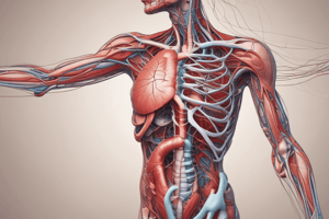
General Heart Structure
-
Four vessels enter the right atrium:
- Cranial vena cava
- Caudal vena cava: The fossa ovalis is a remnant of the foramen ovale that allowed connection between the right and left atria in fetal life.
- Azygos vein: Drains the dorsal thorax and drops into the right atrium in horses, dogs, and cats
- Coronary sinus: Drains blood from the heart itself
-
The inter-venous crest between the cranial and caudal vena cava diverts blood flow to prevent collisions.
-
Both atria have an 'out pouching' called the auricle (ear), located cranially and to the left, which increases the atrium's volume.
-
The main part of the atrium consists of smooth muscle, while the auricle has ridges in the wall called pectinate muscles.
-
The right atrium pumps blood into the right ventricle via the atrio-ventricular valve (tricuspid), with cusps anchored by chorda tendinae (tendinous cords) that attach to papillary muscles and allow the valve to open downwards only.
-
The internal surface of the ventricle has ridges called trabeculae carneae, which may help smooth blood flow.
-
The right ventricle pumps blood around the cranial side of the heart.
-
Coronary vessels are situated in the coronary groove.
-
The left atrio-ventricular valve is bicuspid/mitral.
-
Moderator bands, or trabecula septomarginalis, are bands of heart muscle found in both ventricles between the septum and ventricular wall, serving an electrical rather than physical function.
Heart Base
- Inflows and outflows include the aorta, pulmonary artery, and vena cavae.
- The ductus arteriosus in the fetus bypasses the pulmonary artery and aorta, closing within the first three days of life and remaining as the vestigial ligamentum arteriosum.
- The fibrous skeleton contains valves and vessels and provides insulation.
- Mineralization of two regions forms ossa cordis (bones of the heart) in large ungulates.
- Coronary circulation accounts for approximately 15% of cardiac output.
- Major vessels are positioned outside the myocardium to prevent occlusion from heart pressure.
- Venous drainage occurs into the atria via foramina venarum minimarum and into the ventricles via venae cordis parvae.
- In species with a left azygos vein (pigs, ruminants), it drains diagonally across to the coronary sinus on the right.
- Pigs typically have a left azygos vein, while ruminants may have both.
Pacemaker and Impulse
- The primary pacemaker of the heart is the sinuatrial node (not sinoatrial node) near the intervenous crest in RA.
- The impulse is transferred across the heart muscle in carnivores, and a specialized bundle of His-like bundle transmits the electrical impulse to the atrio-ventricular node near the coronary sinus opening in larger animals.
- The impulse travels down through the bundle of His, which divides into two crura, reaching the apex and moving upwards to cause a wave-like contraction.
- Moderator bands may allow the impulse to jump from the septum to the wall more quickly.
Innervation
- Parasympathetic innervation arises from the vagus nerves (cranial nerve X), descending in the vago-sympathetic trunk (VST) along with the common carotid artery, and innervates the SAN and AVN, reducing heart rate through cardiovagal fibers.
- Sympathetic innervation originates from the thoracic outflow from stellate and middle cervical ganglia, innervating the myocardium and vessels, and cardiosympathetic fibers increase heart rate and force.
Studying That Suits You
Use AI to generate personalized quizzes and flashcards to suit your learning preferences.