Podcast
Questions and Answers
What is the primary role of the gastrointestinal tract (GIT) in relation to nutrients?
What is the primary role of the gastrointestinal tract (GIT) in relation to nutrients?
- Filtering toxins from the blood and eliminating them through feces.
- Synthesizing essential vitamins and storing them for later use.
- Breaking down and transferring nutrients from the external environment into the body. (correct)
- Producing hormones that regulate appetite and satiety.
Which of the following best describes the function of the GIT lumen in host defense?
Which of the following best describes the function of the GIT lumen in host defense?
- It serves as a physical barrier and contains a developed immune system to inactivate harmful microorganisms. (correct)
- It secretes hormones that stimulate the production of antibodies in the blood.
- It facilitates the excretion of immune cells to regulate the body's immune response.
- It directly absorbs beneficial bacteria to enhance the immune system.
How does the composition of the human esophagus change along its length regarding muscle type?
How does the composition of the human esophagus change along its length regarding muscle type?
- It alternates between bands of skeletal and smooth muscle throughout its length.
- It transitions from skeletal muscle in the top third to smooth muscle in the remaining portion. (correct)
- It is primarily composed of skeletal muscle, with a small portion of smooth muscle near the stomach.
- It is entirely composed of smooth muscle from beginning to end.
What structural modification within the intestine significantly increases its surface area?
What structural modification within the intestine significantly increases its surface area?
In polarized epithelial cells lining the GIT, what is the primary characteristic of the apical surface?
In polarized epithelial cells lining the GIT, what is the primary characteristic of the apical surface?
What is the main function of stem cells located within the crypts of the small intestine?
What is the main function of stem cells located within the crypts of the small intestine?
What characterizes the selective transport of nutrients across the intestinal epithelium via the paracellular pathway?
What characterizes the selective transport of nutrients across the intestinal epithelium via the paracellular pathway?
What is the primary function of the muscularis mucosa?
What is the primary function of the muscularis mucosa?
What is the main role of the submucosal nerve plexus within the GIT?
What is the main role of the submucosal nerve plexus within the GIT?
How does the arrangement of the circular muscle layer in the muscularis externa contribute to GIT function?
How does the arrangement of the circular muscle layer in the muscularis externa contribute to GIT function?
Which of the following describes the unique aspect of the liver's blood supply compared to other organs?
Which of the following describes the unique aspect of the liver's blood supply compared to other organs?
What is the significance of the blood in the portal circulation being nutrient-rich?
What is the significance of the blood in the portal circulation being nutrient-rich?
How does the autonomic nervous system influence GI processes such as motility and secretion?
How does the autonomic nervous system influence GI processes such as motility and secretion?
What is the primary difference between short and long neural reflex pathways in the regulation of GI function?
What is the primary difference between short and long neural reflex pathways in the regulation of GI function?
What is the role of mechanoreceptors in the GI tract, and how are they activated?
What is the role of mechanoreceptors in the GI tract, and how are they activated?
Which of the following is an example of endocrine regulation in the context of gastrointestinal activity?
Which of the following is an example of endocrine regulation in the context of gastrointestinal activity?
How does cholecystokinin (CCK) participate in a negative feedback control system related to digestion?
How does cholecystokinin (CCK) participate in a negative feedback control system related to digestion?
What is the primary function of segmentation in the small intestine?
What is the primary function of segmentation in the small intestine?
How do slow waves contribute to the basic electrical rhythm (BER) in the GIT?
How do slow waves contribute to the basic electrical rhythm (BER) in the GIT?
How is the force of contraction in the GIT dictated by the basic electrical rhythm?
How is the force of contraction in the GIT dictated by the basic electrical rhythm?
In the context of gastrointestinal control, how are the cephalic, gastric, and intestinal phases classified?
In the context of gastrointestinal control, how are the cephalic, gastric, and intestinal phases classified?
What role does the hypothalamus play in the control of food intake?
What role does the hypothalamus play in the control of food intake?
How does leptin affect food intake and energy balance?
How does leptin affect food intake and energy balance?
What is the most important factor that stimulates the thirst center in the hypothalamus?
What is the most important factor that stimulates the thirst center in the hypothalamus?
Which component of saliva gives it an alkaline nature?
Which component of saliva gives it an alkaline nature?
What is the role of myoepithelial cells in the salivary glands?
What is the role of myoepithelial cells in the salivary glands?
Why is the initial saliva secretion from acinar cells isotonic?
Why is the initial saliva secretion from acinar cells isotonic?
What role does the parasympathetic nervous system play in the regulation of saliva production?
What role does the parasympathetic nervous system play in the regulation of saliva production?
How does saliva aid the process of swallowing?
How does saliva aid the process of swallowing?
How does the esophageal phase of swallowing move the food bolus toward the stomach?
How does the esophageal phase of swallowing move the food bolus toward the stomach?
What is the function of mucus in the stomach?
What is the function of mucus in the stomach?
What is the role of gastrin in the stomach?
What is the role of gastrin in the stomach?
How does the parietal cell maintain a neutral pH during acidification of the stomach?
How does the parietal cell maintain a neutral pH during acidification of the stomach?
What stimulates S-cells to produce the hormone secretin?
What stimulates S-cells to produce the hormone secretin?
Where does blood go during acid tide?
Where does blood go during acid tide?
What causes liver to remove harmful substances?
What causes liver to remove harmful substances?
What produces hormone insulin?
What produces hormone insulin?
Flashcards
Functions of the GI Tract
Functions of the GI Tract
Digestion and absorption of nutrients, minerals, and water from the external environment into the body.
Excretion in the GI Tract
Excretion in the GI Tract
Non-absorbable food components, bacteria, and hydrophobic molecules excreted by the GI tract.
Host Defence in the GIT
Host Defence in the GIT
The GIT forms a barrier, contains a immune system and inactivates harmful bacteria or other microorganisms
Components of the GI Tract
Components of the GI Tract
Signup and view all the flashcards
Accessory Organs of the GI Tract
Accessory Organs of the GI Tract
Signup and view all the flashcards
Structure of the GI Tract
Structure of the GI Tract
Signup and view all the flashcards
Mucosa
Mucosa
Signup and view all the flashcards
Epithelium
Epithelium
Signup and view all the flashcards
Function of Epithelial Layer
Function of Epithelial Layer
Signup and view all the flashcards
Lamina Propria
Lamina Propria
Signup and view all the flashcards
Muscularis Mucosa
Muscularis Mucosa
Signup and view all the flashcards
Submucosa
Submucosa
Signup and view all the flashcards
Muscularis Externa
Muscularis Externa
Signup and view all the flashcards
Serosa
Serosa
Signup and view all the flashcards
Purpose of GIT Blood Supply
Purpose of GIT Blood Supply
Signup and view all the flashcards
Portal Circulation
Portal Circulation
Signup and view all the flashcards
"In Series" Circulation
"In Series" Circulation
Signup and view all the flashcards
Function of nutrient-rich blood reaching the liver
Function of nutrient-rich blood reaching the liver
Signup and view all the flashcards
What GI Processes Include
What GI Processes Include
Signup and view all the flashcards
What Governs GI Processes
What Governs GI Processes
Signup and view all the flashcards
How Reflexes Are Propagated
How Reflexes Are Propagated
Signup and view all the flashcards
Intrinsic Nerve Regulation
Intrinsic Nerve Regulation
Signup and view all the flashcards
Two Nerve Networks
Two Nerve Networks
Signup and view all the flashcards
Extrinsic Nerve Regulation
Extrinsic Nerve Regulation
Signup and view all the flashcards
What Parasympathetic Stimulates
What Parasympathetic Stimulates
Signup and view all the flashcards
Neurocrine regulation
Neurocrine regulation
Signup and view all the flashcards
CCK release simulated by
CCK release simulated by
Signup and view all the flashcards
Peristalsis - driving force for food moving tract:
Peristalsis - driving force for food moving tract:
Signup and view all the flashcards
Segmentation of Motility
Segmentation of Motility
Signup and view all the flashcards
Pacemaker cells
Pacemaker cells
Signup and view all the flashcards
Cephalic Phase
Cephalic Phase
Signup and view all the flashcards
Stomach Functions:
Stomach Functions:
Signup and view all the flashcards
Stomach Division
Stomach Division
Signup and view all the flashcards
Exocrine Secretions
Exocrine Secretions
Signup and view all the flashcards
Protects the stomach from self-digestion and and breaks down
Protects the stomach from self-digestion and and breaks down
Signup and view all the flashcards
Mucous Cell
Mucous Cell
Signup and view all the flashcards
Gastric Motility
Gastric Motility
Signup and view all the flashcards
Vomiting Causes
Vomiting Causes
Signup and view all the flashcards
Ulcer Treatment Methods
Ulcer Treatment Methods
Signup and view all the flashcards
Exocrine pancreas
Exocrine pancreas
Signup and view all the flashcards
Study Notes
- The gastrointestinal tract (GIT) digests, absorbs, and transfers organic nutrients, minerals, and water from the external environment into the internal environment.
- Digestion involves using GIT motility, pH changes, biologic detergents, and enzymes to form absorbable molecules from food.
- Enzymes are predominantly produced by the pancreas.
- Absorption involves the movement of digestive food from the intestine into the blood or the lymphatic system.
Functions of the GIT
- Excretion: Non-absorbable food components, bacteria, intestinal cells, hydrophobic molecules (drugs), cholesterol, and steroids are excreted.
- Host defense: The lumen of the GIT is considered to be outside the body.
- The GIT forms a barrier with the outside environment and contains a highly developed immune system that can inactivate harmful bacteria or other microorganisms
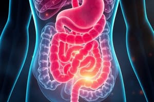
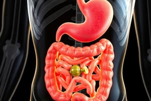
- Components: Mouth, pharynx, esophagus, stomach, small intestine (duodenum, jejunum, ileum), and large intestine.
- Accessory organs: Pancreas, liver, and gallbladder.
Structure of the GIT
- A long muscular tube stretches from the mouth to the anus with a similar composition from mid-esophagus to anus.
- The top third of the human esophagus is made of skeletal muscle, while the rest of the GIT is composed of smooth muscle
- Lumen: The inside of the tube contains many folds and processes to increase the surface area.
- Circular folds: The entire inner surface folds in with villi (singular villus)
- Villi project into the lumen of the tube above a crypt or invagination.
- Layers: Mucosa, submucosa, muscularis externa, and serosal layer (connective tissue layer).
- Mucosa: Contains three different subsections: epithelium, lamina propria, and muscularis mucosa.
- Layers of the Mucosa:
- Epithelium: A layer of cells lining all body cavities and surfaces.
- Epithelial cells: Polarized with a basolateral surface (closest to the blood surface, facing away from the tube; basal surface and the lateral surface) and an apical surface (inserts the inside of the tube or the lumen of the tube) as well as different transport proteins for each.
- Transport proteins: confined to different cell surfaces due to tight junctions.
- Function: Selective uptake of nutrients, electrolytes, and water, prevents passage of harmful substances.
- Surface area: Amplified by villi and crypts.
- Villus: A single layer of epithelial cells containing microvilli. Crypt: A region which invaginates into the lamina propria
- Stem cells: Located within crypts divide and produce daughter cells which differentiate into a variety of cells, migrate up the villus, reach the end of their life, and slough off.
Submucosa
- Located beneath the mucosa layer.
- Contains blood vessels, lymphatic vessels, submucosal nerve plexus (network of nerves), and connective tissue.
- Submucosal nerve plexus relays information to and away from the mucosa.
Muscularis Externa
- Contains circular muscle, myenteric nerve plexus, and longitudinal muscle.
- Circular muscle fibers are orientated in a circular pattern and contract and relax to close and open the tube.
- Myenteric nerve plexus regulates muscle function of the GIT (myo means "of muscle").
- Longitudinal muscle lengthens and shortens to control the length of the tube without changing the diameter.
Serosa
- Connective tissue layer encases the intestine and forms connections with the intestine and the abdominal wall.
Blood Supply to the GIT
- Carries away nutrients and other components absorbed from the diet, transports water-soluble molecules.
- Lacteals (lymphatic system; lamina propria in the mucosa layer) are important for fat absorption.
- Food follows this process: digested in the stomach, absorption and secretion in the small intestine, processing in the colon, then elimination of indigestible material.
- Blood is highly oxygenated entering the GIT but loses oxygen as it perfuses the intestine.
- Blood from GIT flows to the liver before returning to the heart via the portal vein.
Portal Circulation
- This circulation carries the blood from the intestinal tract to the liver.
- The blood is nutrient rich.
- Liver removes harmful substances (acting as a filter with many enzymes) and processes nutrients.
- Other organs are perfused with arterial blood (fully oxygenated).
- The liver receives blood from both arterial and venous sources.
- Hepatic artery contains fully oxygenated blood that perfuses the liver.
- Hepatic portal vein carries blood to the liver that has already perfused the stomach, pancreas, small intestine, and large intestine.
- These two blood supplies mix, liver is perfused with nutrient-rich blood from the GIT organs and has a poor oxygen content
- Most organs are perfused in parallel within the systemic circulation, but the liver is perfused in series.
- Blood flows from the digestive organs to the liver where blood flows from the digestive organs to the liver
Regulation of GI Processes
- Includes secretion, motility, or the movement of the Gl tract
- Processes are governed by the volume and composition of what is inside the tract
- Reflexes are initiated by distension of the GIT wall, osmolarity of the contents, pH of the contents, and the concentration of digestion contents like monosaccharides, fatty acids, peptides, and amino acids
- Digestion contents initiate different regulatory pathways.
- Reflexes are propagated by various receptors in the wall of the tract.
- Mechanoreceptors: Activated by mechanical stimuli (pressure and stretch).
- Osmoreceptors: Activated by a change in osmolarity.
- Chemoreceptors: Activated by specific chemicals.
Intrinsic Neural Regulation
- Controls motility and secretion.
- Intrinsic: Contained wholly within the organ itself.
- Occurs through nerve plexi located in the GIT wall: nerve plexi are branching networks of intersecting nerves.
- Enteric nervous system: Intrinsic nerve regulation. Controls the activity of the secretomotor neurons which play a role in secretion and motility, is contained completely within the walls of the GIT, the large number of neurons classifies it as the "Brain of the gut", can function independently of the CNS, and is critical for involuntary functions. It allows digestion without conscious thought.
- Two main nerve networks: myenteric plexus and the submucosal plexus.
- Myenteric plexus: Found between the two muscle layers, the circular muscle and the longitudinal muscle, of the muscularis externa and influences and regulates smooth muscle.
- Submucosal plexus: Found in the submucosa and predominantly influences secretion.
- Nerves extend from the submucosal plexus to the mucosa to control secretion.
- Neural activity in one plexus influences the activity in the other.
Extrinsic Neuronal Regulation
- Occurs outside of the GIT wall.
- Regulation occurs through the autonomic nervous system (ANS) through the parasympathetic and sympathetic divisions.
- Nerve fibers from the parasympathetic and sympathetic pathways enter the intestinal tract and synapse with neurons in both plexuses.
- Through the ANS, the CNS can influence motility and secretion.
- Smell of food sends signals through the brain to the GIT through the ANS.
- Different emotional states in the brain (CNS) will influence appetite.
Autonomic Pathways and Digestion
- Parasympathetic: Stimulates the flow of watery saliva, stimulates peristalsis (muscle contraction), stimulates secretion, and stimulates bile release from the liver; is considered the "Rest and digest" response.
- Sympathetic: Stimulates the small volume of thick saliva, inhibits peristalsis, inhibits secretion; the “Fright, flight, fight" response.
Neural Reflex Pathway
- Long reflex = extrinsic pathway; short reflex = intrinsic pathway.
- Eating a meal activates receptors in the GIT wall that feed into the nerve plexus and stimulates the smooth muscle to contract or a gland to secrete, causing a response in the GIT (contraction which breaks down food).
- Smell of food when hungry/emotional state stimulates the CNS, efferent autonomic neurons fire and interact with the same nerve plexus, which stimulates the smooth muscle to contract or a gland to secrete, causing a response in the GIT (contraction which breaks down food), creating the same CNS stimulation.
- Responses in the GIT can occur without any input from the CNS.
Chemical Messenger Regulation
- Four categories of chemical messenger regulation:
- Endocrine regulation: A hormone secreting gland cell releases a hormone across its basolateral surface into the blood. The hormone travels to its target cells in one or more distant places in the body.
- Neurocrine regulation: A nerve cell produces an electrical signal resulting in the release of a neurotransmitter which travels across a synapse and acts on a post-synaptic target cell.
- Paracrine regulation: A local cell releases a paracrine substance which diffuses through the interstitial fluid to act on target cells in close proximity to the site of release of the paracrine substance.
- Autocrine regulation: A local cell releases a substance which acts on the cell that released it.
- Hormonal Control of Gl Activity:
- Endocrine cells produce hormones, are found in the epithelium of the stomach and small intestine.
- Enteroendocrine cells release hormones which control GI functions.
GI Hormones
- Secretin, CCK (cholecystokinin), and gastrin are all peptide hormones.
- Each hormone participates in a feedback control system, and most affect more than one type of target cell.
- CCK (an Example): Release is stimulated by the presence of fatty acids and amino acids in the SI and is released into blood. It stimulates the pancreas to increase digestive enzyme secretion and causes contraction of the gallbladder. • Contraction of the gallbladder releases bile acids for fat breakdown. Absorption of fats and amino acids stops the release of CCK, a negative feedback control system
Intestinal Motility
- Stimulated by contraction and relaxation of the two muscle layers in the outer portion of the GIT; causes contents to move along the tract
- Peristalsis: The main force for food moving through the intestinal tract and for propulsion
- Circular muscle contracts on the oral side of a bolus of food (Longitudinal layer relaxes)
- Circular muscle contracted moves toward the anus, propelling the contents of the lumen in that direction, as the ring moves, the circular muscle on the other side of the distended area relaxes (Longitudinal muscle contracts), facilitating smooth passage of the bolus
- Segmentation: Important for the mixing of food which involves contraction and relaxation of intestinal segments with very little net movement of the food towards the large intestine and mostly occurs in the small intestine.
- Allows the mixing of the contents of the GIT with digestive enzymes and slows the transit time to allow for the absorption of nutrients and water.
- Basic Electrical Rhythm:
- Pacemaker cells are cells in the GIT that are distributed throughout the smooth muscle cells and are constantly under spontaneous depolarization-repolarization cycles called slow waves which give the GIT the basic electrical rhythm
- Under all circumstances the pacemaker cells undergo spontaneous depolarization-repolarization cycles and every time this happens, if there is a stimulus, there is the potential for action potential generation and muscle contraction
- Slow waves are propagated through the circular and longitudinal muscle layers through gap junctions.
- In the absence of any neural or hormonal input, the spontaneous slow waves do not result in any contraction, only when there is a stimulation, such as an excitatory hormone or neurotransmitter. The membrane potential is increased enough that threshold is reached, and an action potential is generated, resulting in muscle contraction.
- Slow waves are period fluctuations drift up and down due to regular variations in ion flux across the membrane.
- action potentials that lead to smooth muscle contraction occur when the slow waves are depolarized above threshold,
- Number of action potentials fired is proportional to the force of the contraction
- The frequency of the contraction is dictated by the basic electrical rhythm
- Force is mediated by neuronal and hormonal input.
Gastrointestinal Control
- Neural and hormonal control of the gastrointestinal system is divisible into 3 phases, classified on stimulus initiation.
- The various site of stimulus is named for where they initiate the reflex and not effector activity. Cephalic: Head phase is initiated through stimulation of receptors in the head (sight, smell, taste, chewing of food, emotional state) and predominantly regulated by parasympathetic fibers that activate neurons in the GIT nerve plexuses.
- Gastric: Stomach phase is when receptors in the stomach are stimulated distension, stretching, acidity, amino acids and peptides and is mediated by both short and long neural reflexes: Gastrin (hormone) represents the short reflex and acetylcholine represents the long reflex.
Control of Food Intake
- Essential for maintaining homeostasis, command center for neural and endocrine control coordination and for control of behavior.
- Contains a feeding center in the lateral region, whose Activation increases hunger and with lesions become anorectic and lose weight
- Contains a satiety center in the ventromedial region. Activation makes you feel full with lesions this area overeat and become obese. Factors Influence Food Intake: Orexigenic and Anorexigenic factors
- Orexigenic factor such as Neuropeptide Y, promotes hunger Ghrelin→ synthesized and released from the endocrine cells in the stomach during fasting which stimulates the release of neuropeptide Y to try and increase food intake.
- Anorexigenic promote appetite reduction Leptin → produced by adipose or fat tissue Insulin → produced by the pancreas; stimulates a reduction in food intake Peptide YY → released from the intestine to reduce food intake and Melanocortin → released directly from the hypothalamus
Factors Affecting Food Intake
- Take in more energy/eat more than burned during exercise, deposit fat in tissues increases the secretion of leptin Plasma leptin concentrations increase which travels to the hypothalamus, inhibiting neuropeptide Y release. (neuropeptide stimulates eating) and causes a in appetite and reduced energy intake and an increase in the metabolic rate Lack of leptin results in no appetite regulation, overeating and obesity
- Regulation of Water Intake:
- Hypothalamus: Contains a thirst center, stimulated by increased plasma osmolarity (most important factor)
- Increased blood osmolarity stimulates osmoreceptors, which stimulates thirst & the release of ADH, resulting in water conservation Also stimulated by decreased blood volume and a dry mouth.
Salivary Glands
- Three main pairs of large glands: parotid, submandibular, and sublingual that compose Saliva:
- Saliva: Hypotonic, slightly alkaline, and is made of Water Electrolytes - potassium and bicarbonate (gives alkaline nature); poor in sodium and chloride Digestive enzymes - amylase (breaks down starches into disaccharides and trisaccharides); lipase (breaks down fat into fatty acids) Glycoproteins, such as mucin (when combined with water mucin is called mucus) , Antimicrobial factors Lysozyme - breaks down the bacterial cell wall and Lactoferrin - chelates iron which prevents the multiplication of bacteria as iron is required for bacterial growth Functions: Moistens and lubricates the food to make it easier to swallow. Initiates digestion with digestive enzymes (amylase and lipase) and dissolves a small amount of food too
Anatomy of the Salivary Glands
- Structure of a salivary gland or a salivary duct: Made up of many microscopic ducts which branch out from gross ducts and are Composed of 3 different cell types: acinar cells, ductal cells, myoepithelial cells Acinar cells → secrete the initial saliva Ductal cells → create the alkaline and hypotonic nature of saliva Myoepithelial cells → have characteristics of both smooth muscle (can contract) and epithelial cells Saliva moves from the acinus to the striated duct Myoepithelial cells contract to constrict the acinus end of the duct, component
Formation of Saliva
- Tight junctions: Acinar cells have tight junctions between them - leaky and allow the passage of water and small ions, while ductal cells have tight junctions which do not allow the passage of water through Saliva is hypotonic and alkaline: All of the components are pumped into, or passed into, the lumen of the acinus The primary secretion from the acinar cells is not yet alkaline or hypotonic; it is isotonic with high Na, K, Cl, HCO3 and H2O Enzymes and exocytosis Electrolytes in the primary secretion (bicarbonate, chloride and potassium) are actively secreted with sodium water following through paracellular transport Myoepithelial cells contract to push the saliva into the region where there are ductal cells Ductal cells modify the saliva to form the hypotonic and alkaline saliva Sodium and chloride are actively taken up into the cells from the saliva resulting in a net gain of potassium and bicarbonate; saliva is hypotonic due to loss of sodium. Bicarbonate makes the saliva basic or alkaline
- Regulation:
- Parasympathetic pathways - taste smell. pressure receptors can be inhibited etc
Role of Saliva in Digestion
- Amylase: Found in saliva an enzyme that can breakdown starches, also called ptyalin as well as inhibited by stomach pH
- Plant starch is made up of glucose polymers including amylose and amylopectin Amylose → a straight chain of glucose molecules with alpha-1,4 linkages Amylase → breakdown leads to the formation of maltose. Amylopectin → chain of glucose molecules with alpha-1, 4 linkages as well as alpha-1,6 linkages, Amylase can only cleave the alpha-1,4 linkages which generates glucose molecules in alpha-limit dextrin
- Lingual Lipase: Found in saliva and stable in acid; can remain active in the stomach
Salivary Pathophysiology
- Xerostomia: Conditions where salivary secretion is impaired: Congenital, Sjögrens syndrome (autoimmune disease where system destroys the salivary glands), and side effects of other medicine Treatment: Frequent sips of water and fluoride to combat bacterial infection
- Swallowing: Pharynx → the passage at the back of your throat that is common to air and food Larynx → air passage between the pharynx and the trachea; voice box as it contains the vocal cords Glottis → area in the larynx around the vocal cords, where the air passes through the vocal cords Epiglottis → flap of cartilage that closes off the trachea when swallowing so food cannot enter the lungs Chew food → tongue pushes it to the back of throat Soft palette elevates to stop food from entering nose as a series of relfexes with receptors sensing this, which signals to muslces of the pharynx
Esophagus
- Contains skeletal muscle in the top 1/3 and smooth muscle in the lower 2/3 and is where mucus is secreted Esophageal Phase of Swallowing Upper esophageal sphincter relaxes food passes through glottis opens to close for breathing then waves move food to stomach, followed by a a sphincter being opened
Heart Burn
- Lower esophageal sphincter which normally prevents gasrtric reflux aids in the neutralization of the acid If they are not working effectively The Stomach:
- A muscular organ, a sac located between the esophagus and the small intestine with the main functions of storing and Mech / chemical breakdpwn including secreting pepsinogen Functions: storage of food • Mechanical breakdown of food (breaking food into smaller pieces) • Chemical breakdown of food • Secretes pepsinogen
Stomach Components
- Antrum: Lower region of the stomach (a Lower region of the stomach ( thicker smooth muscle layer), Pyloric sphincter controls emptying of the stomach Anatomy: Fundus and body ( mucus pepsinogen and hydrocholoric acid are secreted ) Secretions: HCL and mucus to allow the breakdown and digestion
Stomach Cells
Generalized gastric gland including ( a Generalized gastric gland ( (mucous/parietal/chief: Endocrine produces histamines etc Parietal Cell has oxyntic features plus secreting HCL as weel as activating the chief Cell Types and their Functions: Chief cell (in regions secrete pepsinogen which requires chemical change, interoendocrine secrete hormone, G cell secrete gastritis , ECL cell secrete hormone: Histamine stin HCl All cells require H that regulate neg / pos with eachother
Acidification of the Stomach Lumen
- Transporters that allow acidity to maintaine cell pH using Na/K ATPase. Chloride channels too Regulartion compenent - chemical meesengers regulate transport etc Hormones are gastrin to stimulate HCL, AcH causes ACH production, with histamine and somatostat
Phase of Gastric Digestion
Regulation phases that occur with secretin phase and intestinal phase where small epithelial insets
Gastric Motility
- Important for breaking down food with contractions allowing mixing down
Vomiting
- Stimulius from infections that feed inot medulla Types are nausea, salivation, followedy glottis closing
- Causes an imbalance of fluid
Duodemun
- Digestion
- Absorbs fe,Ca
- Releqse secretin and ccik
Jejenum
- Digeats and absorbs
Lilium
- Digeats and absorbs
- Absorbs Ba12
Large Intenstinde
- Reabsorbs H2)
- Resovoir for waste
- Absorbs bacterial metaboolite
SI in depth
- Folds on kerkhuring
- Villis on folds with microvilli and cpysts creating hugh surface area and the different stem cells
Intenstinal Cell Types
Parietal , Ecl and cheif cells Brush border
Charbohydrate digestion
- Brish bordes that digest starch like amylse, a limitextrose
Digestion of CHOP
Absrbed by guldt 5
Proteins
- Begins in sotmach with protein activated by hcf by enzymes Cabonox peptidase / Amino
Pancreatc
Bile
Liver
Gallstone
Appendix
Microbillome
Large Iintestine
- Water
7.33 SI
- Mixis
- Mmtilin relqxe
- Iletose / Cholere .
Studying That Suits You
Use AI to generate personalized quizzes and flashcards to suit your learning preferences.