Podcast
Questions and Answers
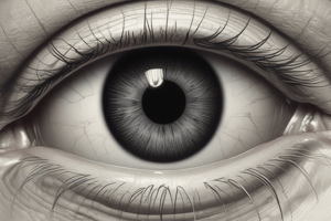
What is the pupil?
What is the pupil?
The opening through which light enters the eye
What is the cornea?
What is the cornea?
The clear tissue that covers the front of the eye
What is the aqueous humor?
What is the aqueous humor?
Fluid in the eye, found between the cornea and the lens
What is the optic nerve?
What is the optic nerve?
What is the optic disc (blind spot)?
What is the optic disc (blind spot)?
What is the ciliary body?
What is the ciliary body?
What is the macula lutea?
What is the macula lutea?
What is the fovea centralis?
What is the fovea centralis?
What is the tapetum lucidum?
What is the tapetum lucidum?
Flashcards
pupil
pupil
The opening through which light enters the eye.
cornea
cornea
The clear tissue that covers the front of the eye.
sclera
sclera
The white outer layer of the eyeball.
iris
iris
Signup and view all the flashcards
aqueous humor
aqueous humor
Signup and view all the flashcards
optic nerve
optic nerve
Signup and view all the flashcards
optic disc
optic disc
Signup and view all the flashcards
vitreous humor
vitreous humor
Signup and view all the flashcards
lens
lens
Signup and view all the flashcards
ciliary body
ciliary body
Signup and view all the flashcards
retina
retina
Signup and view all the flashcards
choroid
choroid
Signup and view all the flashcards
macula lutea
macula lutea
Signup and view all the flashcards
fovea centralis
fovea centralis
Signup and view all the flashcards
tapetum lucidum
tapetum lucidum
Signup and view all the flashcards
Study Notes
Eye Anatomy
- Pupil: The opening in the eye through which light enters.
- Cornea: The clear, front tissue of the eye.
- Sclera: The white outer layer of the eye.
- Iris: The colored portion of the eye that controls pupil size.
- Aqueous Humor: Fluid between the cornea and lens.
- Optic Nerve: Carries visual signals from the eye to the brain.
- Optic Disc (Blind Spot): The point where the optic nerve exits the eye, lacking photoreceptors.
- Vitreous Humor: Jelly-like substance behind the lens, maintaining the eye's shape.
- Lens: Transparent structure behind the pupil that focuses light onto the retina.
- Ciliary Body: Structure around the lens containing muscles to adjust lens shape and secreting aqueous humor.
- Retina: Contains photoreceptor cells (rods and cones) that convert light into neural signals.
- Choroid: Middle, vascular layer between the retina and sclera.
- Macula Lutea: Yellowish area of the retina rich in cones, crucial for detailed vision.
- Fovea Centralis: Part of the macula lutea with high cone density, providing the sharpest vision.
- Tapetum Lucidum: Reflective layer in the eyes of some nocturnal animals, enhancing night vision.
Studying That Suits You
Use AI to generate personalized quizzes and flashcards to suit your learning preferences.


