Podcast
Questions and Answers
What stimulates a blink in both eyes during the corneal reflex?
What stimulates a blink in both eyes during the corneal reflex?
- Contact with a wisp of cotton (correct)
- Bright light exposure
- Touching the eye with a finger
- Sound exposure
Which cranial nerve is responsible for carrying afferent sensation into the brain for the corneal reflex?
Which cranial nerve is responsible for carrying afferent sensation into the brain for the corneal reflex?
- Cranial nerve V (correct)
- Cranial nerve III
- Cranial nerve VII
- Cranial nerve II
What is the function of the iris in the eye?
What is the function of the iris in the eye?
- Transmits nerve impulses to the brain
- Regulates the amount of light entering the eye (correct)
- Refracts light onto the retina
- Produces aqueous humor
What is produced continually by the ciliary body to maintain intraocular pressure?
What is produced continually by the ciliary body to maintain intraocular pressure?
The optic disc is located on which side of the retina?
The optic disc is located on which side of the retina?
What does the lens of the eye primarily do?
What does the lens of the eye primarily do?
What is the main function of the choroid in the eye?
What is the main function of the choroid in the eye?
Which layer of the eye contains the visual receptive layer that converts light into nerve impulses?
Which layer of the eye contains the visual receptive layer that converts light into nerve impulses?
What is considered a normal finding during the Diagnostic Positions Test?
What is considered a normal finding during the Diagnostic Positions Test?
What is a normal characteristic of the eyebrows during inspection?
What is a normal characteristic of the eyebrows during inspection?
What is the expected position of the upper eyelids in relation to the iris?
What is the expected position of the upper eyelids in relation to the iris?
Which finding indicates a potential issue during the examination of the conjunctiva?
Which finding indicates a potential issue during the examination of the conjunctiva?
What color is the sclera typically expected to be?
What color is the sclera typically expected to be?
What variation in appearance can be noted in the sclera of dark-skinned people?
What variation in appearance can be noted in the sclera of dark-skinned people?
During inspection of the lacrimal apparatus, what would an excessive tearing indicate?
During inspection of the lacrimal apparatus, what would an excessive tearing indicate?
Which of the following describes a normal function of the puncta in the lacrimal apparatus?
Which of the following describes a normal function of the puncta in the lacrimal apparatus?
What should be checked when pressing the index finger against the sac just inside the lower orbital rim?
What should be checked when pressing the index finger against the sac just inside the lower orbital rim?
Which of the following describes a normal finding when inspecting the cornea and lens?
Which of the following describes a normal finding when inspecting the cornea and lens?
What appearance should the iris normally have?
What appearance should the iris normally have?
What does the acronym PERRLA stand for?
What does the acronym PERRLA stand for?
In which condition would the positive diopter of an ophthalmoscope be used?
In which condition would the positive diopter of an ophthalmoscope be used?
During the pupillary light reflex test, what is the expected outcome?
During the pupillary light reflex test, what is the expected outcome?
What indicates a normal response to accommodation when testing the eyes?
What indicates a normal response to accommodation when testing the eyes?
What do red numbers on the lenses of an ophthalmoscope represent?
What do red numbers on the lenses of an ophthalmoscope represent?
What is the primary purpose of darkening the room during an ocular fundus examination?
What is the primary purpose of darkening the room during an ocular fundus examination?
What should the diopter be set to for individuals with normal vision during the examination?
What should the diopter be set to for individuals with normal vision during the examination?
Which describes the optimal position to start when examining a person's ocular fundus?
Which describes the optimal position to start when examining a person's ocular fundus?
What should be noted as one approaches the eye during the fundus examination?
What should be noted as one approaches the eye during the fundus examination?
Which color is typically associated with a healthy optic disc?
Which color is typically associated with a healthy optic disc?
What characteristic of the optic disc margins is considered normal?
What characteristic of the optic disc margins is considered normal?
When examining retinal structures, following a blood vessel will help locate which part?
When examining retinal structures, following a blood vessel will help locate which part?
What type of lens should be used for a patient with nearsightedness during the examination?
What type of lens should be used for a patient with nearsightedness during the examination?
What is a common risk factor for glaucoma?
What is a common risk factor for glaucoma?
Which sign indicates esotropia?
Which sign indicates esotropia?
Which test is performed to evaluate extraocular muscle dysfunction?
Which test is performed to evaluate extraocular muscle dysfunction?
What is ptosis?
What is ptosis?
Which condition is associated with dilated and fixed pupils?
Which condition is associated with dilated and fixed pupils?
What type of growth is a pterygium?
What type of growth is a pterygium?
Which condition is characterized by inflammation of the conjunctiva?
Which condition is characterized by inflammation of the conjunctiva?
What is a common treatment for glaucoma?
What is a common treatment for glaucoma?
Flashcards are hidden until you start studying
Study Notes
Corneal Reflex
- Stimulated by contact with a wisp of cotton
- Causes blinking in both eyes
- Afferent sensation carried by the trigeminal nerve (cranial nerve V)
- Efferent message carried by the facial nerve (cranial nerve VII) that stimulates blinking
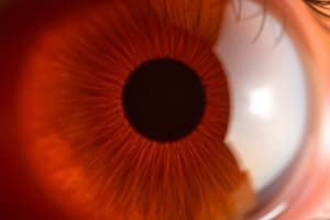
Eye Anatomy
- Sclera: Outermost layer, white, tough, protective
- Choroid: Middle layer, dark pigmentation prevents internal light reflection, heavily vascularized
- Retina: Inner layer, visual receptive layer where light waves change into nerve impulses
- Vitreous body: Clear, jelly-like substance that fills the space between the lens and retina
- Superior rectus muscle: Controls eye movement up
- Conjunctiva: Transparent membrane lining sclera and inner eyelid
- Iris: Functions as a diaphragm, controls pupil size
- Pupil: Opening in the center of the iris, allows light to enter the eye
- Lens: Biconvex, transparent disc, refracts light, keeps objects in focus
- Anterior chamber: Space between cornea and iris, filled with aqueous humor
- Posterior chamber: Space between iris and lens, filled with aqueous humor
- Ciliary body: Produces aqueous humor, controls lens shape
- Inferior rectus muscle: Controls eye movement down
- Macula: Region of central vision, located on the temporal side of the fundus
- Optic disc: Area where optic nerve fibers converge, appears as a pale disc
- Retinal vessels: Blood vessels supplying retina, normally a paired artery and vein in each quadrant
Iris and Pupil Functions
- Muscle fibers in the iris contract the pupil in bright light for near vision.
- Muscle fibers dilate the pupil in dim light for far vision.
- Parasympathetic stimulation constricts the pupil
- Sympathetic stimulation dilates the pupil and elevates the eyelid
Aqueous Humor
- Clear, watery fluid produced by the ciliary body
- Continuously flows to deliver nutrients and drain metabolic wastes
- Intraocular pressure is determined by the balance between production and outflow
Ophthalmoscope Examination
- Enlarges the view of the eye to inspect the media and ocular fundus
- Diopter lenses control focus
- Black numbers represent positive diopter, focusing on nearer objects
- Red numbers represent negative diopter, focusing on farther away objects
- Large round aperture with white light is used for routine examination
- Smaller aperture with white light is used for small pupils
Ocular Fundus Examination
- Darken the room to dilate pupils
- Start 25 cm from the patient at a 15-degree angle
- Observe for red reflex and move closer to the eye
- Adjust lens as needed to bring the fundus into focus
- Use red lens for nearsighted eyes and black lens for farsighted eyes
- Systematically inspect the optic disc, retinal vessels, general background and macula
Optic Disc
- Creamy yellow-orange to pink color
- Round or oval shape
- Distinct margins
- Physiologic cup is brighter yellow-white than the rest of the disc
Glaucoma
- Progressive eye disease
- Risk factors include age, diabetes mellitus, and family history
- Eye specialist screening includes optic nerve fiber thickness measurement, formal visual field testing, intraocular pressure measurement, and stereoscopic optic nerve examination
- Treatment options include eye drop medication, laser trabeculoplasty, and surgery
- Early detection is crucial to prevent further damage
Health Promotion and Patient Teaching
- Glaucoma is a serious eye condition and early detection is key for prevention and treatment.
- Encourage patients to get regular eye exams, especially those with risk factors, and to follow recommendations from their eye care provider, including medication adherence.
Abnormal Findings
Extraocular Muscle Dysfunction (EOM)
- Asymmetric corneal light reflex
- Strabismus
- Esotropia: Inward turning of the eye
- Exotropia: Outward turning of the eye
- Strabismus
- Cover test: Performed on all children
- Diagnostic positions test: Paralysis indicates cranial nerve dysfunction
Eyelid Abnormalities
- Periorbital edema: Edema around the eye
- Exophthalmos: Protruding eyes
- Enophthalmos: Sunken eyes
- Ptosis: Drooping upper lid
- Upward palpebral slant
- Ectropion: Lower lid rolling out
- Entropion: Lower lid rolling in
Lesions on the Eyelids
- Blepharitis: Inflammation of the eyelids
- Chalazion: Red bump on eyelid
- Hordeolum (stye): Infection of oil gland at the eyelid margin
- Dacryocystitis: Inflammation of the lacrimal sac
- Basal cell carcinoma
Pupil Abnormalities
- Unequal pupil size: Anisocoria
- Monocular blindness
- Constricted and fixed pupils: Miosis
- Dilated and fixed pupils: Mydriasis
- Argyll Robertson pupil
- Tonic pupil (Adie's pupil)
- Cranial nerve III damage
- Horner's syndrome
Visual Field Loss
- Retinal damage
- Lesion in globe or optic nerve
- Lesion at optic chiasm
- Lesion of outer uncrossed fibers at optic chiasm
- Lesion of the right optic tract or right optic radiation
Red Eye-Vascular Disorders
- Conjunctivitis: Inflammation of the conjunctivae
- Allergic conjunctivitis
- Uritis: Circumcorneal redness
- Primary angle-closure glaucoma (PACG)
- Subconjunctival hemorrhage
- Herpes simplex virus
Cornea and Iris
- Pterygium: Growth of the conjunctiva over the cornea
Studying That Suits You
Use AI to generate personalized quizzes and flashcards to suit your learning preferences.