Podcast
Questions and Answers
How are organs related to tissues in the organization of eukaryotic cells?
How are organs related to tissues in the organization of eukaryotic cells?
- Organs are composed of multiple organ systems working together.
- Organs are made up of a range of tissues working together to perform a function. (correct)
- Organs are made up of different types of specialized cells.
- Organs and tissues are different names for the same structure.
Which characteristic is exclusive to eukaryotic cells compared to prokaryotic cells?
Which characteristic is exclusive to eukaryotic cells compared to prokaryotic cells?
- Smaller size
- DNA enclosed within a nucleus (correct)
- Lack of membrane-bound organelles
- Presence of DNA
Which of the following features is associated with the cell-surface membrane?
Which of the following features is associated with the cell-surface membrane?
- Site of rRNA production and ribosome synthesis
- Controls the entry and exit of molecules to the cell (correct)
- Contains the genetic code for the cell
- Composed of a single layer of phospholipids
What role does the nucleolus play within the nucleus of a cell?
What role does the nucleolus play within the nucleus of a cell?
How do cristae contribute to the function of mitochondria?
How do cristae contribute to the function of mitochondria?
What is the role of the stroma within chloroplasts?
What is the role of the stroma within chloroplasts?
Which function is performed by the Golgi apparatus and vesicles?
Which function is performed by the Golgi apparatus and vesicles?
What is the primary function of lysosomes in a cell?
What is the primary function of lysosomes in a cell?
Which of the following is true regarding ribosomes found in eukaryotic cells?
Which of the following is true regarding ribosomes found in eukaryotic cells?
What is the key distinction between rough endoplasmic reticulum (RER) and smooth endoplasmic reticulum (SER)?
What is the key distinction between rough endoplasmic reticulum (RER) and smooth endoplasmic reticulum (SER)?
What role does the cell wall play in plant cells?
What role does the cell wall play in plant cells?
How does the tonoplast contribute to the function of a cell vacuole?
How does the tonoplast contribute to the function of a cell vacuole?
Which component is present in the cell walls of fungi?
Which component is present in the cell walls of fungi?
Which feature distinguishes prokaryotic cells from eukaryotic cells?
Which feature distinguishes prokaryotic cells from eukaryotic cells?
What is the composition of the cell wall in prokaryotic cells?
What is the composition of the cell wall in prokaryotic cells?
Which of the following structures is commonly found in prokaryotic cells?
Which of the following structures is commonly found in prokaryotic cells?
What is the function of flagella in prokaryotic cells?
What is the function of flagella in prokaryotic cells?
What components do viruses consist of?
What components do viruses consist of?
What is the primary limitation of using optical microscopes compared to electron microscopes?
What is the primary limitation of using optical microscopes compared to electron microscopes?
Why must samples examined under an electron microscope be in a vacuum?
Why must samples examined under an electron microscope be in a vacuum?
How does a scanning electron microscope (SEM) produce an image?
How does a scanning electron microscope (SEM) produce an image?
What is the purpose of calibrating an eyepiece graticule?
What is the purpose of calibrating an eyepiece graticule?
During cell fractionation, what condition is essential to maintain to prevent damage to organelles?
During cell fractionation, what condition is essential to maintain to prevent damage to organelles?
In cell fractionation, what does the first spin at low speed typically separate?
In cell fractionation, what does the first spin at low speed typically separate?
Which of the following does NOT occur during interphase?
Which of the following does NOT occur during interphase?
What is the key event that occurs during anaphase of mitosis?
What is the key event that occurs during anaphase of mitosis?
What event characterizes telophase in mitosis?
What event characterizes telophase in mitosis?
What is the role of cytokinesis in the cell cycle?
What is the role of cytokinesis in the cell cycle?
What does a high mitotic index indicate about a tissue sample?
What does a high mitotic index indicate about a tissue sample?
What feature contributes to the fluid-mosaic model of cell membranes?
What feature contributes to the fluid-mosaic model of cell membranes?
How do phospholipids arrange themselves to form a cell membrane?
How do phospholipids arrange themselves to form a cell membrane?
What role does cholesterol play within the cell membrane?
What role does cholesterol play within the cell membrane?
What role do glycoproteins and glycolipids perform in cell membranes?
What role do glycoproteins and glycolipids perform in cell membranes?
Which substance would most easily pass through a plasma membrane by simple diffusion?
Which substance would most easily pass through a plasma membrane by simple diffusion?
What is the primary difference between simple and facilitated diffusion?
What is the primary difference between simple and facilitated diffusion?
How do protein channels facilitate the transport of water-soluble ions across the cell membrane?
How do protein channels facilitate the transport of water-soluble ions across the cell membrane?
What defines osmosis as a transport process?
What defines osmosis as a transport process?
In an isotonic solution, how does the water potential compare between the solution and the cell?
In an isotonic solution, how does the water potential compare between the solution and the cell?
Under what solution conditions will an animal cell undergo lysis?
Under what solution conditions will an animal cell undergo lysis?
What is required for a molecule to move against its concentration gradient during active transport?
What is required for a molecule to move against its concentration gradient during active transport?
Describe the function of a co-transporter protein.
Describe the function of a co-transporter protein.
Flashcards
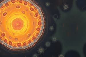
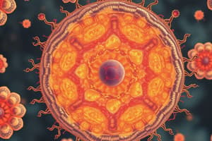
Eukaryotic Cells
Eukaryotic Cells
Cells with DNA in a nucleus, larger than prokaryotic cells. They form multicellular organisms and contain specialised cells.
Organ System
Organ System
A group of organs working together to perform a specific function in an organism.
Organ
Organ
A structure made of different tissues that work together to perform a specific function.
Tissue
Tissue
Signup and view all the flashcards
Nuclear Envelope
Nuclear Envelope
Signup and view all the flashcards
Nuclear Pores
Nuclear Pores
Signup and view all the flashcards
Nucleoplasm
Nucleoplasm
Signup and view all the flashcards
Nucleolus
Nucleolus
Signup and view all the flashcards
Cristae
Cristae
Signup and view all the flashcards
Mitochondrial matrix
Mitochondrial matrix
Signup and view all the flashcards
Thylakoid Membrane
Thylakoid Membrane
Signup and view all the flashcards
Stroma
Stroma
Signup and view all the flashcards
Cisternae
Cisternae
Signup and view all the flashcards
Exocytosis
Exocytosis
Signup and view all the flashcards
Hydrolyse
Hydrolyse
Signup and view all the flashcards
Protein synthesis
Protein synthesis
Signup and view all the flashcards
Endoplasmic Reticulum
Endoplasmic Reticulum
Signup and view all the flashcards
rRNA
rRNA
Signup and view all the flashcards
Cellulose
Cellulose
Signup and view all the flashcards
Chitin
Chitin
Signup and view all the flashcards
Tonoplast
Tonoplast
Signup and view all the flashcards
Plasmid
Plasmid
Signup and view all the flashcards
Capsule
Capsule
Signup and view all the flashcards
Flagella
Flagella
Signup and view all the flashcards
Capsid
Capsid
Signup and view all the flashcards
Binary Fission
Binary Fission
Signup and view all the flashcards
Cell fractionation
Cell fractionation
Signup and view all the flashcards
Ultracentrifugation
Ultracentrifugation
Signup and view all the flashcards
Magnification
Magnification
Signup and view all the flashcards
Resolution
Resolution
Signup and view all the flashcards
Eyepiece Graticule
Eyepiece Graticule
Signup and view all the flashcards
Calibration
Calibration
Signup and view all the flashcards
Stage Micrometer
Stage Micrometer
Signup and view all the flashcards
Isotonic
Isotonic
Signup and view all the flashcards
Buffer
Buffer
Signup and view all the flashcards
Centrifuge
Centrifuge
Signup and view all the flashcards
Nuclear division
Nuclear division
Signup and view all the flashcards
Diploid
Diploid
Signup and view all the flashcards
fibers attach to chromatids
fibers attach to chromatids
Signup and view all the flashcards
Study Notes
- Two cell types needs learning: Eukaryotic and prokaryotic cells, alongside the structure of viruses
Eukaryotic Cells
- Eukaryotic cells are larger than prokaryotic cells, and their DNA is contained in a nucleus
- Most eukaryotic cells are part of complex multicellular organisms containing cells specialized for various functions
- Organisms are made of organ systems which contains an array of organs working together for a common function
- Organs are made of tissues working together to perform a function
- Specialized cells with similar structures and functions organize into tissues
- Animals, plants, and fungi are eukaryotic organisms
- Eukaryotic cells contain a range of organelles
- There are ten organelles to know, including their structure and function:
- Cell-surface membrane
- Nucleus
- Mitochondria
- Chloroplasts
- Golgi apparatus and Golgi vesicles
- Lysosomes
- Ribosomes
- Rough endoplasmic reticulum and smooth endoplasmic reticulum
- Cell wall
- Cell vacuole
Cell-Surface Membrane
- The cell-surface membrane is found in all cells
- It has a structure called Phospholipid bilayer embedded with molecules within and attached on the outside such as proteins, carbohydrates, and cholesterol
- It controls the entrance and exit of molecules
Nucleus
- The nucleus has a double membrane called the nuclear envelope and it contains nuclear pores
- It contains nucleoplasm, which is a granular, jelly-like material
- The nucleus holds chromosomes, which are protein-bound, linear DNA
- The nucleolus is a smaller sphere in the nucleoplasm responsible for rRNA production and ribosome synthesis
- It is the site of DNA replication and transcription to make mRNA
- It contains the genetic code for each cell
Mitochondria
- The mitochondria has a double membrane, including an inner membrane called the cristae
- The fluid center is called the mitochondrial matrix
- It contains 70S ribosomes and circular DNA
- The mitrochondria is the site of aerobic respiration and ATP production
Chloroplast
- Chloroplasts are surrounded by a double membrane
- It contains thylakoids, which are folded membranes embedded with pigment
- The fluid-filled stroma contains enzymes for photosynthesis
- Chloroplasts are found in plants
- It contains 70S ribosomes and circular DNA
- Chloroplast are the site of photosynthesis
Golgi Apparatus and Vesicles
- The Golgi apparatus consist of folded membranes that make up cisternae
- Secretory vesicles pinch off from the cisternae
- The golgi apparatus add carbohydrates to proteins to form glycoproteins
- They are able to produce secretory enzymes
- Golgi apparatus can secrete carbohydrates
- Transport, modify, and store lipids
- The golgi apparatus can forma lysosomes
- Molecules are 'labelled' to reach their destination in the golgi apparatus
- There are finished products transported to the cell surface in Golgi vesicles where they fuse with the membrane and the contents are released via exocytosis
Lysosomes
- Lysosomes are bags of digestive enzymes and can contain 50 different enzymes
- Lysosomes hydrolyse pathogens in phagosomes
- They completely break down dead cells in a process called autolysis
- Lysosomes involves exocytosis to release enzymes outside of the cell
- Digest worn-out organelles for reuse of materials
Ribosome
- Ribosomes are small granules, made up of two sub-units of protein and rRNA
- There are 80S ribosomes which are large ribosomes found in eukaryotic cells (25nm)
- There are also 70S -smaller ribosome found in prokaryotic cells, mitochondria and chloroplasts
- Ribosomes are the site of synthesis of proteins
Endoplasmic Reticulum
- Rough and Smooth ER both have folded membranes called cisternae
- Rough ER have ribosomes on the cisternae
- The rough ER is responsible for protein synthesis
- The smooth ER synthesizes and stores lipids and carbohydrates
Cell Wall
- Cell walls are present in plants and fungal cells
- Plant cell walls are made of microfibrils of the cellulose polymer
- Fungi cell walls are made of chitin, a nitrogen-containing polysaccharide
- Cell walls provide structural strength to the cell
Cell Vacuole
- Cell vacuoles are filled with fluid and surrounded by a single membrane called a tonoplast
- Cell vacuoles function is to make the cell turgid and provides support
- Cell vacuoles are a temporary store of sugars and amino acids
- The pigments in cell vacuoles are responsible for colored petals which attract pollinators
Prokaryotic Cells
- Key differences between prokaryotic cells and eukaryotic cells are:
- Much smaller cells
- Prokaryotic cells have no membrane bound-organelles compared to eukaryotic cells
- Smaller ribosomes
- No nucleus
- Made of cell walls of murein
- Some bacteria also contain three additional features:
- Plasmids, which are rings of DNA containing genes linked to survival such as antibiotic resistance
- Some bacteria have a capsule surrounding their cell wall, which provides protection from other cells and helps bacteria agglutinate
- Flagella are used for locomotion
Viruses
- Viruses are non-living and acellular
- They contain genetic material, a capsid and attachment proteins
- HIV is surrounded by a further lipid envelope which has attachment proteins on the outside that enable the virus to identify which host cells to enter
Microscopy
- There are three key types of microscopes: optical microscopes, transmission electron microscopes and scanning electron microscopes
- Magnification is how many times larger the image appears compared to the object
- Resolution is the minimum distance between two objects in which they can still be viewed separately
- Resolution in an optical microscope is determined by the wavelength of light
- Wavelength beam of electrons determines resolution of electron microscope
Optical vs electron
- Optical (light) microscope:
- A beam of light is condensed to create the image with use of a glass lens
- Poorer resolution due to light having a longer wavelength
- Lower magnification
- Coloured images
- Can view living samples
- Electron microscope (scanning or transmission):
- A beam of electrons is condensed to create the image with electromagnets
- Higher resolving power as electrons have a shorter wavelength
- Higher magnification
- Black and white images
- The sample must be in a vacuum, and therefore non-living
Types of Electron Microscopes
- Transmission Electron Microscopes (TEM):
- Used extremely thin specimens that are stained and placed in a vacuum.
- An electron gun produces a beam of electrons that passes through the specimen, with parts absorbing electrons to appear dark
- Generates a 2D image that shows detailed images of the internal structure of cells
- Scanning Electron Microscope (SEM):
- Specimens not need to be thin
- Electrons are not transmitted through but beamed onto the surface and electrons are scattered in different ways depending on the contours
- Produces a 3D image
Magnification Calculations
- The size of structures viewed under an optical microscope can be measured using the formula:
- Image size = Actual size x Magnification I=AM
- Magnification = Image size / Actual size
- All the units must be the same, so convert as required
Eyepiece Graticule
- Inside optical microscopes, there is a scale on a glass disc called the eyepiece graticule
- The eyepiece graticule is used to measure the size of objects on the microscope
- Each time you change the objective lens, you must calibrate the eyepiece to determine the distance between each division at that magnification
- A stage micrometer is used to calibrate the eyepiece graticule using a glass slide with a scale on it
- To perform callibration, line up the stage micrometer and eyepiece graticule whilst looking through the eyepiece.
- Count the number of divisions on the eyepiece graticule that fit into one division on the micrometer scale
- Each division on the micrometer is 10μm, so can calculate one division on the eyepiece graticule is at the current magnification
- Now the graticule is calibrated, you can measure the size of cells or organelles
Cell Fractionation
- Cell fractionation and ultracentrifugation are used to break down cells and separate organelles for study
- Cells are broken down so that the organelles are free to be separated with a homogeniser (blender)
- During homogenisation, cells are kept in a cold solution to reduce enzyme activity and prevent the breakdown of cell components
- The solution should be isotonic to prevent water movement by osmosis, which could cause organelles to shrivel or burst
- Solution must be buffered to resist pH changes and prevent damage to organelles and enzymes
- Solution is filtered to remove larger debris pieces
Ultracentrifugation Procedure
- Once filtered, the homogenate solution is centrifuged
- Solution is placed into a centrifuge which spins at high speed to separate organelles depending on density due to the centrifugal force
- Supernatant after first spin at low speed- pellet contains the nuclei.
- Supernatant after the second spin at medium speed-pellet contains mitochondria and chloroplasts if a plant cell.
- Supernatant after the third spin at high speed- pellet contains lysosomes and SER/RER.
- Supernatant after the fourth spin at very high speed- the pellet contains ribosomes
Cell Cycle and Mitosis
- Eukaryotic cells enter the cell cycle and divide by mitosis or meiosis
- Prokaryotic cells replicate by binary fission
- Viruses do not undergo cell division as they are non-living
- Binary fission involves the circular DNA and plasmids replicating, then the cytoplasm splits to create two daughter cells.
- Each daughter cell gets one copy of the circular DNA, but a variable number of plasmid copies
- Viral replication occurs inside the host cells where nucleic acid gets injected into the host cell
The Cell Cycle
- The cell cycle comprises three key stages:
- Interphase (G1, S, G2)
- Nuclear division (mitosis or meiosis)
- Cytokinesis
- Interphase is the longest stage:
- Organelles duplicate
- The cell grows
- DNA replicates
- Nuclear division can be either:
- Mitosis: two identical diploid cells are create
- Meiosis: creating four genetically different haploid cells
- Mitosis creates cells with identical DNA for growth and repair
- Meiosis creates gametes
- Cytokinesis:
- the final stage of the cell cycle
- involves division of the cytoplasm to create new daughter cells
Mitosis Stages
- Mitosis involves one round of division and results in two diploid, genetically identical daughter cells
- Mitosis is for growth and repair, for example in the clonal expansion of B cells in the humoral response
- Mitosis has four key stages:
- Prophase
- Metaphase
- Anaphase
- Telophase
Prophase
- Chromosomes condense and become visible and the nuclear envelope disintegrates
- In animal cells, the centrioles separate and move to opposite poles of the cell
- Centrioles create spindle fibres attach to the centromere and chromatids on the chromosome in later stages
- Plants have a spindle apparatus but lack centrioles
Metaphase
- Chromosomes align along the cell's equator
- Spindle fibres released from the poles now attach to the centromere and chromatid
Anaphase
- Spindle fibres start to retract and pull the centromere and chromatids.
- This causes the centromere to divide into two and the individual chromatids are pulled to each opposite pole
- Separated chromatids are now chromosomes
- This stage requires energy in the form of ATP which is provided by respiration in the mitochondria
Telophase and Cytokinesis
- Chromosomes are now at each pole of the cell and become longer and thinner
- The spindle fibres disintegrate, and the nucleus starts to reform
- The final stage in is when the cytoplasm splits creating the two new genetically identical cells
Mitotic Index
- The mitotic index can be calculated by counting how many cells are visible in the field of view and the number of visible cells that are in a stage of mitosis
- The mitotic index is calculated using:
- (The number of cells in mitosis)/(The total number of cells)
Transport Across Cell Membranes
- All cell-surface membranes and organelle membranes have the same structure
- The Membranes are described as a fluid-mosaic model as it made of the mixture and movement of the phospholipids, proteins, glycoproteins and glycolipids
- All these molecules are arranged within the phospholipid bilayer and create a partially permeable membrane, that is the cell-surface and organelle membrane
- The phospholipids align as a bilayer due to the hydrophilic heads being attracted to water and the hydrophobic tails being repelled by water
- Cholesterol is present in some membranes too and will restrict the lateral movement of other molecules in the membrane
- Peripheral proteins provide mechanical support, as well as carbohydrate chains that are connected to proteins or lipids to make glycoproteins and glycolipids and its function is to enable cell recognition, as receptors
- The integral proteins are protein carriers or channel proteins involved in transport of a molecule across the membrane
- Protein channels form tubes that fill with water to enable water-soluble ions to diffuse
- Carrier proteins bind with other ones and larger molecules, such as glucose and amino acids, and change shape to transport them to the inside of the cell or organelle
The Partially Permeable Membrane
- Molecules that can pass through the plasma membrane:
- Lipid soluble substances (e.g. some hormones)
- Very small molecules (e.g. CO2, O2, H₂O)
- Molecules that cannot pass through the membrane:
- Water-soluble (polar) substances (sodium ions)
- Large molecules (glucose)
Transport Across Membranes
- Four key types of transport across cell membranes:
- Simple diffusion
- Facilitated diffusion
- Active transport
- Osmosis
- Adaptations to increase rate of transport in cell includes increasing cell surface area, or increasing the number of protein channels and carrier molecules in the membranes
Simple Diffusion
- Simple difussion is when there is a net movement of molecules from an area of high concentration to an area of lower concentration until equilibrium is reached
- Simple diffusion does not require ATP, and therefore it is a passive process
- Movement by simple diffusion is due to the kinetic energy molecules possess to enable constant movement
- Molecules must be lipid-soluble and small to diffuse
Facilitated Diffusion
- It is a type os passive process
- Membrane proteins are used to transport ions and polar molecules that cannot simply diffuse,
- Protein channels and carrier proteins are used for transport during facilitated diffusion
- Protein channels form tubes filled with water which enables water-soluble ions to pass through the membrane, remaining selective as channels only open if certain ions are present
- Carrier proteins bind with molecules, such as glucose, causing a change in the shape of the protein to enable molecule to be released
Osmosis
- A net movement of molecules from an area of higher water potential to an area of a lower water potential
- The movement occurs across a partially permeable membrane
Water Potential
- Water potential is the pressure created by water molecules
- Water pressure is measured in kPa and represented with the symbol Ψ
- Pure water has a water potential of zero
- When solutes are dissolved in water the water potential will become negative
- The more negative the water potential, the more solute
- Types of solutions includes the water potential of a cell to determine osmosis:
- Isotonic
- water potential of the solution is the same as the water potential of the cell
- Hypotonic
- water potential of a solution is more positive (closer to zero) than the cell
- Hypertonic
- water potential of a solution is more negative than the cell
Types of Cells in Solutions
- Placed in a hypotonic solution, animal cells will move will water into the cell by osmosis as their not constrained by cell wall, and this can cause them to burst
- Due to their strengthed cells walls, plant cells will not burst but become turgid
- Both animal and plant cells will shrink and become shrivelled under hypertonic conditions, as large volumes of water leaving
Active Transport
- Movement of molecules and ions from an area of lower concentration to an area of higher concentration with ATP and carrier proteins
- Carrier proteins act as pumps to move the substance across
- This is very selective, only certain molecules can bind to the carrier proteins to be pumped
Process of ATP Use in Active Transport
- Molecules bind to receptor site on a carrier protein
- ATP will bind to the protein on the inside of the membrane and gets hydrolysed into ADP and Pi
- This causes the protein to change shape, opening it towards the inside of the membrane
- Molecule is released to the the other side causing the Pi molecule to be released from the protein, resulting in reverting protein back to its origional shape
Co-Transport
- Absorption of glucose from the lumen of the intestines into the epithelial cells involves active transport
- Active transport is needed, as there must be a higher concentration of glucose in the lumen than in the epithelial cell
- The co transport of glucose/amino acids process:
- Sodium ions are actively transported out of the epithelial cell into the blood in the capillary
- This reduces the sodium ion concentration of the epithelial cell
- Sodium ions can then diffuse from the lumen down their concentration gradient into the epithelial cell
- A co-transporter protein transports sodium ions to enable glucose or amino acids to attach and transport into the epithelial cell against their concentration gradient
- Glucose moves by facilitated diffusion from the epithelial cell to the blood
The Immune System
- Function is to uses cells to identify the presence of pathogens identify and neutralize foreign substances to prevent harm
- The immune system's cells are called lymphocytes
- The lymphocytes use specific molecules on their surface, usually proteins, to identify anitgens
- Tetiary structure enables these protein molecules to have lots of unique and identifiable shapes to be made, allowing them to bind to antigens specifically
- If a non-self cell is detected a respnce is triggered to destroy the cell
Recognising Self Cells
- In the body, there are 10 million different lymphocytes each able to recognise a different shaped antigen
- Lymphocytes are made during fetal development which means they need to differentiate self cells to other
- Therefore lymphocytes complementary to the antigens on self-cells will die or production will be suppressed
- Prevention of attacking self cells is done with lymphocytes complementary to pathogenic/non-self cells due to bone marrow destruction
- Sometimes a failure in this process occurs with result of the production of autoimmune diseases
Antigens
- Antigens are foreign proteins that generate an immune response by lymphocyte cells when detected in the body and are displayed on the surfaces of cells
- Pathogenic DNA can mutate frequently impacting antigens
- Mutations that occur in antigen-coding genes can change the shape of antigens
- Immunity to previous pathogens gets changed (either naturally or artificially) to prior infection vaccinations
- Memory cells in blood have old antigens remembered which means current antigens cant attack cells
- Antigen variability involves a virus mutating antigens with changes such has influenza
Immune Response
- If a pathogen passes the chemical and physical barriers (skin and stomach acid) and enters the blood then the white blood cells are the second line of defence
- White blood cells have a specific but also non-specific response
- Phagocytes are responsible for the non-specific response and lymphocytes are responsible for the specific response
- Phagocytosis
- A phagocyte is a macrophage (a type of white blood cell) that carries out phagocytosis
- Phagocytes are found in blood and in the tissues
- The process of phagocytosis is a non-specific response which leads to a response destroying the pathogen
The Process of Phagocytosis
- Involves any chemicals/ debris is released by pathogens or abnormal cells attracts the blood/tissue-bound phagocytes towards the cells
- Each surface of a phagocyte has Receptor binding points bind chemicals/antigens on the pathogen
- Cell changes shape enabling it to move around and engulf pathogens
- Once the process is finshed pathogens go into phagosome vesicle
- Within this is a lysozome, which fuses the vesicle and lyzozyme and hydrolyses pathogen using the Lytic enzyme
- The soluble products get absorbed and used by the Macrophage
T Lymphocytes
- Lymphocytes carry a specific white blood cell role from Immune response
- Lymphocytes start in Bome marrow but t cells mature in Thymus
- Involves cell mediated responses and other body cells
Antigen Presenting Cells and Response:
- T-Cells respond to antigen of surfaces meaning the response relates to presentation of A,P,C.s.
- Includes: INFFCTED Body cells.
- Macrophanges, engulf/destroy pathogen to show antigen of cell surface
Cell mediated Response
- T Cells respond to antigens presented of cells not the fluids so cell mediated
- Occurs after engulf with cell surfaced placement by pathogens. (antigen presenting cell)
- helper T-cells gain receptors able interact with antigens.
- Attachment activates and divides to replicate.
- Helps T cells differentiate (helper, B, for memories, cytotoxic)
Cytoxic T cells
- Can destroy cells(Abnormal& infected).
- Does this using Perforin protein, embeds into cell membrane which creates pores which leads to cell death. Most common in viral, which sacrificed body cells to prevent replication)
- Destorys common viral infection symptoms after infectinos in other locations
B-lymohocytes
- The humoral response:
- All lymphocytes located/matured in bones.
- B-cells in blood collide with antigens in body which activate as T cell receptor.
- The B cell goes via clonal expansion through cells differentiating.
- Plasma cells release antibodies which b memories act to collude which activated plasma, makes bodies rapidly
Key Terms:
Active and active immunitisation
Antibodies
- Have quartenary proteins made up 4 polypeptide chains
- The shaped biding differentiates but is a unique variable
- Have flexible bonds which causes clumping. For destruction
Immunity Types
Passive Is antibodies introduced (short time, no memories, pregnancy of breast feed Active IS WHEN immunity created to expose of Pathogens
Types: Natural and Attificle. Via infections to produce b cell/antobies with weakened versions using pathogens Her dinunities. Populated Vaccum causes cannot spread and protects lowered immunities.
Monocloncal Antibodies
- Protein
- Produced to for medical and pregnancy testing with designed binding of antigen cell and cloned antibody
Medical Treament
- Use direct and in-direct, use in cancer treamtns
Inderect - use direct toxic relation to kill cells, delivered in blood which reduces harm to treat radiation These are designed to only affect cancer cells which prevents/reduced affect
Medical Diagnosis
- For pregnancy, inflenza/ and ELISA. For tests, need 3 bodies A B C 1st to test antibodies, use in solution using coloned dyes that links. 3 bind firs to be test/immovable by antibody
Ethics:
Mice are requires used for antibodies, but debated and creates better treamtns
HIV Structure
Is difficult harm cells due replication. Which use no shells. Includes: Core (genetic part) Capsid Envelope ( extra part of host cells)
- Can travel around to find CD4 to help T cells using fuse
Viral replication.
Copies after in cell using DNA and new RNA for cell.
Symptoms.
When positive for HIV, leads to inability in cells being destroyed.
Studying That Suits You
Use AI to generate personalized quizzes and flashcards to suit your learning preferences.