Podcast
Questions and Answers
Which diagnostic test is most reliable for identifying microfilaria in cases of Onchocerciasis?
Which diagnostic test is most reliable for identifying microfilaria in cases of Onchocerciasis?
- Minced skin preparations (correct)
- Histopathology
- Skin scraping
- Impression smear
A horse presents with a non-painful, non-pruritic, raised dermal mass on its neck. Histopathology reveals collagen degeneration with eosinophilic inflammation. Which of the following treatments is most appropriate?
A horse presents with a non-painful, non-pruritic, raised dermal mass on its neck. Histopathology reveals collagen degeneration with eosinophilic inflammation. Which of the following treatments is most appropriate?
- Topical antibiotics
- Intralesional triamcinolone (correct)
- Surgical excision
- Systemic corticosteroids
Which diagnostic finding is most consistent with dermatophilosis?
Which diagnostic finding is most consistent with dermatophilosis?
- Acantholytic cells on histopathology
- Granulomatous inflammation on biopsy
- Septate hyphae on histopathology
- Fine branching, multiseptate hyphae with cocci in a 'railroad track' formation on impression smear (correct)
A horse with lesions consistent with cutaneous drug reaction is being evaluated. Which course of action is LEAST appropriate for initial management?
A horse with lesions consistent with cutaneous drug reaction is being evaluated. Which course of action is LEAST appropriate for initial management?
Which of the following is the most accurate statement regarding equine sarcoids?
Which of the following is the most accurate statement regarding equine sarcoids?
An older gray horse is presented with a firm, black, nodular mass in the perineal region. What diagnostic test would be most useful in determining the prognosis and guiding treatment decisions?
An older gray horse is presented with a firm, black, nodular mass in the perineal region. What diagnostic test would be most useful in determining the prognosis and guiding treatment decisions?
When performing a skin biopsy for suspected scaling/crusting dermatosis, what critical step should be taken during the skin preparation?
When performing a skin biopsy for suspected scaling/crusting dermatosis, what critical step should be taken during the skin preparation?
A horse is diagnosed with pythiosis. What is the significance of 'kunkers' in this disease?
A horse is diagnosed with pythiosis. What is the significance of 'kunkers' in this disease?
Which of the following is the MOST important consideration when treating a horse with insect hypersensitivity due to Culicoides?
Which of the following is the MOST important consideration when treating a horse with insect hypersensitivity due to Culicoides?
A yearling Quarter Horse presents with fragile skin, failure to heal wounds, and areas of loose skin with serum pockets. Histopathology is not helpful. Which condition is most likely?
A yearling Quarter Horse presents with fragile skin, failure to heal wounds, and areas of loose skin with serum pockets. Histopathology is not helpful. Which condition is most likely?
Which ectoparasite is host-specific and most commonly found during winter months?
Which ectoparasite is host-specific and most commonly found during winter months?
What is the underlying mechanism of Pemphigus Foliaceus?
What is the underlying mechanism of Pemphigus Foliaceus?
In cases of suspected Dermatophytosis, what is the preferred method for sample collection for fungal culture?
In cases of suspected Dermatophytosis, what is the preferred method for sample collection for fungal culture?
Why is a skin scrape an unreliable diagnostic tool in cases of Habronemiasis?
Why is a skin scrape an unreliable diagnostic tool in cases of Habronemiasis?
Ideal sites for skin scrapes when evaluating for mange on a horse include which of the following?
Ideal sites for skin scrapes when evaluating for mange on a horse include which of the following?
Which approach would be LEAST effective in preventing insect hypersensitivity in a horse that is very sensitive to Culicoides?
Which approach would be LEAST effective in preventing insect hypersensitivity in a horse that is very sensitive to Culicoides?
What is the MOST likely predisposing factor for a horse to develop Dermatophytosis?
What is the MOST likely predisposing factor for a horse to develop Dermatophytosis?
What is a likely cause of Leukoderma?
What is a likely cause of Leukoderma?
What is the MOST likely cause of Fistulous Withers, which presents as a chronic bacterial infection of the supraspinous bursa?
What is the MOST likely cause of Fistulous Withers, which presents as a chronic bacterial infection of the supraspinous bursa?
In a horse presenting with signs of urticaria and respiratory distress, which treatment is most immediately indicated?
In a horse presenting with signs of urticaria and respiratory distress, which treatment is most immediately indicated?
Flashcards
Major Dermatoses
Major Dermatoses
Differential diagnoses of skin diseases based on lesion characteristics, diagnostic plans and treatments.
Equine Sarcoids
Equine Sarcoids
Acquired skin disease of horses, with lesions varying from flat to aggressive.
Dermatophilosis Definition
Dermatophilosis Definition
A Gram-positive, anaerobic bacteria causing crusting skin lesions.
Dermatophytosis
Dermatophytosis
Signup and view all the flashcards
Pemphigus Foliaceus
Pemphigus Foliaceus
Signup and view all the flashcards
Mange definition
Mange definition
Signup and view all the flashcards
Onchocerciasis
Onchocerciasis
Signup and view all the flashcards
Insect Hypersensitivity
Insect Hypersensitivity
Signup and view all the flashcards
Nodular Necrobiosis
Nodular Necrobiosis
Signup and view all the flashcards
Papillomatosis
Papillomatosis
Signup and view all the flashcards
Sporotrichosis
Sporotrichosis
Signup and view all the flashcards
Pythiosis Definition
Pythiosis Definition
Signup and view all the flashcards
Fistulous Withers
Fistulous Withers
Signup and view all the flashcards
Ehler Danlos
Ehler Danlos
Signup and view all the flashcards
Ulcerative Lymphangitis
Ulcerative Lymphangitis
Signup and view all the flashcards
Urticaria
Urticaria
Signup and view all the flashcards
Fading Arab Syndrome
Fading Arab Syndrome
Signup and view all the flashcards
Genital SCC
Genital SCC
Signup and view all the flashcards
Melanoma
Melanoma
Signup and view all the flashcards
Pruritic Dermatoses
Pruritic Dermatoses
Signup and view all the flashcards
Study Notes
Objectives of Equine Dermatology
- Recognize and list major dermatoses, including pruritic, scaling, and pigment disorders, and formulate diagnostic plans.
- Understand the lifecycle, signs, diagnosis, and treatment of parasitic skin diseases.
- Compare dermatophilosis and dermatophytosis.
- Differentiate between equine sarcoid, habronemasis, pythiosis, squamous cell carcinoma, and exuberant granulation tissue.
- Recognize the types and treatment options for equine sarcoids.
- Compare and contrast equine dermal neoplasia, including prognosis.
Evaluation of a Dermatology Patient
- History includes duration, seasonality (allergies, insects), individual vs. herd (immune vs. infectious), zoonotic evidence, and past treatments.
Physical Examination
- Assess lesion type and configuration: pruritus, excoriation, pustules, lichenification, pigmentation, maculopapular eruption.
- Note lesion distribution: dorsal, ventral, nonpigmented skin, mane, and tail.
- Evaluate the horse's overall health for systemic illness signs.
Diagnostic Approach
- Skin scrapes are performed to identify mites, indicating mange.
- Cultures are obtained to identify fungal and bacterial infections.
- Skin biopsies can help diagnose immune-mediated dermatoses.
- Avoid excessive skin preparation before biopsy.
- Collect crusts in scaling/crusting cases.
- Take multiple samples from different lesions, especially early or demarcated ones, using a 6 mm punch biopsy or elliptical resection.
- Close the skin with 1-2 cutaneous sutures.
- Mince preps can help identify Onchocerca.
- Macerate a fresh 6mm punch biopsy sample in warm saline and incubate for 30 minutes.
- Microscopic examination can reveal moving microfilaria.
Pruritic Dermatoses
- Presence of broken hairs, excoriation, hemorrhagic crust, and alopecia.
Lice (Pediculosis)
- Common ectoparasite, host-specific, more prevalent in winter.
- Sucking louse: Haematopinus asini affects mane and tail and feeds on blood.
- Biting louse: Damalinia equi affects the dorsolateral trunk and feeds on epidermal debris.
- Transmission is through direct or indirect contact (tack, brushes, bedding).
- Clinical signs include a patchy, rough hair coat, pruritus, alopecia, anemia, and debilitation.
- Diagnosis involves demonstrating nits attached to hair.
- Treatment includes Ivermectin, topical pyrethrin or lime-sulfur (repeated), disinfecting tack, and using commercial flea sprays.
Mange (Acariasis)
- Sarcoptes scabiei var equi (head mange) causes intense pruritus on the head and neck, burrowing into the superficial epidermis, resulting in scaling, crusting, excoriation, and lichenification; it's very uncommon and highly contagious, reportable outside of North America.
- Psoroptes equi (body mange) causes intense pruritus on the mane and tail, spreading to the trunk; it's very uncommon, highly contagious, and reportable outside of the US.
- Chorioptes (leg mange) is relatively common, especially in Draft horses, causing intense pruritus on legs and perineum, pastern dermatitis, scaling, hemorrhage, oozing serum, lichenification, and hypersensitivity to insect saliva.
- Trombicula (chiggers) are common but hard to find.
- They are larvae of free-living mites found in grasses, forests, or swamps, and are most common in late summer and fall.
- They cause wheals and papules with orange, red, or yellow dots (larvae) in the center.
- Infections are usually self-limiting but may require lime-sulfur or prednisone.
Diagnosing and Treating Mange
- For diagnosis, ideal skin scrape sites are non-traumatized areas near new lesions, using mineral oil and deep scrapes with a surgical blade.
- Skin biopsy rarely identifies mites.
- Ivermectin is a treatment.
Ticks
- They suck blood, transmit diseases, and cause tick paralysis (primarily in foals) and urticaria.
- They are most common in spring and summer on ears, perineum, axilla, distal limbs, mane, and tail.
- Frontline spray is an effective treatment for legs, mane, and tail.
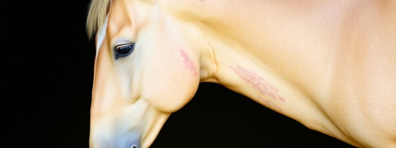
Onchocerciasis (Onchocerca cervicalis)
- Adult nematodes live in the ligamentum nuchae and produce microfilaria that migrate to the ventral midline and head, with Culicoides as the intermediate host.
- Causes leukoderma, alopecia, lichenification, excoriation, and ulceration.
- Commonly affects horses over 4 years old.
- Skin scrapings are ineffective for diagnosis, but histopathology reveals superficial eosinophilic perivascular cuffing; minced skin preparations are more reliable.
- Current anthelmintics do not kill adult worms; periodic ivermectin treatment helps manage exacerbations.
- Pretreatment with prednisolone can prevent reactions to dying microfilaria.
Insect Hypersensitivity
- Flying insects like Culicoides, black flies, horn flies, deer & horse flies, stable flies & mosquitoes can cause hypersensitivity.
- Culicoides (no-see-ums, midges, Queensland Itch) feed at dusk to dawn in pasture.
- Black flies feed in the morning and evening.
- Horn, Deer/Horse & Stable flies feed in bright sunlight and are associated with round bales.
- Hypersensitivity results in pruritic dermatitis, alopecia, crusts, and scaling, especially on the mane, tail, and pectoral region, leading to self-trauma.
- Skin scraping is uninformative.
- Diagnosis is often based on history, season, and lesion distribution; skin biopsy shows superficial eosinophilic perivascular dermatitis and edema.
- Prevention involves knowing fly types, using fans, housing horses in screened areas during peak feeding times (dusk, night, dawn), and applying topical residual insecticides (pyrethrins).
- Reduce inflammation using prednisolone, dexamethasone, or hydroxyzine.
- Immunotherapy/hyposensitization can help horses with atopy, especially if started young (under 4 years old).
Miscellaneous Causes of Pruritus
- Food allergies (potatoes, malt, beet pulp, alfalfa).
- Contact allergies and irritant dermatitis.
- Cutaneous drug reactions (penicillin, sulfa-based antimicrobials).
- Tail rubbing is related to pinworms or needing sheath/mammary glands cleaning and can be due to illegal alcohol injection.
Scaling and Crusting Dermatoses
- Key dermatoses causing scaling and crusting
Dermatophilosis (Dermatophilus congolensis)
- Gram-positive, facultative anaerobic actinomycete that can't penetrate intact skin; causes rain scald or rain rot.
- Forms crusted, moist mats of hair resembling paintbrushes; under fresh crusts, the skin is ulcerated, moist, and yellow to pink, primarily on the dorsal area, needing chronic moisture and macerated skin for transmission through zoospores.
- Carrier animals serve as reservoirs and stay viable for 42 months.
- Diagnose using impression smears to identify branching, multiseptate hyphae with transverse and horizontal cocci (railroad tract formation), bacterial culture, and histopathology.
- Treat by bathing with antibacterial shampoo (chlorhexidine) with sedation for crust removal, systemic penicillin for severe cases, and environmental improvements.
Dermatophytosis
- Dermatophytosis poses a zoonotic concern (Trichophyton equinum, Trichophyton metagrophytes, Microsporum gypseum, and Microsporum canis).
- Contagious skin disease, transmitted by direct contact or fomites; predisposed by debilitation, moisture, and stress.
- Clinical signs: alopecia, crusted papules with circumferential spread on the face, trunk, and pectoral region; pain and pruritus vary based on secondary bacterial infection.
- Diagnosis: Wood’s lamp is rarely successful, skin scrape isn't successful, fungal culture is performed using a dermatophyte test medium, and histopathology detects septate hyphae or oval spores; perform fungal cultures on hairs from lesion periphery.
- Treatment is self-limiting in 1-6 months, contagious with zoonotic potential, and usually treated with topical antifungals; systemic antifungals are costly.
Pemphigus Foliaceus
- Autoimmune skin disease (Type II hypersensitivity) where antibodies target keratinocyte glycocalyx, disrupting intercellular cement.
- Results in non-pruritic scaling, crusting, alopecia, rare subcorneal pustules, and painful skin; lesions start on the head and spread.
- Histopathology reveals subcorneal acantholysis and neutrophilic inflammation; direct immunofluorescence highlights intercellular cement.
- Young horses have an excellent prognosis with prednisolone (1 mg/kg, SID) for a cure.
- Adults are difficult to treat and may need long-term glucocorticoids
Distal Limb Dermatitis
- Also known as mud fever, dew poisoning, or greasy heel, is an infection of the lower leg.
- Often caused by muddy, wet conditions and involves Staph, Dermatophilosis, or fungi.
- Treatment: change environment, bandage, and oral antibiotics.
Chronic Progressive Lymphedema
- Is similar to distal limb dermatitis in draft horses but can progress to a large, abnormal leg.
- Genetics and impaired distal limb lymphatics play a role.
- Secondary infections and parasites are involved.
- Is a manageable problem.
Photosensitization
- Causes crusting and inflamed skin on white areas.
- Primary photosensitization is caused by ingesting certain plants.
- Liver disease can also cause it.
Nodular Skin Disease
- Includes diseases that form nodules on the skin
Urticaria
- Type I hypersensitivity reaction.
- Causes wheals, piloerection, and dermal edema.
- The origin of topical, inhaled, or ingested allergens is usually hard to identify.
- Treat with hydroxyzine HCL, prednisolone, dexamethasone, or epinephrine if there's respiratory distress (10 ml, IV 1:10,000 units).
Nodular Necrobiosis
- Etiology is unknown
- Eosinophilic granulomas form as single or multiple, haired, raised, firm dermal masses (0.5-10 mm); nonpainful, nonpruritic.
- Diagnosed based on collagen degeneration with eosinophilic inflammation.
- Treat with intralesional triamcinolone (not exceeding 20 mg).
Papillomatosis
- Caused by papilloma virus in young horses (12-24 months).
- Presents as multiple, raised, dermal, nonhaired, nonpainful, nonpruritic lesions.
- Diagnosed through visual inspection.
- Treatment: benign neglect, crushing lesions to stimulate antigen response, or immunostimulant therapy (EqStim) to clear in weeks to months.
Sporothrix schenkii (Sporotrichosis)
- Caused by dimorphic aerobic fungus; zoonotic, enters via traumatic wounds.
- Presents as papules in a chain along one limb; firm nodules form and ulcerate along a chain with interspersed draining tracts, often on the medial, proximal limb, with brown to red thick exudates.
- Diagnosed via cytologic exam (cigar-shaped yeast, Wright Giemsa), culture, and biopsy (nodular, granulomatous dermatitis, yeast rarely seen).
- Manage with sodium iodide (20%) IV or oral iodide (EDDI 20 mg/kg, SID); iodides can cause dry skin, tearing, and abortion.
Corynebacterium pseudotuberculosis
- Results in 2 different syndromes
- Ulcerative Lymphangitis: a single limb (often hind) shows chains of nodules that abscess, ulcerate, and drain. Affected limb cycles from swollen painful to periods of remission; the regional lymphatics are corded and dilated on ultrasound.
- Pigeon Breast or Dryland Distemper: deep muscle abscess
- Diagnose with bacterial culture and sensitivity (ultrasound-guided aspirate); treatment involves antibiotics, bandaging, and has a poor prognosis for normal leg function; lance and drain pigeon breast (usually avoid antibiotics); it's contagious, and organisms grow in soil (isolate).
Habronemiasis
- Larvae of Habronemia spp or Draschia megastomata cause hypersensitivity.
- Lesions appear seasonally and regress spontaneously in winter.
- Lesions are on legs, ventrum, eye, prepuce, urethral process, or existing wounds.
- Causes ulcerated, granulomatous, pruritic, and hemorrhagic lesions.
- Wounds may contain calcified granules.
- Diagnosed via signs, skin scraping can be unreliable while histopathology may reveal necrotic larvae.
- Treat with surgical excision, steroids, wound care, fly control, and ivermectin.
Pythiosis
- Pythium insidiosum causes pythiosis.
- Aquatic fungus invades macerated skin after contact with contaminated water in Gulf Coast States.
- Forms circular, ulcerated, granulomatous masses with serosanguinous exudates on legs and ventral midline.
- Lesions are intensely pruritic, and hemorrhage is due to self-trauma; hallmark kunkers develop within draining tracts.
- Differentiate from habronemiasis, SCC, and exuberant granulation tissue.
- Diagnose by biopsy (immunohistochemistry), culture, and cytologic evaluation.
- Treat through surgical excision, topical ketoconazole, DMSO, rifampin (phycofixer), and regional antifungal therapy, but the prognosis is poor.
Dermal Neoplasia
- Includes neoplasias that affect the skin
Sarcoid Skin Tumor
- Most common neoplasia in horses.
- Viral etiology (bovine papilloma virus) and genetic susceptibility.
- Three forms: flat/occult, verrucous, and fibroblastic
- Diagnosis - histopathology: Fibroblastic proliferation of the dermis with concurrent epidermal hyperplasia. Collagen in the derm is whorled, tangled or herringbone pattern; individual cells are spindle-shaped, fusiform to stellate.
- Treatment: BCG, chemotherapy (Cistplatin, 5-FU), Xterra, mistletoe extract, chyrotherapy, radioactive isotope implants, surgical removal with medical adjuncts, electrochemical treatment with cisplatin.
Squamous Cell Carcinoma
- Commonly manifests in Appaloosas, Belgians, Paints, or previous burn sites.
- Lesions are ulcerated, hemorrhagic, and nodular, found on ocular area, vulva, and prepuce.
- Genital SCC is linked to a novel equine papilloma virus.
- Diagnose via histopathology and treat with topical or intralesional 5 fluorouracil.
Melanoma
- Occurs in aged, grey horses, more common in horses with genetics of a black horse, more severe in various breeds; many are benign, but some progress.
- Lesions are firm, nodular, dermal, hairless, and black; located on the perineum, prepuce, and other places.
- Diagnosed via clinical signs and fine needle aspirate.
- Rarely requires treatment, but surgical resection may be needed if disruptive; cimetidine, irradiated autologous vaccine, and canine vaccines are treatment options.
Disorders of Pigmentation
- Includes vitiligo, leukoderma, and Reticulated leukotrichia
Fading Arab Syndrome
- Also called vitiligo, is the development of depigmented skin without trauma.
- Presents as annular areas of depigmentation around the eyes, muzzle, anus.
- It's heritable (breed flaw) and may respond to copper supplementation.
Leukoderma
- Is depigmentation resulting from trauma or disease: pressure sores, warts, onchocerciasis, burns.
- There is no treatment.
Reticulated Leukotrichia
- Is an inherited disorder in Quarter Horses.
- Clinical signs begin in yearlings, with dorsal distribution of linear crusting lesions in a reticulated pattern.
- New hair growth is white; considered a breed flaw.
- Miniature horses have a similar pattern (lacing) without crust stage.
Miscellaneous Conditions
- Includes Fistulous withers and Ehler Danlos
Fistulous Withers
- Is a chronic bacterial infection of supraspinous bursa from Streptococcus zooepidemicus or Brucella spp.
- Diagnose through bacterial culture, serology for brucellosis, and radiograph of dorsal spinous processes.
- Prognosis is poor.
- Treat with en bloc resection, long-term antibiotics, or euthanasia for Brucellosis, due to human health risk.
Ehler Danlos Syndrome
- Also called hyperelastosis cutis, is a heritable, autosomal recessive condition in Quarter Horses.
- Yearlings have fragile skin, failure to heal, and loose skin with pockets of serum.
- Located on the axilla, tuber coxae, saddle sores, and bite wounds.
- Histopathology may not be helpful.
- Affects cornea, ligaments, and tendons as well.
- Diagnose through signalment, clinical signs, and genetic testing performed at UCDavis and Cornell.
- Often, horses are euthanized, and sunlight worsens lesions.
Studying That Suits You
Use AI to generate personalized quizzes and flashcards to suit your learning preferences.