Podcast
Questions and Answers
Which of the following best describes the appearance of microvilli under a light microscope?
Which of the following best describes the appearance of microvilli under a light microscope?
- A wavy, undulating border
- A striated or brush border (correct)
- A border with distinct, individual projections
- A smooth, continuous border
What is a key distinction between cilia and microvilli?
What is a key distinction between cilia and microvilli?
- Both structures are motile and contain the same motor proteins.
- Microvilli are motile, while cilia are generally stationary.
- Cilia are primarily involved in absorption, while microvilli are for movement.
- Cilia contain dynein, which facilitates movement, while microvilli lack contractile proteins. (correct)
What characteristic of epithelial cells is highlighted by the presence of distinct apical surface modifications?
What characteristic of epithelial cells is highlighted by the presence of distinct apical surface modifications?
- Extracellular matrix production
- Cellular adhesion properties
- Polarity (correct)
- Ameboid movement
Which surface modification is primarily associated with increasing surface area for absorption?
Which surface modification is primarily associated with increasing surface area for absorption?
What is the function of dynein in relation to cilia?
What is the function of dynein in relation to cilia?
Flashcards
Microvilli
Microvilli
Finger-like projections of the plasma membrane that increase the surface area of epithelial cells for absorption or secretion.
Stereocilia
Stereocilia
Hair-like structures that extend from the apical surface of cells, but are longer and less numerous than microvilli. They are found in the inner ear and epididymis.
Cilia
Cilia
Hair-like structures that protrude from the apical surface of some epithelial cells. They are longer and more motile than microvilli, and are involved in the movement of fluids, mucus, or debris.
Motile Cilia
Motile Cilia
Signup and view all the flashcards
Dynein
Dynein
Signup and view all the flashcards
Study Notes
Epithelial Tissue Part 2: Apical Surface Modifications
- Epithelial cells exhibit polarity, with three main domains: apical, lateral, basal, and basolateral.
- The apical domain faces the lumen or external environment.
- The lateral domain lies between adjacent cells.
- The basal domain faces the basement membrane.
- The basolateral domain encompasses both the lateral and basal domains.
Apical Domain Modifications
- Modifications of the apical domain include microvilli, stereocilia, and cilia.
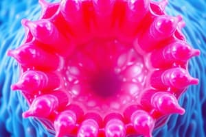
Microvilli
- Microvilli are small, finger-like projections, approximately 1 μm in length, visible under electron microscopy (EM).
- They increase the surface area for absorption.
- The number and shape of microvilli correlate to the absorptive capacity of the tissue.
- Microvilli contain bundles of parallel actin filaments.
- Proteins such as villin and fimbrin hold these actin filaments together in bundles.
- Myosin I and calmodulin connect the actin filament bundle to the plasma membrane.
- Actin filaments in microvilli extend into the apical cytoplasm, interacting with a horizontal network of actin filaments.
- The terminal web consists of actin filaments, spectrin, and myosin II.
- The terminal web may result in decreased diameter of intestinal absorbing cells, increasing the intermicrovillar space.
Stereocilia
- Stereocilia are extremely long, immobile processes extending from the apical cell surface.
- Similar to microvilli, they are supported by actin filaments.
- Stereocilia are found in sensory hair cells of the inner ear.
- Stereocilia facilitate fluid absorption and serve as sensory mechanoreceptors, rather than absorptive structures. Stereocilia aggregate into pointed bundles.
Cilia
- Cilia are hair-like extensions of the apical plasma membrane.
- Cilia contain microtubules or the axoneme.
- The microtubules originate from the basal body, a microtubule organizing center (MTOC) derived from centrioles.
- Cilia are classified as motile or primary.
- Motile cilia actively beat, while primary cilia do not actively move but may bend with fluid flow.
- Microtubules are arranged as a 9+2 array—nine peripheral doublets surrounding two central microtubules.
Motile Cilia
- Motile cilia are present in the respiratory tract and oviduct form a muco-ciliary unit.
- They help to transport ova and fluid as well as clear away mucous and particulate material.
Ciliary Movement
- Ciliary movement is based on the sliding of doublet tubules relative to one another.
- Each doublet contains a pair of arms (dynein).
- Dynein arms extend from the A tubule to temporarily bridge with the B microtubules.
- ATP hydrolysis causes these bridges to slide, causing the cilia to bend.
- Nexin protein connects the doublet tubules and returns the cilium to its straight position.
Disorders Affecting the Muco-ciliary Unit
- Primary Ciliary Dyskinesia (PCD) or Immotile Cilia Syndrome (ICS) is an autosomal recessive disorder that affects ciliary function.
- Kartagener syndrome is a specific form of PCD characterized by situs inversus, chronic sinusitis, and bronchiectasis.
Primary Cilia and Nodal Cilia
- Primary cilia are non-motile and function as signal receptors.
- Nodal cilia are found in the embryonic disc and function in signaling, establishing left-right asymmetry.
Studying That Suits You
Use AI to generate personalized quizzes and flashcards to suit your learning preferences.