Podcast
Questions and Answers
What is the approximate percentage of the population affected by lumbosacral radiculopathy?
What is the approximate percentage of the population affected by lumbosacral radiculopathy?
- 9-10%
- 6-8%
- 3-5% (correct)
- 1-2%
Which of the following is the single most common lumbar radiculopathy?
Which of the following is the single most common lumbar radiculopathy?
- L5 radiculopathy (correct)
- L3 radiculopathy
- L4 radiculopathy
- S1 radiculopathy
What is the most common symptom experienced by patients with lumbosacral radiculopathy?
What is the most common symptom experienced by patients with lumbosacral radiculopathy?
- Pain
- Muscle weakness
- Paresthesia (correct)
- Numbness
Which of the following is a risk factor for lumbosacral radiculopathy?
Which of the following is a risk factor for lumbosacral radiculopathy?
What should be assessed during the history intake for lumbosacral radiculopathy?
What should be assessed during the history intake for lumbosacral radiculopathy?
What is typically worse with increased intradiscal pressure?
What is typically worse with increased intradiscal pressure?
What is an important associated sign or symptom to ask about during the history intake?
What is an important associated sign or symptom to ask about during the history intake?
What is the typical pain quality experienced by patients with lumbosacral radiculopathy?
What is the typical pain quality experienced by patients with lumbosacral radiculopathy?
What is the approximate incidence of cauda equina syndrome?
What is the approximate incidence of cauda equina syndrome?
What is the most common cause of cauda equina syndrome?
What is the most common cause of cauda equina syndrome?
What is the diagnostic gold standard for cauda equina syndrome?
What is the diagnostic gold standard for cauda equina syndrome?
What is the most common primary cancer that metastasizes to the spine?
What is the most common primary cancer that metastasizes to the spine?
What is the typical presentation of vertebral fracture due to osteoporosis?
What is the typical presentation of vertebral fracture due to osteoporosis?
What is the recommended time frame for surgical decompression in cauda equina syndrome?
What is the recommended time frame for surgical decompression in cauda equina syndrome?
What is the estimated number of new cases of cauda equina syndrome per year in the US?
What is the estimated number of new cases of cauda equina syndrome per year in the US?
Which of the following muscle weaknesses is associated with L2, L3, and L4 radiculopathy?
Which of the following muscle weaknesses is associated with L2, L3, and L4 radiculopathy?
Which of the following is a characteristic of L5 radiculopathy?
Which of the following is a characteristic of L5 radiculopathy?
Which of the following reflexes is absent in L2, L3, and L4 radiculopathy?
Which of the following reflexes is absent in L2, L3, and L4 radiculopathy?
Which of the following is a characteristic of chronic L5 radiculopathy?
Which of the following is a characteristic of chronic L5 radiculopathy?
Which of the following motor weaknesses is associated with L5 radiculopathy?
Which of the following motor weaknesses is associated with L5 radiculopathy?
What is the primary goal of counseling patients with radicular symptoms?
What is the primary goal of counseling patients with radicular symptoms?
What is a key aspect of disease education for patients with radiculopathy?
What is a key aspect of disease education for patients with radiculopathy?
What is a common complication of acute radicular pain?
What is a common complication of acute radicular pain?
What is the typical approach to pain management for patients with radiculopathy?
What is the typical approach to pain management for patients with radiculopathy?
What is a red flag symptom that warrants an immediate emergent evaluation and potential surgical consultation?
What is a red flag symptom that warrants an immediate emergent evaluation and potential surgical consultation?
What is the percentage of regression without treatment in Lumbar Disc Herniation, sequestration?
What is the percentage of regression without treatment in Lumbar Disc Herniation, sequestration?
What is the recommended approach to physical activity for patients with radiculopathy?
What is the recommended approach to physical activity for patients with radiculopathy?
What is the typical prognosis for most cases of lumbosacral radiculopathy?
What is the typical prognosis for most cases of lumbosacral radiculopathy?
Which of the following physical exam findings is associated with Lumbar Disc Herniation?
Which of the following physical exam findings is associated with Lumbar Disc Herniation?
What is the diagnostic accuracy of the Hancock Rule based on?
What is the diagnostic accuracy of the Hancock Rule based on?
What is an important aspect of patient education for those with radiculopathy?
What is an important aspect of patient education for those with radiculopathy?
What is the sensitivity of the Straight Leg Raise (SLR) test in diagnosing Lumbar Disc Herniation?
What is the sensitivity of the Straight Leg Raise (SLR) test in diagnosing Lumbar Disc Herniation?
What is the likely presentation of an acute injury in the distribution of L2, L3, and L4 radiculopathies?
What is the likely presentation of an acute injury in the distribution of L2, L3, and L4 radiculopathies?
What is the significance of calf wasting in Lumbar Disc Herniation?
What is the significance of calf wasting in Lumbar Disc Herniation?
What is the likely distribution of pain in L2, L3, and L4 radiculopathies?
What is the likely distribution of pain in L2, L3, and L4 radiculopathies?
What is the likely cause of hematuria in a patient with a recent back trauma?
What is the likely cause of hematuria in a patient with a recent back trauma?
What is the significance of early morning stiffness in a patient with low back pain?
What is the significance of early morning stiffness in a patient with low back pain?
What is the diagnosis of a patient with a history of malignancy and low back pain, who presents with urinary retention and incontinence?
What is the diagnosis of a patient with a history of malignancy and low back pain, who presents with urinary retention and incontinence?
What is the likely cause of muscular spasms in a patient with low back pain?
What is the likely cause of muscular spasms in a patient with low back pain?
What is the diagnostic test for lumbar disc herniation?
What is the diagnostic test for lumbar disc herniation?
What is the significance of fever in a patient with low back pain?
What is the significance of fever in a patient with low back pain?
What is the likely presentation of a patient with L5 radiculopathy?
What is the likely presentation of a patient with L5 radiculopathy?
What is the key aspect of disease education for patients with radiculopathy?
What is the key aspect of disease education for patients with radiculopathy?
What is the primary motor weakness associated with S1 radiculopathy?
What is the primary motor weakness associated with S1 radiculopathy?
What is the most common symptom experienced by patients with lumbosacral radiculopathy, apart from pain?
What is the most common symptom experienced by patients with lumbosacral radiculopathy, apart from pain?
Which of the following is a risk factor for lumbosacral radiculopathy?
Which of the following is a risk factor for lumbosacral radiculopathy?
What is the primary mechanism underlying spinal stenosis?
What is the primary mechanism underlying spinal stenosis?
In S1 radiculopathy, which of the following sensory changes is commonly seen?
In S1 radiculopathy, which of the following sensory changes is commonly seen?
What is the estimated prevalence of spinal stenosis in individuals aged 60-69 years?
What is the estimated prevalence of spinal stenosis in individuals aged 60-69 years?
Which of the following reflexes is typically absent in S1 radiculopathy?
Which of the following reflexes is typically absent in S1 radiculopathy?
What is the likely course of pain in patients with lumbosacral radiculopathy?
What is the likely course of pain in patients with lumbosacral radiculopathy?
What is the term used to describe the slippage of one vertebral body with respect to the adjacent vertebral body?
What is the term used to describe the slippage of one vertebral body with respect to the adjacent vertebral body?
What is the term used to describe a weakness or stress fracture through the pars interarticularis?
What is the term used to describe a weakness or stress fracture through the pars interarticularis?
During the history intake, what is an important aspect to assess?
During the history intake, what is an important aspect to assess?
What is the distribution of pain in S1 radiculopathy?
What is the distribution of pain in S1 radiculopathy?
What is the significance of checking for absent ankle reflexes in patients with lumbosacral radiculopathy?
What is the significance of checking for absent ankle reflexes in patients with lumbosacral radiculopathy?
What is the term used to describe age-related degeneration of the spinal column?
What is the term used to describe age-related degeneration of the spinal column?
What is the typical approach to managing pain in patients with lumbosacral radiculopathy?
What is the typical approach to managing pain in patients with lumbosacral radiculopathy?
What is the presentation of spinal stenosis in terms of symptoms?
What is the presentation of spinal stenosis in terms of symptoms?
What is the significance of asking about saddle anesthesia in patients with lumbosacral radiculopathy?
What is the significance of asking about saddle anesthesia in patients with lumbosacral radiculopathy?
What is the primary reason for the relief of pain in neurogenic claudication?
What is the primary reason for the relief of pain in neurogenic claudication?
What is the percentage of patients who are asymptomatic in spondylolysis?
What is the percentage of patients who are asymptomatic in spondylolysis?
What is the typical presentation of patients with spinal stenosis?
What is the typical presentation of patients with spinal stenosis?
What is the risk factor associated with spondylolysis?
What is the risk factor associated with spondylolysis?
What is the prediction rule for diagnosing spinal stenosis?
What is the prediction rule for diagnosing spinal stenosis?
What is the percentage of patients who eventually undergo repeat surgery for spinal stenosis?
What is the percentage of patients who eventually undergo repeat surgery for spinal stenosis?
What is the primary causative organism of vertebral osteomyelitis?
What is the primary causative organism of vertebral osteomyelitis?
Which of the following is a common risk factor for vertebral infection?
Which of the following is a common risk factor for vertebral infection?
What is the preferred diagnostic imaging modality for vertebral osteomyelitis?
What is the preferred diagnostic imaging modality for vertebral osteomyelitis?
What is the approximate percentage of cases of osteomyelitis that affect the spine?
What is the approximate percentage of cases of osteomyelitis that affect the spine?
Which of the following is a common symptom of vertebral infection?
Which of the following is a common symptom of vertebral infection?
What is the most common site of vertebral infection?
What is the most common site of vertebral infection?
Which of the following occupations is more likely to develop lumbosacral radiculopathy due to repetitive lifting and twisting motions?
Which of the following occupations is more likely to develop lumbosacral radiculopathy due to repetitive lifting and twisting motions?
What is the typical course of pain in lumbosacral radiculopathy?
What is the typical course of pain in lumbosacral radiculopathy?
What is the primary goal of assessing dermatomal distribution during the history intake?
What is the primary goal of assessing dermatomal distribution during the history intake?
Which of the following is not a characteristic of pain experience in lumbosacral radiculopathy?
Which of the following is not a characteristic of pain experience in lumbosacral radiculopathy?
What is the significance of saddle anesthesia in lumbosacral radiculopathy?
What is the significance of saddle anesthesia in lumbosacral radiculopathy?
Which of the following is a common risk factor for lumbosacral radiculopathy?
Which of the following is a common risk factor for lumbosacral radiculopathy?
What is the typical pattern of pain radiation in S1 radiculopathy?
What is the typical pattern of pain radiation in S1 radiculopathy?
Which of the following physical exam findings is associated with S1 radiculopathy?
Which of the following physical exam findings is associated with S1 radiculopathy?
What is the typical sensory change seen in S1 radiculopathy?
What is the typical sensory change seen in S1 radiculopathy?
Which of the following reflexes is typically absent in S1 radiculopathy?
Which of the following reflexes is typically absent in S1 radiculopathy?
What is the primary mechanism underlying neurogenic claudication in spinal stenosis?
What is the primary mechanism underlying neurogenic claudication in spinal stenosis?
What is the primary risk factor associated with spondylolysis?
What is the primary risk factor associated with spondylolysis?
What is the typical presentation of spondylolysis?
What is the typical presentation of spondylolysis?
What is the diagnostic gold standard for spinal stenosis?
What is the diagnostic gold standard for spinal stenosis?
What is the approximate percentage of patients with spondylolysis who are asymptomatic?
What is the approximate percentage of patients with spondylolysis who are asymptomatic?
What is the likelihood of lumbar spinal stenosis in patients with a score ≥ 7 on the prediction rule?
What is the likelihood of lumbar spinal stenosis in patients with a score ≥ 7 on the prediction rule?
What is the significance of a wide-based gait in patients with spinal stenosis?
What is the significance of a wide-based gait in patients with spinal stenosis?
What percentage of bilateral spondylolysis will have spondylolisthesis at diagnosis?
What percentage of bilateral spondylolysis will have spondylolisthesis at diagnosis?
What is the most common cause of spondylolisthesis?
What is the most common cause of spondylolisthesis?
What is the typical presentation of spondylolisthesis in adults?
What is the typical presentation of spondylolisthesis in adults?
What is the recommended initial imaging modality for spondylolisthesis?
What is the recommended initial imaging modality for spondylolisthesis?
What is the purpose of Meyerding's Classification in spondylolisthesis?
What is the purpose of Meyerding's Classification in spondylolisthesis?
What is the typical grade of spondylolisthesis in older patients?
What is the typical grade of spondylolisthesis in older patients?
What is the indication for imaging in patients with low back pain?
What is the indication for imaging in patients with low back pain?
What is a common feature of spondylolisthesis on X-ray?
What is a common feature of spondylolisthesis on X-ray?
Study Notes
Here are the study notes for the text:
Red Flag Findings for Epidural Abscess
- Fecal incontinence or loss of bowel control
- Urinary retention or loss of bladder control
- Saddle anesthesia
- Unexplained fever
- Unexplained weight loss
- Focal neurological deficit, progressive or disabling symptoms
- No improvement after 6 weeks of conservative management
- Immunosuppression, recent infection, chronic steroid use, osteoporosis, significant trauma, or history of cancer
Diagnostic Accuracy of Individual Red Flag Findings
- Age > 50 years: sensitivity 74%, LR+ 1.1, LR- 0.79
- Recent loss of bladder control: sensitivity 22.2%, LR+ 2.31, LR- 0.86
- Recent loss of bowel control: sensitivity 13.9%, LR+ 2.78, LR- 0.91
- Unexplained weight loss: sensitivity 8.2%, LR+ 1.87, LR- 0.96
- Personal history of cancer: sensitivity 32%, LR+ 7.25, LR- 0.71
- Fever, chills, or sweating: sensitivity 11.7%, LR+ 1.71, LR- 0.95
- Recent infection: sensitivity 24.2%, LR+ 9.31, LR- 0.78
Diagnostic Accuracy of Combined Red Flag Findings
- Trauma + Age > 50 years: sensitivity 14.8%, LR+ 2.54, LR- 0.90
- Trauma + Age > 70 years: sensitivity 5.2%, LR+ 4.35, LR- 0.96
- Recent loss of bowel + bladder control: sensitivity 8.3%, LR+ 3.0, LR- 0.94
- Unexplained weight loss + personal history of cancer: sensitivity 2.5%, LR+ 10.25, LR- 0.98
- Fever, chills, or sweating + recent infection: sensitivity 7.5%, LR+ 13.15, LR- 0.93
Cauda Equina Syndrome
- Compression and disruption of function to cauda equina (L3-L5 nerve roots)
- Classical symptoms: new urinary retention or overflow incontinence, fecal incontinence, progressive motor or sensory loss, saddle anesthesia, lower motor neuron weakness, and significant deficits that encompass multiple nerve roots
- Diagnosis: imaging (MRI) is diagnostic gold standard
- Urgent ER referral; requires surgical decompression within 24-48 hours
Spinal Malignancy: Metastases
- Most common tumors of the spine are metastases of other primary cancers (breast, lung, prostate, renal, gastrointestinal, and thyroid)
- Personal history of cancer, back pain, unexplained weight loss, possibly sensory loss, weakness, or radiculopathy
- Diagnosis: imaging (x-ray or MRI) and blood work (incl CBC) and symptoms depend on type of primary cancer
- Urgent referral back to oncologist or palliative care
Vertebral Fracture
- A break in one or more spinal vertebrae that can result from trauma and metastatic disease but, in most cases, are the result of osteoporosis (at T11-L2)
- Low bone density, female > 50 years, prolonged use of corticosteroids, trauma/fall, personal history of vertebral fracture
- Back pain (acute or chronic, localized)
LBP with Radiculopathy: Epidemiology
- Lumbosacral radiculopathy is common: 3-5% of population
- Male > 40 years, female > 50 years, 3:2
- L5 radiculopathy is the single most common lumbar radiculopathy
LBP with Radiculopathy: Risk Factors
- Social history: repetitive lifting and twisting motions, chronic overloading of disc, driving occupations, heavy industry work, military, smoking, overweight, sedentary
- Medical history: prior trauma, multiple pregnancies, history of back pain, chronic cough
LBP with Radiculopathy: History Intake
- Site: ask about the location of the back pain
- Onset: how and when the back pain developed
- Quality: pain is often described as throbbing, aching, sharp, dull, burning, pressure, numbness, tingling, or shooting
- Radiation: assess dermatomal distribution
- Time course: consider how the back pain changed over time
- Severity of pain experience: scale of 1-10
- Pain is typically worse with: increased intradiscal pressure, Valsalva, weight bearing, standing, walking, sitting for prolonged periods
- Pain is typically better with: extension of the lumbar spine, recumbent position (knees flexed)
- Associated signs and symptoms: motor or sensory disturbances, suggestive of nerve root or spinal cord compression
Physical Exam Findings associated with Lumbar Disc Herniation
- Weak ankle dorsiflexion: sensitivity 54%, LR+ 4.9, LR- 0.5
- Calf wasting: sensitivity 29%, LR+ 5.2, LR- 0.8
- Leg sensation abnormal: sensitivity 16%, LR+ NS, LR- NS
- Abnormal ankle reflex: sensitivity 48%, LR+ 4.3, LR- 0.6
-
- Straight Leg Raise (SLR) test: sensitivity 73-98%, LR+ NS, LR- 0.2
-
- Crossed SLR test: sensitivity 23-43%, LR+ 4.3, LR- 0.8
Hancock Rule: Clinical Prediction Rule for Lumbar Disc Herniation
- The diagnostic accuracy of multiple neurologic findings improves clinician ability to determine level of disc herniation if at least 3 of 4 findings are in concordance with a specific nerve root:
- dermatomal pain location
- sensory deficit
- reduced reflex
- motor weakness
- Findings: score ≥ 3 for L3/L4 disc herniation, sensitivity 50%, LR+ 5.0, LR- 0.56
- Findings: score ≥ 3 for L4/L5 disc herniation, sensitivity 37%, LR+ 2.18, LR- 0.76
- Findings: score ≥ 3 for L5/S1 disc herniation, sensitivity 28%, LR+ 4.67, LR- 0.77
LBP with Radiculopathy: Epidemiology
- 3-5% of population has lumbosacral radiculopathy, with males (40+ years) more common than females (50+ years) at a 3:2 ratio
- L5 radiculopathy is the single most common lumbar radiculopathy
- Pain experience: often described as tingling, electric, burning, or sharp
- Paresthesia: 63-72% of patients, radiation of pain into lower limb: 35%, numbness (anesthesia): 27%, and muscle weakness: up to 37%
LBP with Radiculopathy: Risk Factors
- Social History:
- Repetitive lifting and twisting motions
- Chronic overloading of disc: driving occupations, heavy industry work, military
- Lifestyle: smoking, overweight, sedentary
- Medical History:
- Prior trauma (fall, motor vehicle accident)
- Multiple pregnancies
- History of back pain
- Chronic cough
LBP with Radiculopathy: History Intake
- Site: ask about the location of the back pain
- Onset: how and when the back pain developed
- Quality: pain is often described as throbbing, aching, sharp, dull, burning, pressure, numbness, tingling, or shooting
- Radiation: assess dermatomal distribution
- Time Course: consider how the back pain changed over time
LBP with Radiculopathy: Associated Signs and Symptoms
- Motor or sensory disturbances: suggestive of nerve root or spinal cord compression
- Ask specifically about saddle anesthesia if cauda equina syndrome is a possibility
- Urinary retention or incontinence: typical features of cauda equina syndrome
- Hematuria (blood in urine): may occur secondary to back trauma
LBP with Radiculopathy: History Intake
- Associated Signs and Symptoms:
- Fever: typically associated with urinary tract infection, pneumonia, and discitis
- Malaise: associated with a wide range of pathology but in the context of back pain, consider discitis or malignancy
- Weight loss: associated with malignancy
- Early morning stiffness: associated with inflammatory arthritis (e.g., rheumatoid arthritis, ankylosing spondylitis)
- Muscular spasms: may be associated with spinal fracture or primary muscular injury
LBP with Radiculopathy: Key Medical History
- History of malignancy
- Recent bacterial infections
- Recent history of epidural or spinal surgical procedures
- Medications: history of or current corticosteroid use
- Assess ability to function in daily life
Disc Herniation
- Displacement of intervertebral disc material (nucleus pulposus or annulus fibrosis) beyond the intervertebral disc space
- 1-3% of patients with acute LBP; age 30-40, male: female 2:1
- Acute-chronic pain, paresthesia, sensory change, loss of strength or reflexes (depends on affected nerve root)
- Diagnosis: SLR + Hancock rule
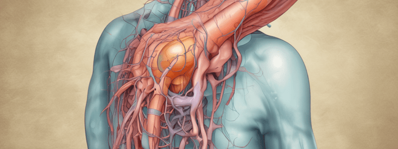
S1 Radiculopathy
- Distribution of Pain:
- S1 radiculopathy will cause radiation of sacral or buttock pain into the posterior aspect of the patient's leg, into the foot, or the perineum
- Physical Exam:
- Motor Weakness: plantar flexion
- Paresthesia/Sensory Changes: sole, lateral foot and ankle, fourth and fifth toes
- Absent Reflexes: ankle reflex (S1)
S1 Radiculopathy: Physical Exam Findings
- Weak ankle plantar flexion: 26-45% sensitivity
- Ipsilateral calf wasting: 43% sensitivity
- Sensory loss in S1 distribution: 32-49% sensitivity
- Asymmetric achilles reflex: 45-91% sensitivity
Serious Disorders Affecting the Lumbar Spine
- Spondylosis: an umbrella term for age-related degeneration of the spinal column (often involves degenerative disc disease and facet arthropathy)
- Spinal stenosis: narrowing of the spinal canal, neural foramen, and lateral recess which can lead to compression of the nerve roots and neurogenic claudication
- Spondylolysis: weakness or stress fracture through the pars interarticularis
- Spondylolisthesis: the slippage of one vertebral body with respect to the adjacent vertebral body
Spinal Stenosis
- Narrowing of the spinal canal, neural foramen, and lateral recess which can lead to compression of the nerve roots and neurogenic claudication
- 3% with mechanical low back pain (LBP), 19.4% aged 60-69 years
- LBP with B/L lower extremity pain, numbness or loss of strength (depends on affected nerve root) aggravated by ambulation, standing, and lumbar extension
- Neurogenic claudication: pain/discomfort with walking or prolonged standing that radiates into one or both lower extremities (relieved by rest/sitting, lumbar flexion)
- Diagnosis: imaging is diagnostic
Vertebral Infection: Osteomyelitis
- Most common vertebral infection, particularly the vertebral body
- Often caused by hematogenous spread of Staphylococcus aureus
- Rare, approx. 4.8 cases per 100,000 in US, 3-5% of all cases of osteomyelitis
- Risk factors: recent spinal procedure/surgery, recent infection, wound in spinal region, Hx IV drug use, immunosuppression
- Symptoms: back pain, fever, sensory loss, weakness or radiculopathy
- Diagnosis: imaging (MRI) is preferred, CBC often normal, ESR elevated, CRP elevated, blood cultures
- Urgent ER referral, requires antibiotic therapy (IV and oral)
LBP with Radiculopathy: Etiology
- L4-L5 (L5 nerve root) and L5-S1 (S1 nerve root) are most susceptible to injury
- Particularly flexible part of lumbar spine, bears more impact than thoracic and cervical spine
LBP with Radiculopathy: Epidemiology
- Lumbosacral radiculopathy is common: 3 - 5% of population
- Male (40yr+) > female (50yr+): 3:2
- L5 radiculopathy is the single most common lumbar radiculopathy
LBP with Radiculopathy: Risk Factors
- Social History: repetitive lifting and twisting motions, chronic overloading of disc, smoking, overweight, sedentary
- Medical History: prior trauma, multiple pregnancies, history of back pain, chronic cough
LBP with Radiculopathy: History Intake
- Site: ask about the location of the back pain
- Onset: how and when the back pain developed
- Quality: pain is often described as throbbing, aching, sharp, dull, burning, pressure, numbness, tingling, or shooting
- Radiation: assess dermatomal distribution
- Time Course: consider how the back pain changed over time
- Pain experience: scale of 1-10, pain is typically worse with increased intradiscal pressure, pressure increases with coughing, sneezing, straining, forward flexion of the lumbar spine
LBP with Radiculopathy: Physical Exam
- Motor Weakness: plantar flexion
- Paresthesia/Sensory Changes: sole, lateral foot and ankle, fourth and fifth toes
- Absent Reflexes: ankle reflex (S1)
Spinal Stenosis
- Pain/discomfort with walking or prolonged standing that radiates into one or both lower extremities (relieved by rest/sitting, lumbar flexion)
- Diagnosis: imaging is diagnostic
- Subjective + Physical Findings associated with Spinal Stenosis:
- No pain when seated
- Unexplained urinary disturbance
- Symptoms improve with bend forward
- Bilateral buttock or leg pain
- Neurogenic claudication
- Wide-based gait
- Abnormal rhomberg test
Spondylolysis
- Unilateral or bilateral defect through the pars interarticularis (most commonly affects L5, 90%)
- 6-18% population (14+); up to 50% young athletes (male:female, 2:1)
- Risk associated with excessive lumbar lordosis, genetics (fHx)
- Asymptomatic (90% of patients); insidious onset, recurrent axial low back pain exacerbated with activity or lumbar hyperextension, +/- radiculopathy
Spondylolisthesis
- Slippage of one vertebral body with respect to the adjacent vertebral body
- 3-4% of patients with mechanical LBP
- Adults, female > male, obesity, fHx (spondylolisthesis, scoliosis, spina bifida)
- Intermittent and localized low back pain that radiates into buttock or posterior thigh; paresthesia, sensory change, loss of strength or reflexes (depends on affected nerve root)
Evaluation - Imaging
- Initial imaging is not indicated in the majority of patients with low back pain
- Conservative management for 6 weeks is typically recommended before considering imaging (radiography, MRI, CT)
- UNLESS presenting with severe symptom intensity or red flag findings for conditions that require timely diagnosis to prevent serious consequences
Studying That Suits You
Use AI to generate personalized quizzes and flashcards to suit your learning preferences.
Related Documents
Description
Identify the presenting symptoms and medical history that indicate an epidural abscess, a serious medical condition. Learn the red flag findings from the American Academy of Family Physicians (AAFP).


