Podcast
Questions and Answers
What two layers are formed when the endometrium erodes due to the syncytiotrophoblast?
What two layers are formed when the endometrium erodes due to the syncytiotrophoblast?
- Hypoblast and cytotrophoblast
- Syncytiotrophoblast and cytotrophoblast
- Epiblast and trophoblast
- Epiblast and hypoblast (correct)
The fetal period spans from the 15th week to the conclusion of the pregnancy.
The fetal period spans from the 15th week to the conclusion of the pregnancy.
False (B)
What is the term for the structure formed when the morula enters the uterus and penetrates fluid through the zona pellucida?
What is the term for the structure formed when the morula enters the uterus and penetrates fluid through the zona pellucida?
Blastocoele/Blastocyst
The inner cell mass of the morula, also known as the trophoblast, eventually forms the ______.
The inner cell mass of the morula, also known as the trophoblast, eventually forms the ______.
Match the following stages of pre-embryonic development with their descriptions:
Match the following stages of pre-embryonic development with their descriptions:
The blastocyst implants into the endometrium using what?
The blastocyst implants into the endometrium using what?
The pre-embryonic period spans from the 4th to the 8th week of development.
The pre-embryonic period spans from the 4th to the 8th week of development.
What cellular structure forms from the fusion of vacuoles within the implanted blastocyst?
What cellular structure forms from the fusion of vacuoles within the implanted blastocyst?
Following implantation, the blastocyst is implanted further into the epithelium, and a ______ forms in the epithelium.
Following implantation, the blastocyst is implanted further into the epithelium, and a ______ forms in the epithelium.
The zygote's characteristics do NOT include:
The zygote's characteristics do NOT include:
Flashcards
What is fertilization?
What is fertilization?
The fusion of male (sperm) and female (ovum) gametes to form a zygote.
What is segmentation?
What is segmentation?
Series of mitotic divisions after fertilization, increasing cell number.
What is a Morula?
What is a Morula?
A solid ball of 16 cells formed during early embryonic development.
What is a Blastocyst?
What is a Blastocyst?
Signup and view all the flashcards
What is Bilaminar Disc Formation?
What is Bilaminar Disc Formation?
Signup and view all the flashcards
What is Gastrulation?
What is Gastrulation?
Signup and view all the flashcards
What is the fetal period?
What is the fetal period?
Signup and view all the flashcards
When is the first trimester?
When is the first trimester?
Signup and view all the flashcards
What is pre-embryonic development?
What is pre-embryonic development?
Signup and view all the flashcards
What is embryonic development?
What is embryonic development?
Signup and view all the flashcards
Study Notes
Pre-embryonic Development (Weeks 1-3)
- Begins with fertilization and lasts until the 3rd week.
Embryonic Development (Weeks 4-8)
- Occurs from the 4th to the 8th week.
Fetal Development (Week 9 to Birth)
- Starts at the 9th week and continues until birth.
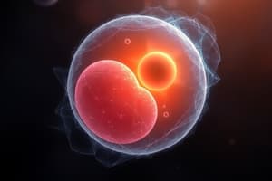
Day 1: Fertilization
- Initialization with Zygote, requires female gamete (secondary oocyte) and male gamete (spermatozoon).
- Occurs in the ampulla of the fallopian tube.
- Zygote's characteristics:
- 46 chromosomes (23 from each parent, XX or XY).
- Pluripotent.
- Mitotic division.
- Takes approximately 24 hours.
Day 2: Segmentation & Compaction
- Formation of blastomeres occurs through mitotic divisions to increase cell number
- 1st division: after 30 hours, resulting in two blastomeres.
- 2nd division: after 40 hours, forming 4 cells.
- 3rd division: forming 8 cells.
- Blastomeres move closer due to glycoproteins.
Day 3: Morula Stage
- The embryo's compacted cells divide to form a morula consisting of 16 cells.
- Cells inside the morula include:
- Inner cell mass (trophoblast): Future placenta.
- Outer cell mass (embryoblast): Future embryo.
Day 4: Morula Enters the Uterus
- The morula enters the uterus and absorbs fluid through the zona pellucida, forming the blastocele.
- The embryo is becomes a blastocyst; the embryoblast positions itself at the embryonic pole.
Day 5: Loss of Zona Pellucida
- The zona pellucida is eliminated to allow adhesion to the endometrium; without adhesion, abortion may occur.
Day 6: Implantation
- Blastocyst attaches to the endometrium via the embryonic pole.
- Upon attaching, the trophoblast divides into two layers.
- Inner layer: Cytotrophoblast (mitotic division).
- Outer layer: Syncytiotrophoblast (produces hCG).
Day 7: Implantation Process
- The syncytiotrophoblast erodes the endometrium, generating two layers.
- Columnar cells (epiblast).
- Cuboidal cells (hypoblast).
- Subsequently, they form the bilaminar germ disc.
Day 8: Complete Endometrial Implantation
- Trophoblast differentiates into:
- Cytotrophoblast.
- Syncytiotrophoblast.
- Embryoblast differentiates into
- Epiblast: Forms the amniotic cavity.
- Hypoblast.
- These give rise to the bilaminar germ disc.
Day 9: Fibrin Coagulum Forms
- The blastocyst is implanted deeper, and a fibrin coagulum forms in the epithelium.
- Vacuoles fuse to form trophoblastic lacunae.
- The exocoelomic membrane, or Heuser's membrane, forms with the hypoblast: Exocoelomic cavity or primary yolk sac.
Days 11-12: Lacunar Network Formation
- The blastocyst is fully immersed; trophoblastic lacunae of the syncytiotrophoblast form an interconnected network.
- Maternal blood enters the lacunar system.
- Maternal blood flows, establishing uteroplacental circulation.
- The extraembryonic mesoderm develops cavities, forming the extraembryonic coelom or chorionic cavity.
- Decidual reaction: The maternal endometrium becomes rich in glycogen, lipids, and edematous.
Day 13: Development of Primary Villi
- Cytotrophoblast cells penetrate the syncytiotrophoblast to form cellular columns called primary villi.
- Primary yolk sac: Hypoblast cells migrate around the primary yolk sac.
- Exocoelomic cysts are formed.
- The extraembryonic mesoderm transitions into the chorionic plate.
Third Week of Gestation - Embryonic Development
-
Gastrulation: Establishes the 3 germ layers (trilaminar disc).
-
Layers from outer to inner:
- Ectoderm
- Mesoderm
- Endoderm
-
Formation is Initiated by the primitive streak
- Primitive node and pit
-
Epiblast cells migrate towards hypoblast and invaginate.
-
Cranial-lateral expansion and growth (broader cephalic part and narrower caudal part).
-
There is also;
- Bucopharyngeal membrane
-
The prechordal plate = prosencephalon
- Formation of the notochord
-
The cephalic part of the embryonic disc is larger than the caudal part, with "Caudal-cephalic growth."
-
Cell migration from the primitive streak continues until the end of the 4th week of gestation, then disappears.
-
Cellular differentiation occurs between the middle of the 3rd and 4th weeks, cephalocaudally.
-
Around the fourth week, the trophoblast subdivides.
- Primary villi: Growth of the mesoderm towards the decidua.
- Secondary villi: Mesoderm differentiates into blood vessels and blood cells.
- Tertiary villi or definitive placenta."
End of the Third Week
- Cytotrophoblast mitotic division. penetrates the endometrium, forming the outer cytotrophoblastic shell, referred to as the "Chorionic Cavity" and increasing its size.
- Days 19-20: Formation of the connecting stalk/future umbilical cord.
Week 4
- Three pharyngeal arches appear between days 24-26:
- 1st arch: Mandibular arch.
- 2nd arch: Hyoid arch.
- The heart produces a large ventral prominence and pumps blood.
- The prosencephalon creates a prominent elevation of the head.
- Limb buds can be identified around days 26-27.
- The fourth pair of pharyngeal arches and the buds of the inferior limbs can be observed at the termination of the 4th week.
End of Week 4
- The fourth pair of pharyngeal arches and the buds of the inferior limbs can be observed at the termination of the 4th week.
- A fetocardia exam can be performed
Week 5
- The head grows larger than other regions due to rapid development of facial and brain prominences
- The face establishes contact with the cardiac prominence
Week 6
- The inferior limbs start to spread when the elbows appear to split from the hands
- Digital rays start to appear on the hands indicating future digit formation.
Week 7
- Notches appear on the hand plates identifying future digits appear on digital hand plates.
Week 8
- Hands and feet approach each other. - Hand digits are free and longer. - Foot digits differentiate although they are still webbed
- The head becomes more rounded and develops distinct facial characteristics at 56 days.
- Eyelids and auricular pavilions are more developed.
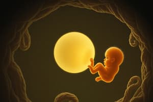
Fetal Development
- Occurs from the 9th week until birth (40-42 weeks)
- Measurements can be taken:
- Fetal Length
- Crown-Rump Length
- Crown-Heel Length
- Weight
Estimating Fetal Age
- First Trimester ( up to 22 weeks): Margin of error is 2 days
- Use the following measurements:
- BIPARIETAL DIAMETER
- CRANIAL CIRCUMFERENCE
- ABDOMINAL CIRCUMFERENCE
- FEMUR LENGTH
- FOOT LENGTH
- Late ultrasound: Not important for age estimation
Week 9
- Measures 45-52 mm/4 to 5 cm.
- Auricular pavilions form at the level of the eyes. -The head makes up 50% of the anatomy
- The Liver enters hematopoietic period
- Nervous system has Mylenation
- Further differentiated Genitals
- Developed kidneys
Weeks 10-13
- Measures 49-112 mm/5 to 11 cm.
- Head makes up 1/3 proportion
- Central hematopoietic spleen
- CNS completes coarse morphogenesis
- The upper limbs are proportionately more mature
- Shorter lower limbs and incipient nails appear
- Anomalies like Sd of down can be detected via:
- Ultrasound
- Chromosomal or Translucence
Weeks 14-16
- Accelerated body Growth
- Measures 100-150 mm/10-15cm
- Lanugo appears
- Definitive Inferior Limb
- Marked ossification (bones present)
- Well difined Genitals.
Weeks 17-20
- Measure 130-195 mm/13-19cm
- More Pronounced Lanugo
- Vernix Caseosa covers skin and Production of melanin
- accumulation of brown fat
- More of the finger nail covers the ungual plate
- Reach Limb Proportions
- Intervention by law
Weeks 21-25
- Fetal Viability is achieved
- Type 2 pneumocyte produces the tension producing Surfactant
- Dense capilaries and superior nail border on fingers
Weeks 26-30
- Greater likelihood Fetal viability
- Eyes voluntarily open and moves respiratory
- bone marrow and blood made
Weeks 31-38
- White Fat, Surfactant peaks 34 week, increases tenso actives and testicles in scrotum
- The testicles are in the scrotum
Birth
Term Product
- Occurs within 37 and 41 weeks
Premature Product
- Occurs before 37 weeks
Post-Term Product
- Occurs after 41+ weekds
Last Menstruation Date
- Last Period + 7 days
- Month -3 months
-
- 1 Year
Legal Pregnancy Interruption
- Occurs by victim of violation
- 24-48 hours and performed in 22-26 weeks
- 800 Mg Misoprostol administered orally and continued with 800 mg
Studying That Suits You
Use AI to generate personalized quizzes and flashcards to suit your learning preferences.