Podcast
Questions and Answers
What embryonic structures contribute to the formation of the diaphragm's crura?
What embryonic structures contribute to the formation of the diaphragm's crura?
- The septum transversum.
- Somites at cervical segments C3-C5.
- The mesentery of the esophagus. (correct)
- The pleuroperitoneal membranes.
Why do the phrenic nerves, which innervate the diaphragm, originate from cervical segments C3-C5?
Why do the phrenic nerves, which innervate the diaphragm, originate from cervical segments C3-C5?
- Because the heart's initial development occurs in the cervical region, influencing nerve pathways.
- Because the diaphragm initially develops in the cervical region during the fourth week of development. (correct)
- Because these segments are closest to the final location of the diaphragm in the fully developed organism.
- Due to the rapid growth of the ventral part of the embryo, stretching the nerve fibers.
The pleuroperitoneal membranes ultimately form which part of the diaphragm?
The pleuroperitoneal membranes ultimately form which part of the diaphragm?
- The crura of the diaphragm.
- The central tendon. (correct)
- The muscular components.
- The phrenic nerves.
Differential growth during the sixth week of development contributes primarily to which of the following regarding the diaphragm?
Differential growth during the sixth week of development contributes primarily to which of the following regarding the diaphragm?
A defect in the formation of the pleuroperitoneal membranes would most likely result in malformation of which diaphragmatic structure?
A defect in the formation of the pleuroperitoneal membranes would most likely result in malformation of which diaphragmatic structure?
During embryonic development, which of the following structures is formed by the rolling up of the ectoderm?
During embryonic development, which of the following structures is formed by the rolling up of the ectoderm?
Which layer of the trilaminar embryonic disc gives rise to the gut tube?
Which layer of the trilaminar embryonic disc gives rise to the gut tube?
The visceral layer of the lateral plate mesoderm is intimately connected to which structure?
The visceral layer of the lateral plate mesoderm is intimately connected to which structure?
What two tissue layers combine to form the somatopleure?
What two tissue layers combine to form the somatopleure?
The space between the visceral and parietal layers of the lateral plate mesoderm ultimately forms what?
The space between the visceral and parietal layers of the lateral plate mesoderm ultimately forms what?
Which component of the intraembryonic mesoderm plays a significant role in the formation of the skull and vertebrae?
Which component of the intraembryonic mesoderm plays a significant role in the formation of the skull and vertebrae?
What two tissue layers combine to form the splanchnopleure?
What two tissue layers combine to form the splanchnopleure?
Which of the following structures contributes to the urogenital system?
Which of the following structures contributes to the urogenital system?
The lateral body wall folds are composed of which of the following?
The lateral body wall folds are composed of which of the following?
What key event occurs with the endoderm layer during the fourth week of embryonic development?
What key event occurs with the endoderm layer during the fourth week of embryonic development?
Closure of the ventral body wall is incomplete at the connecting stalk, which later develops into the:
Closure of the ventral body wall is incomplete at the connecting stalk, which later develops into the:
The visceral layer of lateral plate mesoderm contributes to the formation of which structure?
The visceral layer of lateral plate mesoderm contributes to the formation of which structure?
The dorsal mesentery serves as a pathway for which of the following structures to reach the organs?
The dorsal mesentery serves as a pathway for which of the following structures to reach the organs?
Which structure suspends the gut tube from the posterior body wall into the peritoneal cavity?
Which structure suspends the gut tube from the posterior body wall into the peritoneal cavity?
From which structure does the ventral mesentery arise?
From which structure does the ventral mesentery arise?
During what period does the vitelline duct typically degenerate?
During what period does the vitelline duct typically degenerate?
The septum transversum, a precursor to the diaphragm, originates from which type of mesoderm?
The septum transversum, a precursor to the diaphragm, originates from which type of mesoderm?
What is the fate of the pleuropericardial membranes in the fully developed human body?
What is the fate of the pleuropericardial membranes in the fully developed human body?
Prior to complete separation, the pleural and peritoneal cavities communicate through which structures?
Prior to complete separation, the pleural and peritoneal cavities communicate through which structures?
The pericardioperitoneal canals are located on each side of which structure?
The pericardioperitoneal canals are located on each side of which structure?
What causes the pericardioperitoneal canals to become smaller during development?
What causes the pericardioperitoneal canals to become smaller during development?
Which structures contain the common cardinal veins and phrenic nerves during the formation of the thoracic cavity?
Which structures contain the common cardinal veins and phrenic nerves during the formation of the thoracic cavity?
What is the origin of the pleuroperitoneal folds that contribute to the formation of the diaphragm?
What is the origin of the pleuroperitoneal folds that contribute to the formation of the diaphragm?
The descent of the heart and positional changes of the sinus venosus influence the shift of the common cardinal veins. What is the subsequent effect on the pleuropericardial membranes?
The descent of the heart and positional changes of the sinus venosus influence the shift of the common cardinal veins. What is the subsequent effect on the pleuropericardial membranes?
Flashcards
Neurulation
Neurulation
The process where the ectoderm forms the neural plate, rolling up to create the brain and spinal cord.
Gut Tube Formation
Gut Tube Formation
The ventral layer of the trilaminar embryonic disc which rolls down to form the digestive tube.
Mesoderm's Role
Mesoderm's Role
The mesoderm layer splits into visceral (splanchnic) and parietal (somatic) layers, holding the neural and gut tubes together.
Visceral (Splanchnic) Layer
Visceral (Splanchnic) Layer
Signup and view all the flashcards
Parietal (Somatic) Layer
Parietal (Somatic) Layer
Signup and view all the flashcards
Primitive Body Cavity
Primitive Body Cavity
Signup and view all the flashcards
Somatopleure
Somatopleure
Signup and view all the flashcards
Splanchnopleure
Splanchnopleure
Signup and view all the flashcards
Pleuroperitoneal Membranes
Pleuroperitoneal Membranes
Signup and view all the flashcards
Cervical Segments C3-C5
Cervical Segments C3-C5
Signup and view all the flashcards
Phrenic Nerves
Phrenic Nerves
Signup and view all the flashcards
Diaphragm Migration
Diaphragm Migration
Signup and view all the flashcards
Esophageal Mesentery
Esophageal Mesentery
Signup and view all the flashcards
Lateral Body Wall Folds
Lateral Body Wall Folds
Signup and view all the flashcards
Ventral Body Wall Closure
Ventral Body Wall Closure
Signup and view all the flashcards
Vitelline Duct
Vitelline Duct
Signup and view all the flashcards
Parietal Layer of Serous Membranes
Parietal Layer of Serous Membranes
Signup and view all the flashcards
Visceral Layer of Serous Membranes
Visceral Layer of Serous Membranes
Signup and view all the flashcards
Dorsal Mesentery
Dorsal Mesentery
Signup and view all the flashcards
Mesenteries Function
Mesenteries Function
Signup and view all the flashcards
Septum Transversum
Septum Transversum
Signup and view all the flashcards
Pericardioperitoneal Canals
Pericardioperitoneal Canals
Signup and view all the flashcards
Lung Expansion
Lung Expansion
Signup and view all the flashcards
Pleuropericardial Folds
Pleuropericardial Folds
Signup and view all the flashcards
Pleuropericardial Membranes
Pleuropericardial Membranes
Signup and view all the flashcards
Heart Descent
Heart Descent
Signup and view all the flashcards
Thoracic Cavity Division
Thoracic Cavity Division
Signup and view all the flashcards
Pleuroperitoneal Folds (Diaphragm)
Pleuroperitoneal Folds (Diaphragm)
Signup and view all the flashcards
Study Notes
Tube within a Tube
- During weeks 3 and 4, the top ectoderm layer develops into the brain and spinal chord through neurulation.
- Simultaneously, the endoderm layer folds to create the gut tube.
- The embryo becomes a tube-within-a-tube structure
- The neural tube is positioned dorsally and the gut tube ventrally.
- The mesoderm layer secures the neural and gut tubes.
- The lateral plate of the mesoderm divides into the visceral (splanchnic) and parietal (somatic) layers.
- The visceral layer encloses the gut tube.
- The parietal layer, together with the ectoderm, gives rise to the lateral body wall folds to close the ventral body wall.
- The space between the visceral and parietal layers is the primitive body cavity. -This space is initially continuous without subdivisions into pericardial, pleural, and abdomino-pelvic regions.
Formation of the Body Cavity
- At the third week's end, the intraembryonic mesoderm develops into the paraxial, intermediate, and lateral plate mesoderm. -Paraxial mesoderm plays a key role in the formation of the skull and vertebrae. -Intermediate mesoderm contributes to the urogenital system. -Lateral plate mesoderm is crucial in forming the body cavity.
- Clefts arise in the lateral plate mesoderm and combine, dividing it into parietal and visceral layers.
- (1) the parietal (somatic) layer is next to the surface ectoderm and connects with the extra-embryonic parietal mesoderm across the amnion.
- Together, the parietal layer and the ectoderm are called the somatopleure.
- (2) the visceral (splanchnic) layer is next to the endoderm, which forms the basis of the gut tube, and connects to the visceral layer of extraembryonic mesoderm that covers the yolk sac.
- Together, the visceral layer and the endoderm form the splanchnopleure.
- The space between the two layers becomes the primitive body cavity.
- During the fourth week, the embryo begins to grow ventrally and forms two lateral body wall folds.
Further Body Cavity Development
- The folds consist of the parietal layer of lateral plate mesoderm, ectoderm, and cells that move from nearby somites into the mesoderm layer across the lateral somitic frontier.
- As the folds grow, the endoderm layer also folds ventrally to create the gut tube.
- By the fourth week's end, the lateral body wall folds meet and merge in the midline to shut the ventral body wall under the head and tail regions (folds), causing the embryo to curve into a fetal position.
- The ventral body wall closes completely, except where the connecting stalk exists to be the future umbilical cord.
- The gut tube also closes, but a connection from the midgut to the yolk sac persists.
- The Vitelline (yolk sac) duct is ultimately incorporated into the umbilical cord, narrows extensively, and degenerates along with the yolk sac between the second and third gestation months.
- The parietal and visceral layers of lateral plate mesoderm remain connected where the gut tube meets the posterior body wall during body cavity and gut tube development.
Serous Membranes
- Parietal layer cells of the lateral plate mesoderm turn into mesothelial cells, which form the parietal layer of the serous membranes around the peritoneal, pleural, and pericardial cavities.
- Visceral layer cells of lateral plate mesoderm form the Visceral layer and cover the abdominal organs, lungs, and heart.
- The visceral and parietal layers connect as the dorsal mesentery, suspending the gut tube into the peritoneal cavity from the posterior body wall.
- The dorsal mesentery extends from the end of the foregut to the hindgut.
- The ventral mesentery exists between the caudal foregut and the upper duodenum and arises from mesoderm thinning of the septum transversum.
- The septum transversum is a mesoderm block creating connective tissue in the liver and a the base the diaphragm
- The mesenteries are double peritoneum layers that allow blood arteries, nerves, and lymphatic vessels to reach the organs.
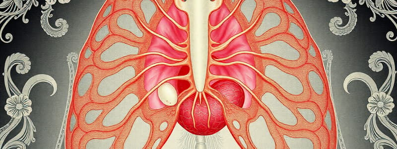
Diaphragm and Thoracic Cavity
- The septum transversum is a solid mesodermal tissue plate sitting in the area involving the thoracic cavity and stalk of the yolk sac.
- The septum develops visceral (splanchnic) mesoderm around the heart.
- The septum settles between the thoracic and abdominal cavities when the top end of the embryo curves.
- The septum only partly separates the thoracic and abdominal cavities, forming pericardioperitoneal canals on each side of the foregut.
- Lung buds grow caudolaterally inside the pericardioperitoneal canals.
- Due to lung development, the pericardioperitoneal canals shrink, so lungs grow into the body section mesenchyme on the dorsal, lateral, and ventral sides.
- Ventral and lateral growth occur behind the pleuropericardial folds.
- These pleuropericardial folds are small ridges projecting into the primitive thoracic cavity.
- As the lungs grow, mesoderm produces the definitive thorax wall and pleuropericardial membranes.
- Descent of the heart and sinus venosus shifts the cardinal veins, drawing the pleuropericardial membranes.
- They merge with each other and the lung roots, splitting the thoracic cavity into the pericardial cavity and pleural cavities.
- In adults, the fibrous pericardium results from pleuropericardial membranes.
Diaphragm Formation
- The pleural and pericardial cavities remain connected to the abdominal cavity through the pericardioperitoneal canals.
- During development, the opening between the pleural and peritoneal space closes partially by pleuroperitoneal folds at the pericardioperitoneal canals.
- In the seventh week, the folds thin into membranes that grow to merge with the esophagus mesentery (the diaphragm crura around the aorta.)
- The pleuroperitoneal membranes grow to coat the septum transversum completely.
- Cervical segments 3-5 (C3-C5) are where muscles originate for the diaphragm.
- The phrenic nerves arise from the ventral primary rami of spinal nerves innervating the diaphragm.
- Diaphragm development begins in the cervical fourth week.
- In the sixth week, differential embryo growth causes the diaphragm to move ventrally and caudally, resulting in phrenic nerves having a long rout.
- The two pleuroperitoneal membranes is what forms the central diaphragm tendon
- Muscular components are derived from somites at cervical segments three to five.
- The crura of the diaphragm are derived from the mesentery of the esophagus.
Studying That Suits You
Use AI to generate personalized quizzes and flashcards to suit your learning preferences.