Podcast
Questions and Answers
The inner cell mass differentiates into two distinct layers: epiblast and ______.
The inner cell mass differentiates into two distinct layers: epiblast and ______.
hypoblast
The amniotic cavity forms within the ______ layer.
The amniotic cavity forms within the ______ layer.
epiblast
The primary yolk sac is formed by the hypoblast and the ______ membrane.
The primary yolk sac is formed by the hypoblast and the ______ membrane.
exocoelomic
Extraembryonic mesoderm forms between the cytotrophoblast and the yolk sac/amniotic ______.
Extraembryonic mesoderm forms between the cytotrophoblast and the yolk sac/amniotic ______.
The chorionic cavity is also referred to as the extraembryonic ______.
The chorionic cavity is also referred to as the extraembryonic ______.
Maternal infections like rubella and cytomegalovirus can lead to severe fetal ______.
Maternal infections like rubella and cytomegalovirus can lead to severe fetal ______.
Exposure to toxic chemicals such as mercury or lead can interfere with fetal ______.
Exposure to toxic chemicals such as mercury or lead can interfere with fetal ______.
Different organs and tissues are most sensitive to teratogens during specific ______ of development.
Different organs and tissues are most sensitive to teratogens during specific ______ of development.
Neural Tube Defects (NTDs) are caused by the failure of the neural tube to close properly during early ______.
Neural Tube Defects (NTDs) are caused by the failure of the neural tube to close properly during early ______.
The genetic makeup of the embryo influences its susceptibility to ______.
The genetic makeup of the embryo influences its susceptibility to ______.
The notochord becomes the nucleus pulposus of the intervertebral discs, while the ______ gives rise to the vertebral bodies and arches.
The notochord becomes the nucleus pulposus of the intervertebral discs, while the ______ gives rise to the vertebral bodies and arches.
The skull develops from both mesodermal tissue and ______ cells.
The skull develops from both mesodermal tissue and ______ cells.
The protective case around the brain is called the ______.
The protective case around the brain is called the ______.
The ______ forms the base of the skull and includes bones like the sphenoid and occipital bones.
The ______ forms the base of the skull and includes bones like the sphenoid and occipital bones.
The bones of the face, such as the maxilla and mandible, are part of the ______.
The bones of the face, such as the maxilla and mandible, are part of the ______.
In newborns, the bones of the skull are separated by ______, which allow for slight deformation during birth.
In newborns, the bones of the skull are separated by ______, which allow for slight deformation during birth.
Ribs develop from costal processes of the vertebrae through ______ ossification.
Ribs develop from costal processes of the vertebrae through ______ ossification.
The sternum develops independently from the vertebrae from a pair of ______ bars.
The sternum develops independently from the vertebrae from a pair of ______ bars.
______ genes regulate the patterning and identity of somites along the cranio-caudal axis.
______ genes regulate the patterning and identity of somites along the cranio-caudal axis.
The manubrium, body, and xiphoid process are parts of the ______.
The manubrium, body, and xiphoid process are parts of the ______.
Flashcards are hidden until you start studying
Study Notes
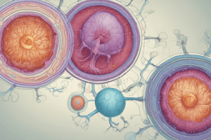
Second Week of Development
- The blastocyst implants fully into the uterine lining.
- The syncytiotrophoblast invades the endometrium and establishes early uteroplacental circulation.
- The inner cell mass differentiates into the epiblast (forming the embryo) and the hypoblast (forming the yolk sac).
- The epiblast forms the amniotic cavity which is filled with amniotic fluid.
- The hypoblast extends to form the primary yolk sac, which is later replaced by the secondary yolk sac.
- The extraembryonic mesoderm forms between the cytotrophoblast and the yolk sac/amniotic cavity.
- The extraembryonic cavities coalesce to form the chorionic cavity, where the embryo, yolk sac, and amniotic cavity are suspended.
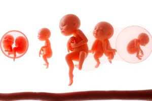
Principles of Teratology
- Different organs are most sensitive to teratogens at specific developmental times. The first trimester is especially crucial for organogenesis, making the embryo highly susceptible to teratogens.
- The severity of the defect often correlates with the dose of the teratogen. Higher doses usually lead to more severe anomalies.
- The genetic makeup of the embryo or fetus plays a role in its susceptibility to teratogens.
Development of the Axial Skeleton
- The vertebral column forms from somites, which differentiate into the sclerotome and dermomyotome.
- The sclerotome gives rise to the vertebral bodies and arches.
- The notochord becomes the nucleus pulposus of the intervertebral discs.
- Vertebrae undergo both endochondral and membranous ossification to form fully formed bony structures.
Development of the Skull
- The skull develops from mesodermal tissue and neural crest cells.
- The neurocranium forms the protective case around the brain.
- Membranous neurocranium gives rise to the flat bones of the skull through intramembranous ossification.
- Cartilaginous neurocranium forms the base of the skull through endochondral ossification.
- The viscerocranium forms the facial bones, primarily derived from neural crest cells.
- Fontanelles are soft spots in the newborn skull, allowing for brain growth during infancy.
Development of Ribs and Sternum
- Ribs develop from the costal processes of the vertebrae, undergoing endochondral ossification.
- The sternum develops independently of the vertebrae from a pair of sternal bars in the ventral body wall, which fuse to form the manubrium, body, and xiphoid process.
Molecular Regulation of Axial Skeleton Development
- Hox genes regulate the patterning and identity of somites along the cranio-caudal axis. Mutations can cause homeotic transformations.
- SHH promotes the formation of sclerotome cells from somites.
- BMP and FGF influence the differentiation of mesodermal cells and neural crest cell migration.
Overview of Muscle Development
- Muscle tissue develops from the mesodermal germ layer.
- Somites give rise to skeletal muscle, while splanchnic mesoderm gives rise to smooth and cardiac muscle.
- Muscle formation involves myogenic progenitor cells, growth factors, and transcription factors.
Skeletal Muscle Development
- Skeletal muscles originate from paraxial mesoderm, which forms somites in the trunk and somitomeres in the head.
- The myotome portion of the dermomyotome gives rise to skeletal muscles.
- The epaxial division forms back muscles.
- The hypaxial division forms limb, body wall, and ventral neck muscles.
- Muscles of the head and neck develop from pharyngeal arches.
Overview of Limb Development
- Limb development occurs between weeks 4 and 8 of gestation.
- Limb buds form from mesoderm covered by ectoderm.
- The apical ectodermal ridge (AER) controls limb growth and patterning.
Origins of Limb Components
- Lateral plate mesoderm forms the bones, blood vessels, and connective tissues of the limbs.
- Paraxial mesoderm (somites) provides the muscle precursors.
- Ectoderm contributes to the skin, epidermis, and sensory innervation pathways.
Development of the Limb Skeleton
- Limb bones form through endochondral ossification.
- Proximal-to-distal bone formation occurs from shoulder to fingers in the upper limb and hip to toes in the lower limb.
- Joint interzones form between adjacent skeletal elements, differentiating into synovial joints.
Studying That Suits You
Use AI to generate personalized quizzes and flashcards to suit your learning preferences.