Podcast
Questions and Answers
What is the most significant feature in the development of the head and neck?
What is the most significant feature in the development of the head and neck?
- The stomodeum
- The foliate papillae
- The maxillary prominences
- The pharyngeal arches (correct)
During which weeks do the pharyngeal arches appear?
During which weeks do the pharyngeal arches appear?
- Weeks 1 and 2
- Weeks 6 and 7
- Weeks 3 and 4
- Weeks 4 and 5 (correct)
What forms the center of the face at the end of the fourth week?
What forms the center of the face at the end of the fourth week?
- The maxillary prominences
- The stomodeum (correct)
- The mandible
- The frontonasal prominence
How many mesenchymal prominences can be recognized at four weeks of development?
How many mesenchymal prominences can be recognized at four weeks of development?
Which of the following statements is true about foliate papillae in humans?
Which of the following statements is true about foliate papillae in humans?
Study Notes
Pharyngeal Arches
- Appear during the fourth and fifth weeks of development
- Contribute to the formation of the neck and face
- Form the center of the face surrounded by the stomodeum by the end of the fourth week
- Five mesenchymal prominences form by the end of week four: two mandibular prominences, two maxillary prominences, and one frontonasal prominence.
Sensory Nerve Supply of the Tongue
- Lingual nerve from the first branchial arch supplies the tongue.
- Chorda tympani, a branch of the facial nerve from the second arch contributes to the nerve supply.
- Glossopharyngeal nerve from the third arch supplies the posterior third of the tongue.
Papillae of the Tongue
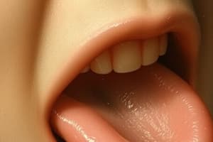
- Four papillae are found on the dorsal surface of the tongue: fungiform, circumvallate, foliate, and filiform.
- Fungiform, circumvallate, and foliate contain taste buds and develop before the filiform papillae.
- Taste buds develop before papillae, initiating taste sensation during embryonic development.
- Fungiform papillae develop around 11 weeks of gestation due to an elevation of the surface epithelium.
- Circumvallate papillae develop around 16 weeks of gestation by moving downward and toward the anterior surface.
- Foliate papillae develop around 17 weeks of gestation and move downward toward the formation of the epithelium.
Advanced Bell Stage
- The lateral dental lamina disappears during this stage due to the invasion of surrounding mesenchyme.
- Remnants of the lamina may be present as isolated groups of epithelial cells (rests of Serres) or Serres pearls.
- The enamel organ changes during this stage, with the outer enamel epithelium (OEE) becoming flattened and developing microvilli on its outer surfaces.
- The basement membrane folds during this stage, increasing the surface area for nutrient uptake.
- OEE cells become active in transporting nutrients through mitochondria and cytoplasmic membranes.
- The cervical loop is formed where the inner enamel epithelium (IEE) meets the OEE at the cervical end of the enamel organ.
- The cervical loop is important for root formation, remains stable, and has no stellate reticulum or stratum intermedium.
Development of Multi-Rooted Teeth
- As the epithelial diaphragm grows, it may make contact and fuse with the developing teeth.
- This process may result in the formation of single, double, or triple-rooted teeth.
- The origin of the root is from the dental papilla (HWR), while the epithelium is of oral origin (HWR).
- The enamel organ originates from ectoderm.
- The dental papilla originates from mesenchyme.
- The epithelial extensions form in the first stages and might contact to develop one or multi-rooted structures.
- The zone where roots begin to separate is labeled as "Furcation".
Enamel Organ
- The crown shape is determined by morphological changes in the cells as they form dentin and enamel.
- The enamel organ is composed of four layers:
- Outer enamel epithelium
- Inner enamel epithelium
- Stellate reticulum
- Stratum intermedium
Stratum Intermedium
- Located between the inner enamel epithelium and the stellate reticulum.
- Composed of 2-3 layers of flat cells.
- Rich in the enzyme alkaline phosphatase, which is necessary for enamel mineralization.
- The stratum intermedium and the inner enamel epithelium are considered the functional unit for enamel formation.
Dental Papilla and Dental Sac
- The dental papilla differentiates into dentin and pulp.
- There is increased vascularity and cellularity of the dental papilla under the influence of the enamel organ.
- Differentiation of odontoblasts occurs between the odontoblasts and the basement membrane.
Dental Follicle (or Dental Sac)
- It presents a circular arrangement around the tooth resembling a fibrous capsule.
- The inner layer faces the enamel organ and dental papilla, and the outer layer faces the bony crypt
- Fibers in the follicle may merge with fibers in the vascular tissue.
Fate and Function of the Dental Lamina
- The dental lamina starts appearing during the second month of intrauterine life.
- The dental lamina has broad and thick connections.
- In the advanced bell stage, the lateral dental lamina degenerates and is invaded by mesenchyme.
- The main dental lamina proliferates in the connective tissue, forming the successional dental lamina.
- The successional lamina forms successors from 5 months (incisors) to 8 months (premolars).
- The main dental lamina grows posteriorly to give rise to the enamel organ for permanent teeth, starting from 1 year for second molars and 4 years for the third molars.
- The lifespan of the dental lamina is approximately 5 years.
Stages of Tooth Development
- Initiation is the starting point of tooth development.
- Proliferation involves further development and growth of initiated structures.
- Histo-differentiation is the formation of different tissues and their specific properties.
- Morpho-differentiation is the development of shape and form.
- Apposition represents a further step in the development of shape.
Proliferation
- Active proliferation occurs in the dental bud, cap, and early bell stages.
- Proliferation also occurs in certain areas of the advanced bell stage, where dental tissues are not yet formed.
Initiation
- Mesenchymal cells from the oral epithelium initiate dental tissue formation.
- The newly developed bud stage is formed in the designated oral region.
- The initiation process is triggered by cells.
Histo- / Morpho-differentiation
- Specific cells undergo changes in morphology and form during this stage.
- Cells involved include: epithelial cells, bell stage cells, and ameloblast (late)/odontoblast cells.
- Proliferation occurs in stages, including the early and late stages of initiation.
Apposition / Maturation (Mineralization)
- Maturation involves the gradual accumulation of mineralized materials, which is a key stage in tooth development.
Root Formation
- Mesenchymal cells of the dental papilla reach the enamel-cemental junction.
- The epithelial root sheath (Hertwig's root sheath) forms from the cervical loop.
- The cervical loop proliferates and forms the epithelial root sheath.
- The epithelial diaphragm forms in the first part of the epithelial root sheath.
- The inner enamel epithelium of the epithelial root sheath induces undifferentiated mesenchymal cells of the dental papilla to differentiate into odontoblasts.
Stages of Root Formation
- Early stage (bell stage):
- Advanced stage.
- Dentin and enamel matrix formation takes place.
- A rhythmic deposition of dentin and enamel occurs.
- Late stage (late bell stage):
- Mineralization of the tooth is mature.
- Post-appositional deposition.
Cells Involved
- Odontoblasts form dentin.
- Ameloblasts form enamel.
- Cementoblasts form cementum.
Studying the Structure of Enamel
- Ground section: organic substance is burnt, and the inorganic substance remains
- Decalcified section: inorganic substance is dissolved, and the organic substance remains
Enamel Structure
- The fundamental units of enamel are rods and inter-rod enamel.
- The rod is shaped like a cylinder, composed of a wide head portion, neck, and a thinner tail portion.
- Interrod enamel comprises crystals that surround each rod.
- Enamel rods start straight at the dentino-enamel junction (DEJ) and then have a wavy course until near the outer surface of the enamel where they become straight once more.
- The rods are formed nearly perpendicular to the DEJ and curve slightly towards the tip.
- They follow a wavy course as they traverse from the DEJ to the surface of the enamel.
- Ground section: suitable for the study of enamel
- Decalcified section: suitable for the study of dentin
Studying That Suits You
Use AI to generate personalized quizzes and flashcards to suit your learning preferences.
Description
Explore the fascinating development of pharyngeal arches and their roles in forming the neck and face during embryonic growth. This quiz also delves into the sensory nerve supply of the tongue and the formation of its papillae, highlighting both their anatomical significance and functional aspects. Test your knowledge on these critical topics in embryology!