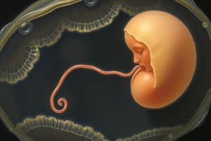
![[HD 201] E01-T07-Development of Embryo_compressed](https://assets.quizgecko.com/cdn-cgi/image/width=800,height=300,fit=crop,quality=75,format=webp/quiz/7529cf918e7491f7bdb66679ee093ae1.jpg)
Podcast
Questions and Answers
What event must precede the acrosome reaction for fertilization to occur?
What event must precede the acrosome reaction for fertilization to occur?
- Capacitation of the sperm (correct)
- Completion of the second meiotic division by the oocyte
- Fusion of the sperm and oocyte cell membranes
- Penetration of the corona radiata
During fertilization, what layer or structure does the sperm initially penetrate?
During fertilization, what layer or structure does the sperm initially penetrate?
- Cell membrane of the oocyte
- Corona radiata (correct)
- Zona pellucida
- Perivitelline space
The syncytiotrophoblast secretes which hormone to maintain the corpus luteum during early pregnancy?
The syncytiotrophoblast secretes which hormone to maintain the corpus luteum during early pregnancy?
- Human chorionic gonadotropin (hCG) (correct)
- Lactogen
- Progesterone
- Estrogen
Which of the following is derived from the ectoderm?
Which of the following is derived from the ectoderm?
The formation of which structure signifies the beginning of gastrulation?
The formation of which structure signifies the beginning of gastrulation?
What structure develops from the connecting stalk?
What structure develops from the connecting stalk?
Which of the following events occurs during the second week of human development?
Which of the following events occurs during the second week of human development?
What is the primary role of the trophoblast during early embryonic development?
What is the primary role of the trophoblast during early embryonic development?
Which of the following structures is derived from the mesoderm?
Which of the following structures is derived from the mesoderm?
What is the significance of the Carnegie stages in the context of early human development?
What is the significance of the Carnegie stages in the context of early human development?
During neurulation, what structure induces the formation of the neural plate?
During neurulation, what structure induces the formation of the neural plate?
Which event characterizes the morula stage of development?
Which event characterizes the morula stage of development?
In embryonic folding, what structure is formed when the endoderm moves towards the midline and fuses?
In embryonic folding, what structure is formed when the endoderm moves towards the midline and fuses?
How does the uteroplacental circulation establish the exchange of gases and metabolites?
How does the uteroplacental circulation establish the exchange of gases and metabolites?
In what stage of early embryonic development does the fluid penetrate the morula, creating a cavity?
In what stage of early embryonic development does the fluid penetrate the morula, creating a cavity?
The amniotic cavity forms within which layer of the bilaminar disc?
The amniotic cavity forms within which layer of the bilaminar disc?
What part of the human anatomy does the oropharyngeal membrane eventually become?
What part of the human anatomy does the oropharyngeal membrane eventually become?
What is the clinical significance of measuring alpha-fetoprotein (AFP) levels during pregnancy?
What is the clinical significance of measuring alpha-fetoprotein (AFP) levels during pregnancy?
What is the expected outcome if the midgut fails to return to the abdominal cavity between weeks 6-10?
What is the expected outcome if the midgut fails to return to the abdominal cavity between weeks 6-10?
What is the underlying cause of sacrococcygeal teratoma?
What is the underlying cause of sacrococcygeal teratoma?
Which of the following represents the correct sequence of early development stages?
Which of the following represents the correct sequence of early development stages?
What is the initial event in the formation of the uteroplacental circulation during the second week of development?
What is the initial event in the formation of the uteroplacental circulation during the second week of development?
What is the result of lateral folding during the fourth week of development?
What is the result of lateral folding during the fourth week of development?
Following the establishment of the three germ layers, what process occurs next in embryonic development?
Following the establishment of the three germ layers, what process occurs next in embryonic development?
In the context of teratogenesis, what does the "peak sensitivity" of an organ refer to?
In the context of teratogenesis, what does the "peak sensitivity" of an organ refer to?
What event marks the end of the embryonic period and the beginning of the fetal period?
What event marks the end of the embryonic period and the beginning of the fetal period?
What structure gives rise to the dermis of the skin?
What structure gives rise to the dermis of the skin?
What happens to the caudal eminence by the eighth week of development?
What happens to the caudal eminence by the eighth week of development?
Which of the following is the correct timeframe for the highest risk of major birth defects due to teratogens?
Which of the following is the correct timeframe for the highest risk of major birth defects due to teratogens?
Which cells are responsible for forming the exocoelomic membrane, which lines the inner surface of the cytotrophoblast during early development?
Which cells are responsible for forming the exocoelomic membrane, which lines the inner surface of the cytotrophoblast during early development?
What is the primary function of Human Chorionic Gonadotropin (hCG) in early pregnancy?
What is the primary function of Human Chorionic Gonadotropin (hCG) in early pregnancy?
What event characterizes the end of implantation during the second week of development?
What event characterizes the end of implantation during the second week of development?
Which primary germ layer gives rise to the epithelial components of the trachea, bronchi, and lungs?
Which primary germ layer gives rise to the epithelial components of the trachea, bronchi, and lungs?
What developmental defect is associated with maternal exposure to thalidomide between 34 and 50 days of gestation?
What developmental defect is associated with maternal exposure to thalidomide between 34 and 50 days of gestation?
Where does the process of sperm capacitation typically occur?
Where does the process of sperm capacitation typically occur?
During the third week of development, which structure forms at the cranial end of the embryonic disc and consists of a slightly elevated area surrounding the primitive pit?
During the third week of development, which structure forms at the cranial end of the embryonic disc and consists of a slightly elevated area surrounding the primitive pit?
Which of the following is a characteristic feature of the blastocyst?
Which of the following is a characteristic feature of the blastocyst?
Flashcards
Fertilization
Fertilization
Fusion of sperm and oocyte, occurring in the ampullary region of the uterine tube.
Capacitation
Capacitation
Period of sperm conditioning in the female reproductive tract, lasting about 7 hours.
Acrosome Reaction
Acrosome Reaction
An exocytotic event leading to the eversion of the acrosomal vesicle, enabling sperm penetration.
Cleavage
Cleavage
Signup and view all the flashcards
Morula
Morula
Signup and view all the flashcards
Blastomere
Blastomere
Signup and view all the flashcards
Blastocyst Formation
Blastocyst Formation
Signup and view all the flashcards
Blastocoel
Blastocoel
Signup and view all the flashcards
Trophoblast
Trophoblast
Signup and view all the flashcards
Embryoblast
Embryoblast
Signup and view all the flashcards
Implantation
Implantation
Signup and view all the flashcards
Embryonic mesoderm
Embryonic mesoderm
Signup and view all the flashcards
Chorion/Chorionic membrane
Chorion/Chorionic membrane
Signup and view all the flashcards
Trophoblast
Trophoblast
Signup and view all the flashcards
Cytotrophoblast
Cytotrophoblast
Signup and view all the flashcards
Syncytiotrophoblast
Syncytiotrophoblast
Signup and view all the flashcards
Human chorionic gonadotropin (hCG)
Human chorionic gonadotropin (hCG)
Signup and view all the flashcards
Hypoblast
Hypoblast
Signup and view all the flashcards
Epiblast
Epiblast
Signup and view all the flashcards
Amniotic cavity
Amniotic cavity
Signup and view all the flashcards
Developing Villi
Developing Villi
Signup and view all the flashcards
Exocoelomic membrane
Exocoelomic membrane
Signup and view all the flashcards
Uteroplacental Circulation
Uteroplacental Circulation
Signup and view all the flashcards
Connecting Stalk
Connecting Stalk
Signup and view all the flashcards
Oropharyngeal & Cloacal Membrane
Oropharyngeal & Cloacal Membrane
Signup and view all the flashcards
Gastrulation
Gastrulation
Signup and view all the flashcards
Definitive endoderm
Definitive endoderm
Signup and view all the flashcards
A-P, D-V, and L-R Axes
A-P, D-V, and L-R Axes
Signup and view all the flashcards
Notochord
Notochord
Signup and view all the flashcards
Neurulation
Neurulation
Signup and view all the flashcards
Somitomeres
Somitomeres
Signup and view all the flashcards
Myotome (muscle tissue), sclerotome (cartilage and bone), and dermatome (dermis of the skin)
Myotome (muscle tissue), sclerotome (cartilage and bone), and dermatome (dermis of the skin)
Signup and view all the flashcards
Approximate Age of Embryo
Approximate Age of Embryo
Signup and view all the flashcards
Folding of Embryo
Folding of Embryo
Signup and view all the flashcards
Head Fold
Head Fold
Signup and view all the flashcards
Tail Fold
Tail Fold
Signup and view all the flashcards
Embryo in the Horizontal Plan
Embryo in the Horizontal Plan
Signup and view all the flashcards
Fifth Weeks of Development
Fifth Weeks of Development
Signup and view all the flashcards
Sixth Week of Development
Sixth Week of Development
Signup and view all the flashcards
Seventh Weeks of Development
Seventh Weeks of Development
Signup and view all the flashcards
Study Notes
The First 8 Weeks
- This period is an exciting area for biologic research but one of the least understood
- Itis the most sensitive period to system abnormalities generated by teratogens
- Miscarriages often happen during the first trimester
- Development is divided into 23 stages by Carnegie
Carnegie Stages
- Not usually sensitive during weeks 0-2; high rate of lethality may occur
- Period of greatest sensitivity to teratogens is weeks 3-8
- Each organ also has a peak sensitivity
First Week: Ovulation to Implantation
- This week includes fertilization, cleavage, blastocyst formation, and implantation
Fertilization
- Mechanism in which the sperm joins the oocyte
- Sperm must have capacitation first for ovum fertilization
- Capacitation involves the biochemical and biophysical changes for fertilization competence
- Fertilization occurs in the ampullary region of the uterine tube
Phases of Oocyte Penetration
- Phase 1: Penetration of the corona radiata
- Phase 2: Penetration of the zona pellucida
- Phase 3: Fusion of the cell membranes of sperm and oocyte
- Out of billions of spermatozoa, only about 300 to 500 get to the oocytes
- Capacitation is a conditioning period in the female reproductive tract that lasts about 7 hours
- Capacitation destabilizes the acrosomal sperm head, enabling penetration of the outer oocyte layer
Acrosome Reaction
- Occurs after sperm capacitation
- An exocytotic event leads to the eversion of the acrosomal vesicle
Fusion of Male and Female Gametes
Letter | Description
- -- | --- A | Oocyte immediately after ovulation, showing the spindle of the second meiotic division B | Spermatozoon has penetrated the oocyte, which has finished its second meiotic division; the chromosomes of the oocyte arranged in a vesicular nucleus, the female pronucleus C | Male and female pronuclei D & E | Chromosomes become arranged on the spindle, split longitudinally, and move to opposite poles F | Two-cell stage
Cleavage
- The embryo undergoes repeated mitotic divisions after fertilization
- Results in a 2-cell, 4-cell, 8-cell, and 16-cell (morula) stages
- Each cell of a morula is a blastomere
Morula
- The 16-cell stage divides into two parts
- Inner cell mass becomes the tissues of the embryo proper
- Outer cell mass forms the trophoblast, which later contributes to the placenta
- The morula is totipotent
Blastomere
- Cells become smaller with each cleavage division, but the total cell volume doesn't change
Blastocyst Formation
- Fluid penetrates the morula as it enters the uterine cavity through the zona pellucida
- This forms a cavity as intercellular spaces become confluent, called Blastocoel
- The cavity of the blastocyst separates the blastomeres into two parts
- Occurs when morula reaches the uterine cavity and forms a blastocoel
Trophoblast
- Corresponds to the outer cell mass and forms the epithelial wall of the blastocyst
- It becomes the membrane that will protect the embryo
Embryoblast
- Corresponds to the inner cell mass
- Blastocyst implants in the endometrium along the anterior or posterior wall of the uterus
- More common in the posterior wall
- Embryonic pole of the blastocyst attaches to the uterine endometrium at the 6th or 7th day after fertilization
Location
- The location of its the inner cell mass
- Uterus is in the secretory phase during implantation
Second Week: Bilaminar Germ Disc
- Known as the "Week of Two's" because a lot of divisions happen
Extraembryonic Membrane
- Also known as Embryonic mesoderm it divides into the somatic and splanchnic layers
Chorion/Chorionic Membrane
- A collective term for extraembryonic membrane
- Composed of the cytotrophoblast and syncytiotrophoblast
Two Cavities
- Amniotic and Yolk Sac
- Epiblast has a cavity known as the amniotic cavity
- The outer cavity is the primary yolk sac. This pinches off to form the secondary yolk sac, and then the chorionic cavity
Nutrients
- The syncytiotrophoblast invades the surrounding uterine blood vessels
- This forms channels that will nourish the embryo
- The chorion or chorionic membrane is composed of the extraembryonic membrane and trophoblast
Trophoblast
- Divides into cytotrophoblast and syncytiotrophoblast
Cytotrophoblast
- This is the inner layer of mononucleated, cuboidal cells, with distinct cell boundaries
- Mitotically active and migrates to syncytiotrophoblast
Syncytiotrophoblast
- The outer multinucleated zone without distinct boundaries
- Invades the endometrial epithelium and underlying stroma
- It rapidly expands as it erodes the uterine wall
Secretes hormones
- Lactogen
- Estrogen
- Progesterone
- Human chorionic gonadotropin (hCG)
Human Chorionic Gonadotropin (hCG)
- This maintains and stimulates the corpus luteum of pregnancy to produce progesterone
- Progesterone maintains the secretory endometrium to maintain pregnancy
- Secreted by the syncytiotrophoblast as early as 8 days post-ovulation
- It is the basis of pregnancy tests
Embryoblast
- A bilaminar disc
Hypoblast
- Contains small cuboidal cells adjacent to the blastocyst cavity
Epiblast
- Contains high columnar cells and epiblast cells adjacent to the cytotrophoblast, called amnioblasts
- Cavity within is the amniotic cavity
Primitive Yolk Sac
- Trophoblast will proliferate and syncytiotrophoblast and cytotrophoblast
- The two cavities present are amniotic cavity lined by amnioblasts, and primitive yolk sac
- Flattened cells from the hypoblast form the exocoelomic membrane, which lines the inner surface of the cytotrophoblast
- Enlarged blood vessels help nourish the developing embryo
Uteroplacental Circulation
- Syncytiotrophoblasts penetrate deeper into the stroma of the uterus
- They encounter capillaries and erode the endothelial lining of these maternal capillaries
- Congested and dilated capillaries are known as sinusoids
- The syncytial lacunae become continuous with the sinusoids, and maternal blood enters the lacunar system
Provides
- nourishment to the developing embryo
Extraembryonic Mesoderm
- Extraembryonic since it is outside the developing embryo
- Divided into Splanchnic and Somatic Mesoderm
- Splanchnic - GI Tract, etc.
- Somatic - External Environment, Muscles, etc.
- Cells appear between the inner surface of the cytotrophoblast and the outer surface of the exocoelomic cavity
- It is derived from yolk sac cells and form a fine, loose connective tissue layer
Hypoblast
- Initially forms the exocoelomic membrane, then it will migrate again, forming the extraembryonic mesoderm
- Surrounds the yolk sac and amniotic cavity
- Initially, they have spaces: The splanchnic component (inner) and somatic component (outer)
- The connecting stalk is the future umbilical cord
- The spaces coalesce to form the chorionic cavity
Secondary Yolk Sac
- The hypoblast produces cells migrating inside the exocoelomic membrane, gradually forming the secondary yolk sac.
- The primitive yolk sac pinches off to become the secondary yolk sac.
- The extraembryonic coelom expands and forms the chorionic cavity.
- Look for chorionic cavity when doing an ultrasound for confirm viability of pregnancy during the first trimester.
- At around 3-5 weeks of gestation, if the chorionic cavity is found, pregnancy is viable.
- Three cavities should be present: Amniotic cavity, Secondary yolk sac, Chorionic cavity
Second Week of Development
- During the second week, implantation of the blastocyst is completed and blastocyst undergoes morphological changes
- This will produces a bilaminar embryonic disc, giving rise to the germ layers
Third Week: Gastrulation
- Process forming the three germ layers in the embryo: endoderm, ectoderm, and mesoderm
Germ Layers
- Epiblast is the source germ layer
- Unlike totipotent morula, are destined to differentiate into specific/specialized structures
- Used to understand tumor formation (tumors arise from germ layers)
Ectoderm Derivatives: The Outside
- Epidermis
- Hair
- Nails
- Brain
- Spinal Cord
Mesoderm Derivatives: The Insides
- Muscles
- Bones
- Connective Tissue
- Heart
- Vasculature
Endoderm Derivatives: The Innards
- GI Tract
- Respiratory Tract
- Liver and Pancreas
The Primitive Streak
- Begins when the primitive streak appears on the epiblast
Begins gastrulation
- Primitive node forms at the cephalic end, containing a slightly elevated area surrounding the primitive pit
Invagination
- Epiblast cells migrate towards the primitive streak, detach and slip beneath, forming germ layers
Formation of Germ Layers
- Epiblast cells proliferate inward and displace hypoblast cells, forming the definitive endoderm
- Remaining epiblast cells give rise to the ectoderm, and the mesoderm is formed when some epiblast cells remain in between
- Once germ layers are formed, the migrating cells from the epiblast
- Ectoderm continues to expand.
Body Axis Establishment
- Occurs early in embryogenesis, probably during morula stage
- A-P (Anterior-Posterior) and D-V (Dorsal-Ventral) axes specified before L-R (Left-Right)
The Notochord
- The notochord underlies the neural tube
- It is a signaling center for inducing the axial skeleton, and persists as the nucleus pulposus of the intervertebral disc
Formation of the Notochord
- Prenotochordal cells invaginate in the primitive node.
- They move forward cranially in the midline until the prechordal plate.
- These cells intercalate in the endoderm and proliferate.
- They eventually detach from the endoderm, forming a solid cord of cells.
Oropharyngeal and Cloacal Membranes
- The oropharyngeal membrane is at the cranial end and the cloacal membrane is formed at the caudal end
- No intervening mesoderm exists in these membranes
The Allantois
- This is a small diverticulum at the posterior end of the yolk sac, extending into the connecting stalk
- Serves as a reservoir for excretion products (urine) of the renal system in lower vertebrates
- Rudimentary in humans but may be involved in bladder development
Neurulation
- A process that marks the beginning of CNS formation, whereby the neural plate forms into a neural tube
- Steps of Neurulation:*
- The notochord induces the formation of a thickened area of cells
- Edges of neural plate elevate to form neural folds
- Neural folds fuse together to form neural tube
- Fusion begins at the middle of the embryo and goes cranial and caudal with the closure of the neural tube and creates neural crest cell
- These neural crest cells contribute to the PNS by migrating ventrally and laterally
Development of Somites
- Paraxial mesoderm forms somitomeres, which give rise to mesenchyme of the head and organize into somites in occipital and caudal segments
- The somites give rise to the myotome (muscle tissue), sclerotome (cartilage and bone), and dermatome (dermis of the skin)
- **
- Somite Numbers/Approximate Age In Days*:
- The specific age numbers can be defined using Table 4. from the text.
Fourth to Eighth Weeks
- Development of the organs happens during this period, and all major external and internal structures are established
- Exposure to teratogens during this period may cause major birth defects and the time of intake determines birth defect that can be observed
Folding of Embryo
- Occurs due to rapid growth resulting in distinct head and tail formations
- Causes constriction between embryo and umbilical vesicle, resulting in cranial and caudal regions moving ventrally
Folding Planes
- Median: creates head and tail folds and moves cranial and caudal regions ventrally leading to brain growth
- Horizontal: creates right and left lateral folds
The Three Layers
- Endoderm: moves to midline, becomes primitive gut. It forms the GI tract
- Mesoderm: middle layer
- Ectoderm: Is the outermost layer
Primitive Gut Tube
- As embryonic folding continues, endoderm moves towards midline forming a primitive gut tube
- Foregut is temporarily closed by the oropharyngeal membrane (ruptures at week 4), the midgut is connected to yolk sac via the vitelline duct, and the hindgut is closed by the cloacal membrane
Folding Result
- Major body plan is established where three germ layers differentiate into specific tissues and organ systems
Other Facts
- Embryo between the amniotic cavity and secondary yolk sac
- The heart is superior to the brain
- Endoderm and ectoderm merge at oropharyngeal/cloacal membranes.
- Folding of the embryo turns flat trilaminar disc into a cylinder
Folding Directions
- Head Fold involves the ventral and caudal movements of the developing forebrain
- Septum transversum moves closer to heart,
- Tail Fold involves growth and folding which causes the connecting stalk is attached the ventral surface of the embryo
- Lateral Folding causes endoderm to form the omphalomesenteric duct, that is attached the dorsal surface of the embryo
Events in the 4th Week
- Starts at Day 24 marks the appearance of pharyngeal arches, and a heart starts to form
- On Days 26+ upper and lower limps are recognizable
- Both the Otic pits, and lens placodes also are visible
Events in the 5th Week
- Rapid development in the brain and the face
- Five areas of the brain are fully developed
Events in the 6th Week
- Is easily identifiable by limbs, and is now reflex based
- Elbows and plates are recognizable, trunks and limbs recognizable
Events in the 7th Week:
- Notches define digits
Events in the 8th Week
- Recognizable face, with eyelids, mouth and nose fully formed
Clinical Correlates
- Omphalocele:* Jarred bowels covered in membranes due to midgut herniation
- Gastroschisis:* Herniated intestinal loops where embryonic lateral body folding did not fuse
- Sacrococcygeal Teratoma:* Cells did not die.
- Thalidomide:* Interferes with limb development
Studying That Suits You
Use AI to generate personalized quizzes and flashcards to suit your learning preferences.