Podcast
Questions and Answers
What structures arise from the ectoderm layer during embryogenesis?
What structures arise from the ectoderm layer during embryogenesis?
- Gastrointestinal tract
- Nervous system and skin (correct)
- Lungs and heart
- Muscles and bones
Which embryonic structure is primarily associated with the development of the face and palate?
Which embryonic structure is primarily associated with the development of the face and palate?
- Pharyngeal arches (correct)
- Mesoderm
- Somites
- Neural crest cells
In which week of embryogenesis does the maturation of the fetus begin?
In which week of embryogenesis does the maturation of the fetus begin?
- Week 9 (correct)
- Week 4
- Week 1
- Week 8
What is the primary function of neural crest cells in embryonic development?
What is the primary function of neural crest cells in embryonic development?
Which of the following cranial nerves is NOT typically associated with the pharyngeal arches?
Which of the following cranial nerves is NOT typically associated with the pharyngeal arches?
Which tissue layer is primarily responsible for forming muscles and bones in the human body?
Which tissue layer is primarily responsible for forming muscles and bones in the human body?
What is the role of stem cells in mesenchyme during embryogenesis?
What is the role of stem cells in mesenchyme during embryogenesis?
Which phase of embryogenesis involves the greatest development of tissues such as the face and palate?
Which phase of embryogenesis involves the greatest development of tissues such as the face and palate?
What are the foundational layers of the embryo that develop into various organs and tissues?
What are the foundational layers of the embryo that develop into various organs and tissues?
Which of the following structures is derived from neural crest cells during embryonic development?
Which of the following structures is derived from neural crest cells during embryonic development?
During weeks 3-4 of embryogenesis, which germ layer is primarily responsible for developing the neural structures?
During weeks 3-4 of embryogenesis, which germ layer is primarily responsible for developing the neural structures?
What is one role of stem cells derived from neural crest cells in the developing embryo?
What is one role of stem cells derived from neural crest cells in the developing embryo?
Which embryonic structures are involved in the formation of structures associated with the head and neck?
Which embryonic structures are involved in the formation of structures associated with the head and neck?
What anatomical structure is primarily formed from the ectoderm during early embryonic development?
What anatomical structure is primarily formed from the ectoderm during early embryonic development?
Neural crest cells contribute to the formation of which of the following?
Neural crest cells contribute to the formation of which of the following?
Which process describes the transition from a bilaminar to a trilaminar embryonic disc?
Which process describes the transition from a bilaminar to a trilaminar embryonic disc?
What critical development occurs in the embryo during week 4 related to gut formation?
What critical development occurs in the embryo during week 4 related to gut formation?
Which pharyngeal arch is associated with the facial nerve and muscles of facial expression?
Which pharyngeal arch is associated with the facial nerve and muscles of facial expression?
What is the nerve supply for the 1st pharyngeal arch?
What is the nerve supply for the 1st pharyngeal arch?
Which structure is NOT formed from the 2nd pharyngeal arch?
Which structure is NOT formed from the 2nd pharyngeal arch?
Which embryonic tissue type is involved in forming the pharyngeal arches?
Which embryonic tissue type is involved in forming the pharyngeal arches?
What is the main muscle associated with the 3rd pharyngeal arch?
What is the main muscle associated with the 3rd pharyngeal arch?
Which part of the body is NOT derived from the 4th pharyngeal arch?
Which part of the body is NOT derived from the 4th pharyngeal arch?
Which cranial nerve is associated with swallowing and originates from the 3rd pharyngeal arch?
Which cranial nerve is associated with swallowing and originates from the 3rd pharyngeal arch?
During which week does the fusion of the frontonasal and maxillary prominences occur in face development?
During which week does the fusion of the frontonasal and maxillary prominences occur in face development?
What is formed from the palatine shelves during the secondary palate formation?
What is formed from the palatine shelves during the secondary palate formation?
Which of the following structures is formed by the mesoderm during embryogenesis?
Which of the following structures is formed by the mesoderm during embryogenesis?
Neural crest cells are essential for the formation of which of the following?
Neural crest cells are essential for the formation of which of the following?
What developmental process occurs during week 4 that is crucial for the formation of cranial structures?
What developmental process occurs during week 4 that is crucial for the formation of cranial structures?
Which layer of the trilaminar embryonic disc directly gives rise to parts of the teeth, excluding enamel?
Which layer of the trilaminar embryonic disc directly gives rise to parts of the teeth, excluding enamel?
Which of the following statements accurately describes apoptosis during tongue development?
Which of the following statements accurately describes apoptosis during tongue development?
What is the focus of the fetal development stage that begins after week 8?
What is the focus of the fetal development stage that begins after week 8?
What is the primary cause of cleft lip?
What is the primary cause of cleft lip?
Which of the following bones develops by endochondral ossification?
Which of the following bones develops by endochondral ossification?
In which part of development does the alveolar bone begin to form?
In which part of development does the alveolar bone begin to form?
What condition can result from the failure of the tongue to separate from the floor of the mouth?
What condition can result from the failure of the tongue to separate from the floor of the mouth?
What is the role of sutures in the infant skull?
What is the role of sutures in the infant skull?
What initiates the process of tooth eruption?
What initiates the process of tooth eruption?
Which facial bone derives partly from Meckel's cartilage?
Which facial bone derives partly from Meckel's cartilage?
Which phenomenon occurs if the palatine shelves do not fuse properly?
Which phenomenon occurs if the palatine shelves do not fuse properly?
What structure protects the brain and is formed by intramembranous ossification?
What structure protects the brain and is formed by intramembranous ossification?
Which age range is typical for the closure of fontanelles in infants?
Which age range is typical for the closure of fontanelles in infants?
Study Notes
Early Embryogenesis
- The three germ layers, ectoderm, mesoderm, and endoderm, are the building blocks of all tissues and organs in the body.
- The neural crest cells, derived from the neuroectoderm, are crucial for the development of the head, face, and oral cavity.
Week 3-4 of Embryonic Development
- The neural plate folds to form the neural tube.
- Somites form from paraxial mesoderm and are essential for the development of the vertebrae and associated muscles.
- Neural crest cells migrate extensively throughout the embryo, forming a variety of structures.
Neural Crest Cell Derivatives
- Cranial and sensory ganglia and nerves
- Peripheral nervous system
- Connective tissues in the head, face, and oral cavity
- Dentine, pulp, and connective tissue of the teeth
Pharyngeal Arches
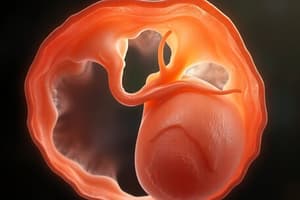
- Pharyngeal arches are bulges that form during week 4 of embryonic development.
- Each arch has a unique nerve supply, blood supply, and cartilage.
- These arches are fundamental for the development of facial structures and the oral cavity.
Pharyngeal Arch Derivatives
- 1st (Mandibular) Arch: Muscles of mastication, Merkel's cartilage, bones of the mandible, maxilla, zygomatic, and temporal bone, malleus, and incus of the ear.
- 2nd (Hyoid) Arch: Muscles of facial expression, Reichert's cartilage, part of the hyoid bone, styloid process, and stapes.
- 3rd Arch: Stylopharyngeal muscle, part of the hyoid bone, connective tissue of the thymus, and inferior parathyroid.
- 4th Arch: Laryngeal muscles, cartilage of the larynx, thyroid, corniculate, and cuneiform.
Key Phases of Embryogenesis and Development
- Early Embryogenesis (Weeks 1-4): Blastocyst formation, germ layer formation, neural crest cell migration, and pharyngeal arch development.
- Embryogenesis (Weeks 5-8): Development of facial structures, palate tongue, jaws, skull, etc.
- Fetal Development (Weeks 9 - Term): Growth and maturation of the fetus.
Overview of the Process
- Early Embryogenesis Weeks 2-4: Formation of the bilaminar embryonic disc.
- Embryogenesis Weeks 4-6: Formation of pharyngeal arches and the development of the oral cavity.
Oral Embryology
- The study of the development of oral tissues.
- Embryogenesis: The process of embryological development, forming tissues and organs.
- Embryonic Origins: The three tissue layers (ectoderm, mesoderm, endoderm) in the trilaminar embryonic disc that give rise to all human tissues.
Key Phases of Embryogenesis:
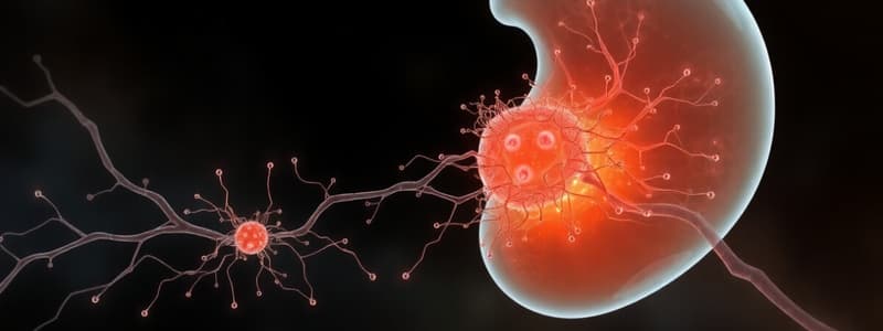
- Week 1: Formation of the blastocyst (70-100 cells) from a single cell zygote.
- Blastocoel forms the primary yolk sac
- Embryoblast forms the inner cluster of cells
- Trophoblast lines the cavity
- Week 2-3: Development of the bilaminar embryonic disc followed by the trilaminar embryonic disc
- Bilaminar embryonic disc consists of ectoderm and endoderm
- Trilaminar embryonic disc adds the mesoderm
- These three layers form all the tissues and organs in the body
- Week 4: Development of the neural crest cells and pharyngeal arches (branchial arches)
- Neural crest cells are crucial in the formation of cranial nerves, the peripheral nervous system, and connective tissues of the head, face, and oral cavity
- Pharyngeal Arches develop into various structures of the face and oral cavity
- Each pharyngeal arch has its own nerve and blood supply
Facial Development
- Weeks 4-6: Fusion of the frontonasal, maxillary, and mandibular prominences
- Primary Palate Formation (Weeks 6-7): Formed by the fusion of the frontonasal and medial nasal prominences
- Secondary Palate Formation (Weeks 7-8): The palatine shelves fuse with the primary palate as the tongue retracts, allowing space for the secondary palate to form
- Tongue Development (Weeks 4-7): Formed from the 1st, 2nd, and 4th pharyngeal arches
- Apoptosis separates the tongue from the floor of the mouth, leaving the frenulum as an anchor
Embryonic Origins & Structures
- Ectoderm: Forms oral epithelial cells, neuroectoderm, cranial nerves, sensory ganglia, and parts of the teeth (except enamel)
- Mesoderm: Forms connective tissue throughout the body, including the head, face, and oral cavity
- Endoderm: Contributes to the formation of the primitive gut
Pharyngeal Arches and Their Derivatives
- The pharyngeal arches form specific bones, muscles, nerves, and blood vessels
Developmental Anomalies
- Cleft Lip and Palate: The most common facial cleft, affecting about 1 in 700 births
- Results from the failure of fusion of facial prominences or palatine shelves
- Types:
- Cleft Lip: Failure of fusion of the medial nasal processes
- Cleft Palate: Failure of the palatine shelves to fuse
- Cleft Lip and Palate: Combination of the two
- Management: Early diagnosis and treatment involving multidisciplinary teams reduce complications. Patients may have increased risks of oral diseases.
Clinical Significance
- Interruptions in embryological development can lead to significant anomalies, such as:
- Cleft Lip/Palate: Can affect feeding, speech, and hearing if not treated
- Tongue Tied (Ankyloglossia): Failure of the tongue to separate properly from the floor of the mouth can affect feeding and speech
Development of the Skull
- The skull develops from neural crest cells and mesoderm, divided into three parts
- Cranial vault (brain case)
- Cranial base
- Facial bones
Cranial Vault
- Consists of flat bones that protect the brain.
- Bones are formed by intramembranous ossification, which involves the direct transformation of mesenchymal cells into bone without a cartilage intermediate
- The bones of the cranial vault (frontal, parietal, occipital) continue to grow and do not fully fuse until the child is around 6-7 years old, allowing the brain to grow
Cranial Base
- Forms the floor of the skull and supports the brain
- Develops via endochondral ossification, where cartilage is first laid down and then replaced by bone
- Bones like the sphenoid, ethmoid, and occipital bones originate from this process
Facial Bones
- Includes the maxilla, mandible, zygomatic bones, and parts of the nasal cavity
- These bones derive from the first and second pharyngeal arches
- Facial bones develop via intramembranous ossification, with the exception of parts of the mandible, which have contributions from Meckel's cartilage
Sutures and Fontanelles
- Sutures are fibrous joints between the bones of the skull that allow for movement and brain growth
- Fontanelles are soft spots on the infant's skull where sutures have not yet closed (e.g., the anterior fontanelle)
- These areas close around 18-24 months after birth
Development of the Alveolar Bone
- The alveolar bone forms the part of the maxilla and mandible that supports and surrounds the roots of the teeth.
- Its development is crucial for the proper anchorage and function of teeth
Formation of the Alveolar Process
- The alveolar bone develops from the maxillary and mandibular processes, starting around week 7 of embryonic development
- It forms by intramembranous ossification, with mesenchymal cells transforming into bone tissue
- The alveolar process creates sockets (alveoli) to house the roots of developing teeth
Tooth Eruption and Bone Remodeling
- As teeth begin to erupt, the alveolar bone undergoes continuous remodeling to accommodate the growing teeth
- During tooth eruption, bone resorption and deposition occur to allow the teeth to move into their correct positions within the jaws
- The periodontal ligament connects the alveolar bone to the teeth, a key role in stabilizing them during function
Alveolar Bone Loss
- Alveolar bone resorption occurs in conditions like periodontal disease, where inflammation leads to the loss of bone supporting the teeth, often resulting in tooth mobility or loss
- Proper oral hygiene and treatment can help prevent or slow the progression of bone loss
Studying That Suits You
Use AI to generate personalized quizzes and flashcards to suit your learning preferences.
Related Documents
Description
Explore the intricacies of early embryogenesis, focusing on the three germ layers: ectoderm, mesoderm, and endoderm. Learn about the important roles of neural crest cells, somite formation, and pharyngeal arches in embryonic development. This quiz covers pivotal concepts relevant to understanding the foundation of body structures.