Podcast
Questions and Answers
If a wavefront of depolarization moves directly away from a recording electrode, what will be the recorded ECG deflection?
If a wavefront of depolarization moves directly away from a recording electrode, what will be the recorded ECG deflection?
- A large positive deflection
- A large negative deflection
- No deflection (correct)
- A small deflection
Which of the following statements accurately describes the relationship between the orientation of the wavefront of depolarization and the recorded ECG deflection?
Which of the following statements accurately describes the relationship between the orientation of the wavefront of depolarization and the recorded ECG deflection?
- A wavefront moving parallel to the lead axis will result in the smallest deflection.
- A wavefront moving perpendicular to the lead axis will result in the largest deflection.
- A wavefront moving parallel to the lead axis will result in the largest deflection. (correct)
- The size of the deflection is independent of the wavefront orientation.
The normal activation sequence of the heart begins at the _______ and then progresses through the _______ to the ventricles.
The normal activation sequence of the heart begins at the _______ and then progresses through the _______ to the ventricles.
- SA node; AV node (correct)
- His-Purkinje system; AV node
- Ventricular muscle; atria
- AV node; SA node
What is the significance of the negative charges moving toward the positive electrode in the context of ECG recordings?
What is the significance of the negative charges moving toward the positive electrode in the context of ECG recordings?
Consider a wavefront of depolarization traveling directly towards the recording electrode (parallel to the lead axis). If the electrode is negative and the other electrode is positive, what will be the direction of ECG deflection?
Consider a wavefront of depolarization traveling directly towards the recording electrode (parallel to the lead axis). If the electrode is negative and the other electrode is positive, what will be the direction of ECG deflection?
What is the unit of measurement on the y-axis of an electrocardiogram (ECG)?
What is the unit of measurement on the y-axis of an electrocardiogram (ECG)?
What type of cellular event is recorded as an upward deflection on the ECG?
What type of cellular event is recorded as an upward deflection on the ECG?
What is the correct order of cardiac activation, starting with the pacemaker?
What is the correct order of cardiac activation, starting with the pacemaker?
Which part of the cardiac conduction system is characterized by the slowest conduction velocity?
Which part of the cardiac conduction system is characterized by the slowest conduction velocity?
Which of the following is NOT a piece of information that can be obtained from an ECG?
Which of the following is NOT a piece of information that can be obtained from an ECG?
What is the purpose of recording a base-apex ECG in horses?
What is the purpose of recording a base-apex ECG in horses?
What cellular event does the QRS complex on an ECG represent?
What cellular event does the QRS complex on an ECG represent?
Which of the following statements regarding cardiac tissue is FALSE?
Which of the following statements regarding cardiac tissue is FALSE?
Which of the following structures has the fastest conduction velocity?
Which of the following structures has the fastest conduction velocity?
What is the normal pacemaker rate of the SA node?
What is the normal pacemaker rate of the SA node?
Which of the following is a function of the AV node?
Which of the following is a function of the AV node?
Which of the following is NOT a characteristic of the His-Purkinje system?
Which of the following is NOT a characteristic of the His-Purkinje system?
Which of the following is a true statement about terminal Purkinje fibers?
Which of the following is a true statement about terminal Purkinje fibers?
In the standard lead II electrocardiogram, which electrode is placed on the right forelimb?
In the standard lead II electrocardiogram, which electrode is placed on the right forelimb?
What is the orientation of the lead axis in the base-apex lead?
What is the orientation of the lead axis in the base-apex lead?
What does the P wave on an electrocardiogram represent?
What does the P wave on an electrocardiogram represent?
Which of the following accurately describes the PR interval?
Which of the following accurately describes the PR interval?
What is the normal QRS complex duration in dogs?
What is the normal QRS complex duration in dogs?
Which of the following is the key difference between the QRS complex in small animals and large animals?
Which of the following is the key difference between the QRS complex in small animals and large animals?
What does the ST segment on an electrocardiogram represent?
What does the ST segment on an electrocardiogram represent?
Which of the following is a true statement about the T wave?
Which of the following is a true statement about the T wave?
What is the purpose of using a grounding electrode in electrocardiography?
What is the purpose of using a grounding electrode in electrocardiography?
When is the use of a base-apex lead particularly advantageous?
When is the use of a base-apex lead particularly advantageous?
What occurs when a resting myocyte is stimulated?
What occurs when a resting myocyte is stimulated?
What is the primary function of the atrial internodal tracts?
What is the primary function of the atrial internodal tracts?
How does depolarization spread from one heart cell to another?
How does depolarization spread from one heart cell to another?
What is the significance of the PR interval being prolonged?
What is the significance of the PR interval being prolonged?
What is necessary for the depolarization process to occur repeatedly?
What is necessary for the depolarization process to occur repeatedly?
When measuring the electrical field with ECG electrodes, where should the negative electrode be placed in relation to the wave of depolarization?
When measuring the electrical field with ECG electrodes, where should the negative electrode be placed in relation to the wave of depolarization?
What effect does a wavefront of negative extracellular charges moving toward the positive electrode have on the ECG reading?
What effect does a wavefront of negative extracellular charges moving toward the positive electrode have on the ECG reading?
What type of wave of electrical activity is primarily recorded by the electrocardiogram?
What type of wave of electrical activity is primarily recorded by the electrocardiogram?
In the context of the electrocardiogram, what does the positive portion of the electrical field represent?
In the context of the electrocardiogram, what does the positive portion of the electrical field represent?
What role do extracellular currents play in the depolarization process as measured by an electrocardiogram?
What role do extracellular currents play in the depolarization process as measured by an electrocardiogram?
What does a bifid (notched) P wave indicate in horses?
What does a bifid (notched) P wave indicate in horses?
Which of the following best describes the pattern of wave depolarization in the cardiac cycle?
Which of the following best describes the pattern of wave depolarization in the cardiac cycle?
In horses, what represents the negative electrode during an ECG?
In horses, what represents the negative electrode during an ECG?
What characterizes the QRS complex in healthy horses?
What characterizes the QRS complex in healthy horses?
What is a misleading characteristic of normal sinus rhythms in dairy cows?
What is a misleading characteristic of normal sinus rhythms in dairy cows?
During ECG, what represents the normal electrical activity of the heart?
During ECG, what represents the normal electrical activity of the heart?
What is NOT true regarding ventricular repolarization?
What is NOT true regarding ventricular repolarization?
What is the correct sequence of electrical activity in a normal cardiac cycle?
What is the correct sequence of electrical activity in a normal cardiac cycle?
Flashcards
Electrocardiogram (ECG)
Electrocardiogram (ECG)
A recording of the heart's electrical activity as seen from the surface of the body.
Wave/Complex in ECG
Wave/Complex in ECG
An upward or downward deflection on the ECG representing a change in electrical potential.
Depolarization
Depolarization
The moment when a heart cell's inside becomes more positive than the outside.
Repolarization
Repolarization
Signup and view all the flashcards
Voltage (mV) Axis
Voltage (mV) Axis
Signup and view all the flashcards
Time (sec) Axis
Time (sec) Axis
Signup and view all the flashcards
Limitations of ECG
Limitations of ECG
Signup and view all the flashcards
ECG and Heart Chamber Size
ECG and Heart Chamber Size
Signup and view all the flashcards
What is depolarization wavefront?
What is depolarization wavefront?
Signup and view all the flashcards
What is the role of the SA node?
What is the role of the SA node?
Signup and view all the flashcards
How does wavefront orientation affect ECG deflection?
How does wavefront orientation affect ECG deflection?
Signup and view all the flashcards
What is the normal activation sequence of the heart?
What is the normal activation sequence of the heart?
Signup and view all the flashcards
What is the role of the recording lead axis in an ECG?
What is the role of the recording lead axis in an ECG?
Signup and view all the flashcards
Wave of Depolarization
Wave of Depolarization
Signup and view all the flashcards
Wavefront Direction
Wavefront Direction
Signup and view all the flashcards
Upward Deflection on the ECG
Upward Deflection on the ECG
Signup and view all the flashcards
Electrical Field of the Heart
Electrical Field of the Heart
Signup and view all the flashcards
Repolarization is Essential
Repolarization is Essential
Signup and view all the flashcards
Cardiac Depolarization
Cardiac Depolarization
Signup and view all the flashcards
Ventricular Depolarization
Ventricular Depolarization
Signup and view all the flashcards
Ventricular Repolarization
Ventricular Repolarization
Signup and view all the flashcards
Atrial Depolarization
Atrial Depolarization
Signup and view all the flashcards
Normal Sinus Rhythm
Normal Sinus Rhythm
Signup and view all the flashcards
Bifid P Waves
Bifid P Waves
Signup and view all the flashcards
Negative QRS Complexes
Negative QRS Complexes
Signup and view all the flashcards
Base-Apex Lead
Base-Apex Lead
Signup and view all the flashcards
SA node pacemaker rate
SA node pacemaker rate
Signup and view all the flashcards
Conduction Velocity
Conduction Velocity
Signup and view all the flashcards
Normal activation sequence
Normal activation sequence
Signup and view all the flashcards
Pacemaker
Pacemaker
Signup and view all the flashcards
Pacemaker Rate
Pacemaker Rate
Signup and view all the flashcards
AV node
AV node
Signup and view all the flashcards
His-Purkinje system
His-Purkinje system
Signup and view all the flashcards
Atrial Muscle
Atrial Muscle
Signup and view all the flashcards
Bundle of His/Bundle Branches
Bundle of His/Bundle Branches
Signup and view all the flashcards
Terminal Purkinje Fibers
Terminal Purkinje Fibers
Signup and view all the flashcards
Electrode Placement
Electrode Placement
Signup and view all the flashcards
Lead
Lead
Signup and view all the flashcards
Lead II
Lead II
Signup and view all the flashcards
Base-to-Apex lead
Base-to-Apex lead
Signup and view all the flashcards
P-wave
P-wave
Signup and view all the flashcards
PR Interval
PR Interval
Signup and view all the flashcards
Study Notes
Electrocardiography Principles
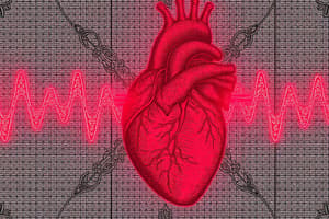
- Electrocardiogram (ECG or EKG) records the heart's electrical activity from the body surface.
- ECG records extracellular signals caused by the movement of depolarization/repolarization waves through cardiac myocytes.
- The graph displays voltage (mV, y-axis) over time (seconds, x-axis).
- Changes in voltage are recorded as waves/complexes (P, QRS, T).
Learning Objectives
- Label the x- and y-axes of an ECG.
- Describe cell surface events associated with upward deflections on an ECG.
- List the normal cardiac activation sequence from memory.
- For each component of the cardiac conduction system, describe relative conduction velocity and expected pacemaker activity.
- Describe the method for recording a 6-lead ECG in a dog and a base-apex ECG in a horse.
- Label individual waves on a normal ECG and explain the cellular events they represent.
Physiology Review: Basic Cardiac Electrophysiology
- Cardiac tissue is excitable.
- At rest, cardiac myocytes are polarized (negatively charged inside versus outside the membrane).
- When stimulated, resting myocytes depolarize (membrane polarity reverses).
- Depolarization spreads as a wave, triggering adjacent cells to depolarize.
- Extracellular currents associated with the depolarization wave are detected by the ECG.
- Cells must repolarize for the cycle to repeat.
Electrocardiography - (re)introduction
- ECG evaluation provides insights into heart rate, rhythm disturbances, conduction abnormalities, and relative chamber sizes (in smaller animals).
- ECG does not measure mechanical activity (contraction strength, etc).
Physiology Review: Normal Cardiac Conduction System
- The heart's electrical activity originates from the sinoatrial (SA) node (pacemaker).
- The sequence: SA node → internodal tracts → atrioventricular (AV) node → Bundle of His → right and left bundle branches → Purkinje fibers → ventricular muscle.
- Conduction velocities and pacemaker rates vary throughout these components.
- The SA node has a rate of 60-250 bpm.
- AV node has a rate of 40-60 bpm.
- His-Purkinje system has a rate of 20-40 bpm.
Physiology Review: Normal Cardiac Conduction System (Special Notes)
- Atrial internodal tracts transmit impulses rapidly between the SA and AV nodes, relatively resistant to hyperkalemia.
- The Bundle of His divides into the right and left bundle branches, which rapidly conduct impulses to terminal Purkinje fibers.
- Terminal Purkinje fibers rapidly conduct impulses throughout the endocardium of the ventricles.
Method for Recording a 6-Lead Surface ECG in Small Animals
- Patient in right lateral recumbency.
- Limbs parallel to one another, perpendicular to trunk.
- Calm environment, non-conducting surface to minimize artifacts.
- Use white/black electrodes on forelimbs and red/green electrodes on hindlimbs.
- Avoid contact between electrodes and trunk, and avoid electrode contact with each other.
Method for Recording a Surface "Base-to-Apex" ECG in Horses and Ruminants
- No universal lead system in large animals.
- Set machine to record in lead I (RA to LA).
- Place a white electrode on the right jugular furrow, a black electrode on the cardiac left apex.
- Lead axis is cranial-to-caudal, right-to-left.
The Normal (Lead II) ECG Tracing
- Depolarization is represented by upward deflections in Lead II.
- Depolarization proceeds from the right atrium to the left ventricle.
- Depolarized tissue is blue.
- Pink arrow signifies the direction of the depolarization wave.
Electrical Correlates of ECG Waves/Intervals
- P wave: Atrial depolarization, positive in lead II, frequently bifid (M-shaped) in horses.
- PR (PQ) interval: Atrial, AV node, and Bundle of His depolarization, <0.13 seconds (dog), < 0.09 seconds (cat).
- QRS complex: Ventricular depolarization, generally upright and narrow in small animals, <0.06 seconds (dog), <0.04 seconds (cat).
- ST segment: Isoelectric line connecting S and T waves, indicating ventricular cell depolarization is complete and no current flows in lead II.
- T wave: Ventricular repolarization, a complex process, important for function of the heart.
Normal Base-Apex ECG in Horses/Ruminants
- Bifid (notched) P waves are normal in horses.
- Negative QRS complexes are normal in horses and cattle.
Studying That Suits You
Use AI to generate personalized quizzes and flashcards to suit your learning preferences.
Related Documents
Description
Test your knowledge of electrocardiography principles with this quiz! It covers the basics of ECG interpretation, including the labeling of the x- and y-axes, the cardiac activation sequence, and individual wave representation. Additionally, you will explore methods for recording ECGs in different animals.