Podcast
Questions and Answers
Electricity plays an important role in medicine.
Electricity plays an important role in medicine.
True (A)
What are the two aspects of electricity and magnetism in medicine?
What are the two aspects of electricity and magnetism in medicine?
- Applications of electricity and magnetism to the surface of the body. (correct)
- Applications of electricity and magnetism to the interior of the body.
- Electrical and magnetic effects generated inside the body. (correct)
- Electrical and magnetic effects generated outside the body.
What serves as the control and operation of nerves, muscles, and organs?
What serves as the control and operation of nerves, muscles, and organs?
Electricity generated inside the body
What is the fundamental unit of the nervous system?
What is the fundamental unit of the nervous system?
What are the two parts that the nervous system can be divided into?
What are the two parts that the nervous system can be divided into?
What does the central nervous system consist of?
What does the central nervous system consist of?
What are the two types of nerve fibers in the peripheral nervous system?
What are the two types of nerve fibers in the peripheral nervous system?
The control of the autonomic nervous system is voluntary.
The control of the autonomic nervous system is voluntary.
What are the functions of a neuron?
What are the functions of a neuron?
Which part of the neuron receives electrical messages from other neurons?
Which part of the neuron receives electrical messages from other neurons?
The soma of a neuron contains the nucleus.
The soma of a neuron contains the nucleus.
Dendrites are specialized extensions of the cell body that carry information to the cell body from other cells.
Dendrites are specialized extensions of the cell body that carry information to the cell body from other cells.
What is the function of the axon?
What is the function of the axon?
Axons are typically 1 meter long.
Axons are typically 1 meter long.
Where are the signals from a neuron transmitted?
Where are the signals from a neuron transmitted?
What is the basic unit of communication in the brain?
What is the basic unit of communication in the brain?
A chemical neurotransmitter is released from one cell and binds to receptors on the second cell.
A chemical neurotransmitter is released from one cell and binds to receptors on the second cell.
What is the name of the potential difference created between the two environments of a cell?
What is the name of the potential difference created between the two environments of a cell?
The bio-potential can be measured using electrodes.
The bio-potential can be measured using electrodes.
What are the two types of bio potential?
What are the two types of bio potential?
The inside of a cell is typically 60 to 90 mV more positive than the outside.
The inside of a cell is typically 60 to 90 mV more positive than the outside.
What is the term used to describe the large momentary change in resting potential that occurs when a neuron is stimulated?
What is the term used to describe the large momentary change in resting potential that occurs when a neuron is stimulated?
The action potential propagates along the axon.
The action potential propagates along the axon.
The action potential is the major method of transmission of signals within the body.
The action potential is the major method of transmission of signals within the body.
What is the record of the potentials from muscles during movement called?
What is the record of the potentials from muscles during movement called?
A muscle is made up of many motor units.
A muscle is made up of many motor units.
What does a motor unit consist of?
What does a motor unit consist of?
Muscle action is initiated by an action potential that travels along an axon and is transmitted across the motor end plates into the muscle fibers.
Muscle action is initiated by an action potential that travels along an axon and is transmitted across the motor end plates into the muscle fibers.
It is easy to isolate a single muscle fiber during an EMG examination.
It is easy to isolate a single muscle fiber during an EMG examination.
What are the two methods of measuring electrical signals during an EMG examination?
What are the two methods of measuring electrical signals during an EMG examination?
Action potential appears in EMG after a latency period.
Action potential appears in EMG after a latency period.
EMGs of symmetrical muscles are compared to each other, or to those of normal individuals, to determine the action potential and latency periods.
EMGs of symmetrical muscles are compared to each other, or to those of normal individuals, to determine the action potential and latency periods.
Nerve damage may result in a decrease in the velocity of the action potential.
Nerve damage may result in a decrease in the velocity of the action potential.
What is the recording of signals from the brain called?
What is the recording of signals from the brain called?
EEG signals are primarily due to the electrical activity of the neurons in the cortex of the brain.
EEG signals are primarily due to the electrical activity of the neurons in the cortex of the brain.
Electrodes for recording EEG signals are attached to the head at locations that are independent of what part of the brain is to be studied.
Electrodes for recording EEG signals are attached to the head at locations that are independent of what part of the brain is to be studied.
The reference electrode in an EEG recording is usually attached to the ear (A1 or A2).
The reference electrode in an EEG recording is usually attached to the ear (A1 or A2).
Asymmetrical brain activity between the left and right sides is often a sign of brain disease.
Asymmetrical brain activity between the left and right sides is often a sign of brain disease.
The amplitude of EEG signals is high (about 50 μV)
The amplitude of EEG signals is high (about 50 μV)
Interference from external electrical signals often causes serious problems in EEG signal processing.
Interference from external electrical signals often causes serious problems in EEG signal processing.
The frequencies of the EEG signals seem to be dependent upon the physical activity of the subject.
The frequencies of the EEG signals seem to be dependent upon the physical activity of the subject.
As a person becomes drowsy, the frequencies from 8 to 12 Hz (alpha wave) dominate the EEG.
As a person becomes drowsy, the frequencies from 8 to 12 Hz (alpha wave) dominate the EEG.
The amplitude of the alpha wave decreases as a person moves from light sleep to deeper sleep.
The amplitude of the alpha wave decreases as a person moves from light sleep to deeper sleep.
Paradoxical sleep is characterized by rapid eye movement (REM) and is associated with dreaming.
Paradoxical sleep is characterized by rapid eye movement (REM) and is associated with dreaming.
What is the sensorimotor rhythm (SMR)?
What is the sensorimotor rhythm (SMR)?
The SMR appears in recordings of EEG, MEG, and ECG over the sensorimotor cortex.
The SMR appears in recordings of EEG, MEG, and ECG over the sensorimotor cortex.
Evoked responses are signals that result when the brain receives external stimuli.
Evoked responses are signals that result when the brain receives external stimuli.
EEGs typically show responses to all pulses of a stimulus.
EEGs typically show responses to all pulses of a stimulus.
What is the recording of potential changes produced by the eye when the retina is exposed to a flash of light called?
What is the recording of potential changes produced by the eye when the retina is exposed to a flash of light called?
The ERG is more complicated than a nerve axon signal because it is the sum of many effects taking place within the eye.
The ERG is more complicated than a nerve axon signal because it is the sum of many effects taking place within the eye.
The B wave of the ERG is the most interesting clinically, arising in the retina.
The B wave of the ERG is the most interesting clinically, arising in the retina.
The B wave is absent in the ERG of patients with inflammation of the retina.
The B wave is absent in the ERG of patients with inflammation of the retina.
What is the recording of potential changes due to eye movements called?
What is the recording of potential changes due to eye movements called?
A pair of electrodes is attached near the eye to measure the EOG.
A pair of electrodes is attached near the eye to measure the EOG.
The EOG provides information on the orientation of the eye and its angular velocity.
The EOG provides information on the orientation of the eye and its angular velocity.
The heart's electrical system controls all the events that occur when the heart pumps blood.
The heart's electrical system controls all the events that occur when the heart pumps blood.
What is the heart's electrical system also known as?
What is the heart's electrical system also known as?
What is the heart test called that is a graphical picture of the heart's electrical activity?
What is the heart test called that is a graphical picture of the heart's electrical activity?
What are the three main parts of the heart's electrical system?
What are the three main parts of the heart's electrical system?
The SA node is located in the left atrium.
The SA node is located in the left atrium.
The AV node is located on the interatrial septum close to the tricuspid valve.
The AV node is located on the interatrial septum close to the tricuspid valve.
The His-Purkinje system is located along the walls of the heart's atria.
The His-Purkinje system is located along the walls of the heart's atria.
A heartbeat is a complex cycle of electrical conductive events.
A heartbeat is a complex cycle of electrical conductive events.
Each heartbeat has two basic parts: diastole and systole.
Each heartbeat has two basic parts: diastole and systole.
During diastole, the heart's atria and ventricles contract.
During diastole, the heart's atria and ventricles contract.
At the end of diastole, the heart's atria contract and pump blood into the ventricles.
At the end of diastole, the heart's atria contract and pump blood into the ventricles.
The ventricles contract first, followed by the atria, during each heartbeat.
The ventricles contract first, followed by the atria, during each heartbeat.
Ventricular systole involves the contraction of the ventricles, pumping blood out of the heart.
Ventricular systole involves the contraction of the ventricles, pumping blood out of the heart.
The SA node and the rest of the conduction system are at rest during the heartbeat.
The SA node and the rest of the conduction system are at rest during the heartbeat.
The SA node initiates the action potential, which causes depolarization of nerves and muscles in both atria.
The SA node initiates the action potential, which causes depolarization of nerves and muscles in both atria.
The SA node is also known as the pacemaker of the heart.
The SA node is also known as the pacemaker of the heart.
The delay of approximately 100 ms after the electrical signal reaches the AV node allows the atria to complete pumping blood before the impulse is transmitted to the atrioventricular bundle.
The delay of approximately 100 ms after the electrical signal reaches the AV node allows the atria to complete pumping blood before the impulse is transmitted to the atrioventricular bundle.
The impulse travels through the atrioventricular bundle, bundle branches, and Purkinje fibers, reaching the contractile fibers of the ventricle.
The impulse travels through the atrioventricular bundle, bundle branches, and Purkinje fibers, reaching the contractile fibers of the ventricle.
Ventricular contraction forces blood into the two systems, then ventricles are repolarized, completing the heartbeat cycle.
Ventricular contraction forces blood into the two systems, then ventricles are repolarized, completing the heartbeat cycle.
The normal heart rate at rest ranges between 60 and 100 beats per minute on average.
The normal heart rate at rest ranges between 60 and 100 beats per minute on average.
The SA node is responsible for adjusting the heart rate to meet the body's needs.
The SA node is responsible for adjusting the heart rate to meet the body's needs.
During periods of exercise, the heart rate decreases to conserve energy for physical exertion.
During periods of exercise, the heart rate decreases to conserve energy for physical exertion.
During periods of rest or sleep, the heart rate increases to meet the reduced oxygen demands of the body.
During periods of rest or sleep, the heart rate increases to meet the reduced oxygen demands of the body.
Changes in heart rate are a normal part of the heart's effort to meet the needs of the body.
Changes in heart rate are a normal part of the heart's effort to meet the needs of the body.
In a normal ECG waveform, the P wave represents ventricular depolarization
In a normal ECG waveform, the P wave represents ventricular depolarization
The QRS complex represents ventricular depolarization.
The QRS complex represents ventricular depolarization.
The T wave represents atrial repolarization.
The T wave represents atrial repolarization.
The P-R interval is measured from the start of the P wave to the start of the QRS complex.
The P-R interval is measured from the start of the P wave to the start of the QRS complex.
The Q-T interval is measured from the start of the QRS complex to the end of the T wave.
The Q-T interval is measured from the start of the QRS complex to the end of the T wave.
The S-T segment is measured from the end of the QRS complex to the start of the T wave and represents the period when all cells are normally depolarized during that phase.
The S-T segment is measured from the end of the QRS complex to the start of the T wave and represents the period when all cells are normally depolarized during that phase.
The P-R interval is typically between 0.12-0.2 seconds (3-5 small squares) on a standard ECG paper.
The P-R interval is typically between 0.12-0.2 seconds (3-5 small squares) on a standard ECG paper.
The QRS complex duration is typically less than or equal to 0.1 seconds (2.5 small squares) on a standard ECG paper.
The QRS complex duration is typically less than or equal to 0.1 seconds (2.5 small squares) on a standard ECG paper.
The Q-T interval corrected for heart rate (QTc) is typically less than or equal to 0.44 seconds.
The Q-T interval corrected for heart rate (QTc) is typically less than or equal to 0.44 seconds.
The location of the electrodes used for ECG recording does not significantly affect the measurements obtained.
The location of the electrodes used for ECG recording does not significantly affect the measurements obtained.
The most common electrode placement for ECG recording is in the left arm (LA), right arm (RA), and left leg (LL).
The most common electrode placement for ECG recording is in the left arm (LA), right arm (RA), and left leg (LL).
A standard ECG graphing typically includes 6 sections, all of which are in the frontal plane.
A standard ECG graphing typically includes 6 sections, all of which are in the frontal plane.
The potential difference between any two electrodes provides information about the relative amplitude and direction of the electric dipole vector in the frontal plane.
The potential difference between any two electrodes provides information about the relative amplitude and direction of the electric dipole vector in the frontal plane.
What does lead I measure in an ECG?
What does lead I measure in an ECG?
The aVR augmented lead measures the potential difference between the right arm and the center of the resistance between the left leg and left arm.
The aVR augmented lead measures the potential difference between the right arm and the center of the resistance between the left leg and left arm.
The aVL augmented lead measures the potential difference between the left arm and the center of the resistance between the left leg and right arm.
The aVL augmented lead measures the potential difference between the left arm and the center of the resistance between the left leg and right arm.
The aVF augmented lead measures the potential difference between the left leg and the center of the resistance between the right arm and left arm.
The aVF augmented lead measures the potential difference between the left leg and the center of the resistance between the right arm and left arm.
The chest electrodes are typically placed in the 4th intercostal space.
The chest electrodes are typically placed in the 4th intercostal space.
V1 is placed in the 4th intercostal space, right of the sternum.
V1 is placed in the 4th intercostal space, right of the sternum.
V3 is placed between V1 and V3.
V3 is placed between V1 and V3.
V4 is placed in the fifth intercostal space in the nipple line.
V4 is placed in the fifth intercostal space in the nipple line.
V6 is placed in the mid axillary line on the same height as V4, but not necessarily in the 5th intercostal space.
V6 is placed in the mid axillary line on the same height as V4, but not necessarily in the 5th intercostal space.
The ECG can be used to diagnose problems with the heart, including arrhythmias.
The ECG can be used to diagnose problems with the heart, including arrhythmias.
Arrhythmias are an irregular pumping pattern which is quite common in young people and often exacerbated by exercise.
Arrhythmias are an irregular pumping pattern which is quite common in young people and often exacerbated by exercise.
An ECG can reveal blockages in the blood supply to the heart, which can lead to myocardial infarction (heart attack).
An ECG can reveal blockages in the blood supply to the heart, which can lead to myocardial infarction (heart attack).
Fibrillation is a condition where there is no coordinated pumping activity, which can lead to death if left untreated.
Fibrillation is a condition where there is no coordinated pumping activity, which can lead to death if left untreated.
Flashcards
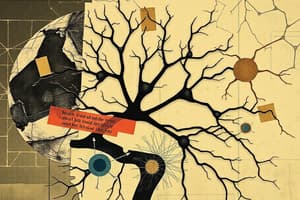
Electricity in medicine
Electricity in medicine
Electricity plays a crucial role in operating nerves, muscles, and organs.
Nervous System
Nervous System
The system that controls nearly every body function through signaling from the brain and nerves.
Neuron
Neuron
aashjhfd
Central Nervous System
Central Nervous System
Signup and view all the flashcards
Autonomic Nervous System
Autonomic Nervous System
Signup and view all the flashcards
Dendrites
Dendrites
Signup and view all the flashcards
Soma (Cell body)
Soma (Cell body)
Signup and view all the flashcards
Axon
Axon
Signup and view all the flashcards
Axon Terminals
Axon Terminals
Signup and view all the flashcards
Synapse
Synapse
Signup and view all the flashcards
Bio-potential
Bio-potential
Signup and view all the flashcards
Resting Potential
Resting Potential
Signup and view all the flashcards
Action Potential
Action Potential
Signup and view all the flashcards
Electromyogram (EMG)
Electromyogram (EMG)
Signup and view all the flashcards
Latency Period in EMG
Latency Period in EMG
Signup and view all the flashcards
Electroencephalogram (EEG)
Electroencephalogram (EEG)
Signup and view all the flashcards
Alpha Waves
Alpha Waves
Signup and view all the flashcards
Evoked Responses
Evoked Responses
Signup and view all the flashcards
Electroretinogram (ERG)
Electroretinogram (ERG)
Signup and view all the flashcards
Electrooculogram (EOG)
Electrooculogram (EOG)
Signup and view all the flashcards
Electrocardiogram (ECG)
Electrocardiogram (ECG)
Signup and view all the flashcards
SA Node
SA Node
Signup and view all the flashcards
Atrial Systole
Atrial Systole
Signup and view all the flashcards
Ventricular Systole
Ventricular Systole
Signup and view all the flashcards
Normal Heart Rate
Normal Heart Rate
Signup and view all the flashcards
P Wave in ECG
P Wave in ECG
Signup and view all the flashcards
QRS Complex in ECG
QRS Complex in ECG
Signup and view all the flashcards
T Wave in ECG
T Wave in ECG
Signup and view all the flashcards
Arrhythmia
Arrhythmia
Signup and view all the flashcards
Myocardial Infarction
Myocardial Infarction
Signup and view all the flashcards
Fibrillation
Fibrillation
Signup and view all the flashcards
Study Notes
Electricity Within The Body
- Electricity and magnetism have been observed since ancient times, playing a crucial role in medicine
- Two aspects of electricity and magnetism in medicine:
- Electrical and magnetic effects generated inside the body
- Applications of electricity and magnetism to the surface of the body
- Electricity generated inside the body controls and operates nerves, muscles, and organs
- The nervous system is fundamental to nearly every body function, acting as a central computer processing internal and external signals
- The nervous system is divided into two parts:
- Central nervous system (brain, spinal cord, peripheral nerves)
- Autonomic nervous system (controls internal organs)
The Neuron
- Neuron: the basic structural unit of the nervous system; specialized for receiving, interpreting, and transmitting electrical signals
- Structure:
- Dendrites: short, branched, unmyelinated parts that receive signals from other neurons
- Soma (cell body): contains the nucleus and associated intracellular structures; receives signals from dendrites
- Axon (nerve fiber): carries electrical signals away from the cell body to axon terminals; typically about 1 meter long
- Axon terminals: transmit signals to other neurons or muscle/glands
- Synapses: the basic unit of communication in the brain; chemical neurotransmitters relay signals between neurons
Electrical Potentials of Nerves
- A cell membrane separates the intracellular and extracellular environments, each with different concentrations of ions (chemicals)
- This difference in ion concentration leads to a potential difference (biopotential, measured through electrodes and amplifiers); necessary for studying heart, brain, muscles, etc.
- Two types of biopotentials:
- Resting potential: a stable voltage difference across the neuron membrane
- Action potential: a temporary, significant change in membrane voltage that rapidly propagates along the axon
Electrical Signals From Muscles (EMG)
- EMG: the recording of potentials from muscles during movement
- Muscles are made of motor units, each comprising a neuron and many muscle fibers
- Muscle contraction is initiated by action potentials that travel along the neuron to motor end plates, causing muscles to contract
- EMG electrodes measure electrical activity from several fibers to study muscle function. There are surface and concentric needle types.
Electrical Signals From The Brain (EEG)
- EEG: measures electrical activity from neurons in the brain's cortex, resulting in complex electrical signals.
- Electrodes are used on the scalp to collect the electrical signals for analysis.
- EEG can be used to detect brain diseases or abnormalities (e.g., asymmetrical activity)
- External electrical interference can be a problem during EEG signal processing.
Electrical Signals From The Eye (ERG)
- ERG: measures the electrical signals produced when the retina in the eye is exposed to light.
- Two electrodes are used (one in a contact lens and the other external), to measure the electrical potential change.
- ERG waves are important for retinal health analysis.
- Clinical uses help detect inflammatory diseases such a inflammation of the retina
Electrical Signals From The Eye (EOG)
- EOG: measures electrical signals produced due to eye movement.
- Two electrodes placed around the eyes; used to measure eye movement related activity.
- EOG used in clinical diagnosis of eye related conditions.
Electrical Signals From The Heart (ECG)
- ECG: provides a graphical record of the heart's electrical activity, showing the cardiac conduction system activity.
- The heart has three main parts to its electrical system: Sinoatrial (SA) node, atrioventricular (AV) node and His-Purkinje system
- The different waves in an ECG represent different phases of the heart's electrical activity (e.g., P wave for atrial depolarization, QRS complex for ventricular depolarization, T wave for ventricular repolarization)
- Heart rate can adjust according to body's needs/physical activity
Heartbeat Conduction Cycle
- A heartbeat is a complete cycle of electrical events, involving the heart's chambers relaxation and contraction to pump blood
- This cycle includes diastole (relaxation) for filling of chambers and systole (contraction) for pumping
ECG Normal Values
- Specific time frames for different components of the ECG are characteristic of a healthy heart
- Certain measurement values of ECG waves should follow certain norms, otherwise medical interventions are necessary
Surface Electrodes
- ECG electrodes measure the potential differences between different parts of the body to create a graphical representation of the heart's electrical activity
- Several electrode positions (frontal plane and chest electrodes)
- The amplitude/direction of the electric dipole vector between different electrodes provides important signal information; the ECG measurement is not only an absolute value, but also relative between electrodes.
Other Diagnostic Notes
- ECG can be used to detect medical conditions such as arrhythmia, heart attack and fibrillation.
Studying That Suits You
Use AI to generate personalized quizzes and flashcards to suit your learning preferences.