Podcast
Questions and Answers
What is the recommended initial approach to manage a complex or unstable elbow dislocation?
What is the recommended initial approach to manage a complex or unstable elbow dislocation?
- Immediate passive range of motion exercises
- Open reduction with possible internal fixation (correct)
- Closed reduction without fixation
- Application of a splint for 8 weeks
Which technique is utilized for the reduction of a posterior elbow dislocation?
Which technique is utilized for the reduction of a posterior elbow dislocation?
- Grasp the patient's hand with fingers interlocked (correct)
- Utilize a traction device on the forearm
- Push upward on the patient's distal humerus
- Position the patient face down
What is the recommended duration for splinting after a simple elbow dislocation?
What is the recommended duration for splinting after a simple elbow dislocation?
- 10 to 14 days
- 2 to 4 weeks
- 3 days
- 5 to 7 days (correct)
Why should passive range of motion exercises be avoided in the early stages of rehabilitation for an elbow dislocation?
Why should passive range of motion exercises be avoided in the early stages of rehabilitation for an elbow dislocation?
When can strengthening exercises typically commence after an elbow dislocation?
When can strengthening exercises typically commence after an elbow dislocation?
What is the primary cause of radial head fractures?
What is the primary cause of radial head fractures?
What is a common sign of a radial head fracture?
What is a common sign of a radial head fracture?
Which movements should be avoided during the first two weeks of rehabilitation after an elbow dislocation?
Which movements should be avoided during the first two weeks of rehabilitation after an elbow dislocation?
What percentage of all elbow injuries do elbow dislocations constitute?
What percentage of all elbow injuries do elbow dislocations constitute?
Which type of elbow dislocation is most commonly associated with fractures?
Which type of elbow dislocation is most commonly associated with fractures?
In the context of elbow dislocations, what is the 'terrible triad'?
In the context of elbow dislocations, what is the 'terrible triad'?
What is the recommended treatment for a simple posterior elbow dislocation?
What is the recommended treatment for a simple posterior elbow dislocation?
What primarily causes posterior elbow dislocations in children under 10 years?
What primarily causes posterior elbow dislocations in children under 10 years?
Which of the following is NOT typically associated with a complex elbow dislocation?
Which of the following is NOT typically associated with a complex elbow dislocation?
What is the classification of simple dislocations based on the direction of the dislocated ulna?
What is the classification of simple dislocations based on the direction of the dislocated ulna?
Which nerve injury might be associated with a complex elbow dislocation?
Which nerve injury might be associated with a complex elbow dislocation?
What is the recommended treatment for a Type I radial head fracture?
What is the recommended treatment for a Type I radial head fracture?
Which classification of radial head fractures involves a complete articular fracture with severe comminution?
Which classification of radial head fractures involves a complete articular fracture with severe comminution?
What is the main risk associated with starting physical therapy too early after a radial head fracture?
What is the main risk associated with starting physical therapy too early after a radial head fracture?
What indicates the need for ORIF in the treatment of a Monteggia fracture?
What indicates the need for ORIF in the treatment of a Monteggia fracture?
In a Galeazzi fracture-dislocation, which anatomical area is primarily involved?
In a Galeazzi fracture-dislocation, which anatomical area is primarily involved?
During which phase of rehabilitation for a 'both bone forearm fracture' is active and active-assisted ROM of the elbow, forearm, and wrist started?
During which phase of rehabilitation for a 'both bone forearm fracture' is active and active-assisted ROM of the elbow, forearm, and wrist started?
What should a patient avoid doing during Phase II of rehabilitation for a forearm fracture?
What should a patient avoid doing during Phase II of rehabilitation for a forearm fracture?
What is the minimal time frame recommended before starting passive movement in the elbow or radio ulnar joint after a radial head fracture?
What is the minimal time frame recommended before starting passive movement in the elbow or radio ulnar joint after a radial head fracture?
What is a characteristic feature of complex regional pain syndrome 1 (CRPS 1)?
What is a characteristic feature of complex regional pain syndrome 1 (CRPS 1)?
Which of the following is NOT a clinical symptom of CRPS?
Which of the following is NOT a clinical symptom of CRPS?
What is the main goal of physical therapy in treating CRPS?
What is the main goal of physical therapy in treating CRPS?
Which type of physical therapy technique encourages the use of the unaffected limb's reflection?
Which type of physical therapy technique encourages the use of the unaffected limb's reflection?
Which treatment modality is indicated for an undisplaced fracture?
Which treatment modality is indicated for an undisplaced fracture?
What is the typical cast position for an undisplaced fracture?
What is the typical cast position for an undisplaced fracture?
What does desensitization therapy consist of?
What does desensitization therapy consist of?
What is the focus during the early rehabilitation phase following a fracture?
What is the focus during the early rehabilitation phase following a fracture?
What is the standard initial treatment for an undisplaced scaphoid fracture?
What is the standard initial treatment for an undisplaced scaphoid fracture?
What is the primary risk associated with fractures of the proximal pole of the scaphoid?
What is the primary risk associated with fractures of the proximal pole of the scaphoid?
In the treatment of a minimally displaced scaphoid fracture, what follows the initial long arm thumb spica cast immobilization?
In the treatment of a minimally displaced scaphoid fracture, what follows the initial long arm thumb spica cast immobilization?
Which imaging method is most reliable for confirming the healing of a scaphoid fracture?
Which imaging method is most reliable for confirming the healing of a scaphoid fracture?
Which treatment is recommended for displaced or unstable scaphoid fractures?
Which treatment is recommended for displaced or unstable scaphoid fractures?
What complication is most associated with lunate fractures?
What complication is most associated with lunate fractures?
Which method is NOT commonly used in the treatment of scaphoid fractures?
Which method is NOT commonly used in the treatment of scaphoid fractures?
What is a characteristic of a Bennett's fracture?
What is a characteristic of a Bennett's fracture?
What is the typical management approach for a Smith fracture?
What is the typical management approach for a Smith fracture?
What is the primary distinguishing characteristic of a Barton’s Fracture?
What is the primary distinguishing characteristic of a Barton’s Fracture?
What is the recommended early mobilization timeframe after surgery for a wrist fracture managed with internal fixation?
What is the recommended early mobilization timeframe after surgery for a wrist fracture managed with internal fixation?
Which of the following correctly describes the mechanism of injury for a Radial Styloid Fracture?
Which of the following correctly describes the mechanism of injury for a Radial Styloid Fracture?
What is a typical symptom of a Scaphoid fracture?
What is a typical symptom of a Scaphoid fracture?
What is the expected range of time for regaining the majority of wrist range of motion post-operatively?
What is the expected range of time for regaining the majority of wrist range of motion post-operatively?
What type of wrist movements should be prioritized during rehabilitation after internal fixation?
What type of wrist movements should be prioritized during rehabilitation after internal fixation?
Which of the following statements regarding a Colles' fracture is accurate?
Which of the following statements regarding a Colles' fracture is accurate?
Flashcards
Anterior Elbow Dislocation
Anterior Elbow Dislocation
This type of elbow dislocation occurs when the radius and ulna are pushed forward (anteriorly) relative to the humerus.
Posterior Elbow Dislocation (PED)
Posterior Elbow Dislocation (PED)
This type of elbow dislocation is much more common and occurs when the radius and ulna are pushed backward (posteriorly) relative to the humerus.
Simple vs. Complex Elbow Dislocations
Simple vs. Complex Elbow Dislocations
Elbow dislocations can be categorized as simple or complex. Simple dislocations involve ligament damage without any associated fractures. Complex dislocations, however, involve both ligament damage and fractures.
Posterior Elbow dislocation (PED)
Posterior Elbow dislocation (PED)
Signup and view all the flashcards
Terrible Triad Elbow Dislocation
Terrible Triad Elbow Dislocation
Signup and view all the flashcards
Treatment of Simple Posterior Elbow Dislocation
Treatment of Simple Posterior Elbow Dislocation
Signup and view all the flashcards
Common Mechanism of PED (PED)
Common Mechanism of PED (PED)
Signup and view all the flashcards
Complex Elbow Dislocations
Complex Elbow Dislocations
Signup and view all the flashcards
Palm-Palm Technique
Palm-Palm Technique
Signup and view all the flashcards
Early Active ROM for Elbow Dislocation
Early Active ROM for Elbow Dislocation
Signup and view all the flashcards
Hinged Elbow Brace
Hinged Elbow Brace
Signup and view all the flashcards
Radial Head Fracture
Radial Head Fracture
Signup and view all the flashcards
Valgus Stress on the Elbow
Valgus Stress on the Elbow
Signup and view all the flashcards
Strengthening Exercises for Elbow Dislocation
Strengthening Exercises for Elbow Dislocation
Signup and view all the flashcards
Elbow Flexion and Extension Recovery
Elbow Flexion and Extension Recovery
Signup and view all the flashcards
Monteggia fracture-dislocation
Monteggia fracture-dislocation
Signup and view all the flashcards
Galeazzi fracture-dislocation
Galeazzi fracture-dislocation
Signup and view all the flashcards
Type I Radial Head Fracture
Type I Radial Head Fracture
Signup and view all the flashcards
Type III Radial Head Fracture
Type III Radial Head Fracture
Signup and view all the flashcards
Type IV Radial Head Fracture
Type IV Radial Head Fracture
Signup and view all the flashcards
Immobilization
Immobilization
Signup and view all the flashcards
Open Reduction and Internal Fixation (ORIF)
Open Reduction and Internal Fixation (ORIF)
Signup and view all the flashcards
Radial Head Resection
Radial Head Resection
Signup and view all the flashcards
Complex Regional Pain Syndrome (CRPS)
Complex Regional Pain Syndrome (CRPS)
Signup and view all the flashcards
Type 1 CRPS
Type 1 CRPS
Signup and view all the flashcards
Type 2 CRPS
Type 2 CRPS
Signup and view all the flashcards
Mirror Therapy
Mirror Therapy
Signup and view all the flashcards
Desensitization
Desensitization
Signup and view all the flashcards
Auto-passive Exercises
Auto-passive Exercises
Signup and view all the flashcards
Transcutaneous Electrical Nerve Stimulation (TENS)
Transcutaneous Electrical Nerve Stimulation (TENS)
Signup and view all the flashcards
Colles Cast
Colles Cast
Signup and view all the flashcards
Smith Fracture
Smith Fracture
Signup and view all the flashcards
Barton's Fracture Dislocation
Barton's Fracture Dislocation
Signup and view all the flashcards
Radial Styloid Fracture (Chauffeur's Fracture)
Radial Styloid Fracture (Chauffeur's Fracture)
Signup and view all the flashcards
Scaphoid Fracture
Scaphoid Fracture
Signup and view all the flashcards
Early Mobilization after Wrist Fracture Surgery
Early Mobilization after Wrist Fracture Surgery
Signup and view all the flashcards
Wrist ROM Restoration
Wrist ROM Restoration
Signup and view all the flashcards
Strengthening Exercises after Wrist Fracture
Strengthening Exercises after Wrist Fracture
Signup and view all the flashcards
Radial Artery Blood Supply to Scaphoid
Radial Artery Blood Supply to Scaphoid
Signup and view all the flashcards
Scaphoid Necrosis: Why?
Scaphoid Necrosis: Why?
Signup and view all the flashcards
Immobilization for Scaphoid Fractures
Immobilization for Scaphoid Fractures
Signup and view all the flashcards
Scaphoid Fracture Healing Times
Scaphoid Fracture Healing Times
Signup and view all the flashcards
Lunate Fracture & Avascular Necrosis
Lunate Fracture & Avascular Necrosis
Signup and view all the flashcards
Assessing Lunate Fractures
Assessing Lunate Fractures
Signup and view all the flashcards
Lunate Fracture Treatment
Lunate Fracture Treatment
Signup and view all the flashcards
Bennett's Fracture: Intra-articular
Bennett's Fracture: Intra-articular
Signup and view all the flashcards
Bennett's Fracture: Mechanism & Description
Bennett's Fracture: Mechanism & Description
Signup and view all the flashcards
Study Notes
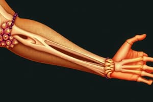
Upper Limb Fractures & Dislocations
- Elbow dislocations account for 10% to 25% of elbow injuries in adults; second only to shoulder dislocations.
- Simple elbow dislocations involve only ligament damage without fractures.
- Posterior elbow dislocations are the most common and are further categorized by the direction of the dislocated ulna (posterior, posteromedial, posterolateral, direct lateral).
- Complex elbow dislocations include associated bone fractures, commonly the radial head, coronoid process of the ulna, and olecranon.
- The "terrible triad" elbow dislocation damages the coronoid process, radial head, and posterior lateral elbow joint.
- Nerve or blood vessel injuries are possible complications of elbow dislocations (ulnar/median neuropraxia, possible brachial artery injury).
- Ulnar collateral ligament tears can accompany elbow dislocations.
Elbow Dislocation Mechanisms
- In children under ten, posterior elbow dislocations (PEDs) are most frequent, often caused by falls on outstretched hands.
- Forceful axial compression, valgus stress, arm abduction, and forearm supination contribute to posterior dislocations.
- Anterior dislocations result from direct force to the posterior forearm with a flexed elbow.
Elbow Dislocation Treatment
- Simple posterior dislocations typically involve closed reduction under sedation, followed by plaster cast or posterior splint immobilization at 90 degrees for 2-3 weeks.
- Early active range of motion exercises are important.
- Complex or unstable elbow dislocations, with severe soft tissue or bony entrapment, require open reduction with or without internal fixation, usually requiring ulnar collateral ligament repair.
Posterior Elbow Reduction Techniques
- Palm-palm technique: Examiner grasps patient's hand with palms touching and interlocked.
- Position examiner's elbow in patient's antecubital fossa.
- Distract dislocation by pushing downward on patient's distal humerus.
- Pull dislocated elbow posteriorly back into anatomical position.
Elbow Rehabilitation
- Extended casting and prolonged immobilization may cause post-traumatic stiffness.
- Early active range of motion (ROM) is crucial for simple dislocations.
- Soft tissue swelling management involves compressive dressings and ice.
- Elbow brace use is typically from 5-7 days extending to up to 3 to 5 months increasing ROM by 10-15 degrees weekly.
- Passive ROM, valgus stress, abduction, external rotation, and forced terminal extension should be avoided for two weeks post injury to allow for proper healing.
- Strengthening and resistive exercises are not prescribed for two weeks and can be initiated after 6-8 weeks, allowing the ligamenous structures time to heal.
Radial Head Fracture
- Occur as a result of falls on an outstretched arm with minimal to moderate elbow flexion (0-80 degrees).
- The history is often a fall onto an outstretched arm.
- An uncommon but possible mechanism of injury is a direct blow to the elbow.
- The fracture occurs when the radial head is forcefully pressed against the capitulum of the humerus.
- Often accompanied by elbow dislocation.
- Commonly results in a 10-15 degree limit in ROM, maximal tenderness at the radial head.
- The most common complication is limited range of motion.
Radial Head Fracture treatment
- Type I: Immobilization in a plaster cast for 3 weeks.
- Type II: Open reduction and internal fixation (ORIF) and immobilization in a plaster cast for 2 weeks.
- Type III: ORIF or excision of the radial head and immobilization in a plaster cast for 2 weeks.
- Type IV: Radial head resection or replacement.
- No passive movement for the elbow or radio-ulnar joint for 14 to 21 days post-injury to reduce myositis osficans risk. Active and active-assisted ROM exercises should begin early.
Forearm Bone Shaft Fracture (Monteggia and Galeazzi)
- Monteggia: Fracture of the upper third of the ulna with anterior displacement of the upper ulna fragment and anterior dislocation of the radius. Requires ORIF or it will redisplace.
- Galeazzi: Fracture of the distal one-third of the radius with dislocation or subluxation of the inferior radioulnar joint. Conservative treatment may cause redisplacement. ORIF may be necessary.
Both Bone Forearm Fracture Rehabilitation
- Phase 1(0-2 weeks): Immobilization in a splint, Sutures or staples removed at week two, Elevation of extremity encouraged, Edema control, ROM of fingers
- Phase 2(2-6 weeks): Active and active-assisted ROM of elbow, forearm, and wrist, No repetitive forearm twisting
- Phase 3(6+ weeks): Lifting and twisting restrictions lifted once union is achieved, Work on regaining pre-operative motion, Communication with surgeon is critical.
Distal Radial Fractures
- Colle's fracture: Extra-articular fracture of the distal radius, typically affects older adults and is linked to falls on dorsiflexed wrists. Results in a Dinner Fork deformity.
- Smith fracture: Fracture of the distal radius with palmar displacement. The typical deformity is a Garden Spade deformity
- Barton's fracture: Intra-articular distal radius fracture with volar displacement. The typical deformity looks like a Smith fracture.
- Radial styloid fracture: Results from compression of the scaphoid against the styloid process.
Carpal Bone Fractures (Scaphoid)
-
Scaphoid fractures: 50-80% of all carpal fractures, commonly caused by falls on outstretched hands.
-
Deep, dull ache typically in the radial part of the wrist, with pain elicited by gripping and pinching. Swelling, bruising, and possible fullness in the anatomical snuffbox may also be present.
-
Proximal portion has no direct blood supply.
-
Distal pole fracture heals in 2 to 3 weeks, whereas waist and proximal fractures may require 8 to 12 weeks immobilization.
-
Non-displaced scaphoid fractures are typically treated with cast immobilization, whereas minimally displaced fractures may need closed reduction, followed by long arm thumb spica casts or short arm thumb spica casts. These may be maintained for 2-4 weeks before progressing to removal of cast or use of a thumb spica splint if union is delayed. Displaced fractures require operative intervention.
Lunate Fracture
-
Typically occurs via a fall onto an outstretched hand (FOOSH).
-
Risk of avascular necrosis due to its blood supply entering at distal / proximal fracture fragment
-
Tenderness along the 3rd metacarpal, elicited by axial compression, may indicate injury, but is not confirmed by radiography.
-
Palpate the area just distal to the center of the distal radius. Wrist flexion exacerbates tenderness by pressing the lunate against the examiner's finger
-
Non-displaced: Short arm cast for 4-6 weeks
-
Displaced: Surgical fixation
Metacarpal Fractures (Bennett's & Boxer's)
- Bennett's: intra-articular base of the first metacarpal with dislocation; triangular or Y-shaped fracture (Rolando).
- Boxer's: fracture of distal 4th or 5th metacarpal from striking a closed fist, resulting in a need for initial immobilization.
Metacarpal Fracture Treatment
- Non-displaced fractures: Immobilization using splints, casts, or buddy taping (taping little finger to the ring finger).
- Significantly displaced or misaligned fractures: Open reduction and internal fixation (ORIF) with wires and screws.
- All splint programs for metacarpal and phalangeal fractures should position joints in flexion to avoid extension contractures
- Thumb metacarpophalangeal joints should be positioned in flexion
Studying That Suits You
Use AI to generate personalized quizzes and flashcards to suit your learning preferences.