Podcast
Questions and Answers
In EEG, what physiological phenomenon primarily generates the electrical potentials that are recorded?
In EEG, what physiological phenomenon primarily generates the electrical potentials that are recorded?
- Summated activity of glial cells
- Cerebrospinal fluid pulsations
- Action potentials propagating along axons
- Summated excitatory and inhibitory postsynaptic potentials (EPSPs and IPSPs) of cortical pyramidal cells (correct)
Why is a minimum cortical area required for detection of epileptiform potentials using scalp EEG?
Why is a minimum cortical area required for detection of epileptiform potentials using scalp EEG?
- To prevent interference from other brain regions
- To ensure that the potentials are synchronous
- To allow for accurate localization of the seizure focus
- To generate a sufficiently large electrical field that can be detected through the scalp (correct)
What arrangement of neurons is most critical for generating EEG signals?
What arrangement of neurons is most critical for generating EEG signals?
- Random arrangement of neurons throughout the cortex
- Parallel arrangement of pyramidal neurons with apical dendrites extending to superficial cortical layers (correct)
- Horizontal arrangement of neurons in deep cortical layers
- Clustered arrangement of inhibitory interneurons
In EEG, what does a superficial cortical 'sink' typically represent?
In EEG, what does a superficial cortical 'sink' typically represent?
According to the chapter 'Introduction to EEG for nonepileptologists working in seizure prediction and dynamics', what is the primary distinction between oscillatory activity and sharp transients as recorded by EEG?
According to the chapter 'Introduction to EEG for nonepileptologists working in seizure prediction and dynamics', what is the primary distinction between oscillatory activity and sharp transients as recorded by EEG?
In EEG recording, what is the significance of 'negative up' convention for polarity?
In EEG recording, what is the significance of 'negative up' convention for polarity?
In EEG montages, what is the primary difference between bipolar and referential montages?
In EEG montages, what is the primary difference between bipolar and referential montages?
What is the '10-20 system' in EEG?
What is the '10-20 system' in EEG?
When using referential montages for EEG, what is the ideal characteristic of a reference electrode?
When using referential montages for EEG, what is the ideal characteristic of a reference electrode?
What is a key limitation of scalp EEG in the context of epilepsy studies?
What is a key limitation of scalp EEG in the context of epilepsy studies?
In section 2.2.5, 'Artifacts and Reference Electrodes' it is stated that scalp EEG studies analyzing what type of brain activity may actually be contaminated by muscle artifacts?
In section 2.2.5, 'Artifacts and Reference Electrodes' it is stated that scalp EEG studies analyzing what type of brain activity may actually be contaminated by muscle artifacts?
What does the analysis of intracranial EEG recordings offer that scalp EEG recordings typically do not?
What does the analysis of intracranial EEG recordings offer that scalp EEG recordings typically do not?
Considering the limitations of scalp EEG, which anatomical structures do spontaneous activities need to occur in, for EEG to properly exhibit them?
Considering the limitations of scalp EEG, which anatomical structures do spontaneous activities need to occur in, for EEG to properly exhibit them?
Referential montages are also referred to as:
Referential montages are also referred to as:
What is 'phase reversal' referred to as?
What is 'phase reversal' referred to as?
Flashcards
EEG Signals
EEG Signals
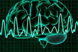
EEG represents voltage fluctuations over time, reflecting summated excitatory and inhibitory postsynaptic potentials, mainly from cortical pyramidal cells.
Cortex Area Needed for EEG Detection
Cortex Area Needed for EEG Detection
At least 6 cm² of cortex must be active for detection. Pathological epileptiform potentials typically need larger cortical areas to be detected.
Cortical Dipole Layer
Cortical Dipole Layer
Parallel arrangement of pyramidal neurons with apical dendrites extending to superficial cortical layers.
Current Flow & EEG Polarity
Current Flow & EEG Polarity
Signup and view all the flashcards
Types of Oscillatory EEG Activity
Types of Oscillatory EEG Activity
Signup and view all the flashcards
Normal EEG Transients
Normal EEG Transients
Signup and view all the flashcards
Non-Cerebral Electrical Potentials
Non-Cerebral Electrical Potentials
Signup and view all the flashcards
Interictal Epileptiform Potentials
Interictal Epileptiform Potentials
Signup and view all the flashcards
Electrode Placement
Electrode Placement
Signup and view all the flashcards
Bipolar Montages
Bipolar Montages
Signup and view all the flashcards
Referential Montages
Referential Montages
Signup and view all the flashcards
Ideal Monopolar Reference Electrode
Ideal Monopolar Reference Electrode
Signup and view all the flashcards
Limitations of Scalp EEG
Limitations of Scalp EEG
Signup and view all the flashcards
Scalp EEG 'Blind Spots'
Scalp EEG 'Blind Spots'
Signup and view all the flashcards
Benefits of Intracranial EEG
Benefits of Intracranial EEG
Signup and view all the flashcards
Study Notes
Introduction to EEG for Nonepileptologists Working in Seizure Prediction and Dynamics
- This chapter provides a brief introduction to electroencephalography (EEG)
- It is specifically oriented toward nonclinicians working in seizure prediction and dynamical analysis.
- The chapter focuses on the generation of potentials, types of potentials, EEG recording conventions, artifacts, limitations, and intracranial EEG.
- Limited attention is given to the clinical interpretation of EEG in the diagnosis and treatment of epilepsy syndromes.
Generation of Potentials Recorded with EEG
- EEG signals are representations of voltage fluctuations in space and time.
- These electrical potentials come from summated excitatory and inhibitory postsynaptic potentials.
- Cortical pyramidal cells mainly generate these potentials.
- Synchronous neural activity involving at least 6 cm² of cortex is needed for detection with scalp EEG, and epileptiform potentials usually involve larger cortical areas extending over at least 10-20 cm².
- The parallel arrangement of pyramidal neurons in the cortical mantle gives rise to the "cortical dipole layer" which dominates the signal recorded with EEG.
- Sufficiently summated excitatory postsynaptic potentials (EPSPs) at the apical dendrites cause inward current flow and a superficial cortical "sink."
- Superficial sinks arise from either summated apical EPSPs or summated deep-cell body layer inhibitory postsynaptic potentials (IPSPs)
- Superficial sources can result from summated apical IPSPs or deep EPSPs.
- It is the extracellular polarity of the superficial cortical field recorded with EEG that is negative for a superficial sink and positive for a superficial source.
Types of Potentials Recorded with EEG
- Potentials recorded with EEG can be classified into two types: oscillations and transients.
- Oscillatory activity recorded with EEG is generated in the cortex.
- Oscillatory activity is dependent on thalamocortical reciprocity
- Recorded oscillations can be normal (alpha, beta, gamma, and mu rhythms, sleep spindles, and delta activity in sleep) or abnormal (seizures, burst-suppression).
- EEG sharp transients are usually of less interest for seizure prediction and dynamical analyses but are important for diagnostic purposes.
- Normal transients include a variety of sleep potentials (vertex waves, K-complexes, positive occipital sharp transients of sleep, benign epileptiform transients of sleep)
- Noncerebral electrical potentials are detected with EEG (eye blinks, cardiac impulses (EKG), and muscle activity (EMG)).
- Abnormal EEG sharp transients often form the basis for the diagnosis of epilepsy and are referred to as interictal epileptiform potentials.
- Interictal epileptiform potentials are identifiable by their particular morphological characteristics, which may be in the form of spikes, polyspikes, and spike and wave.
EEG Recording Conventions
- Uses a long-established convention for head measurement and EEG scalp electrode placement and labeling known as the international 10-20 system
- Electrodes on the left are assigned odd numbers
- Electrodes on the right are assigned even numbers
- Midline electrodes are given the suffix “Z.”
- Electrodes over frontal regions are denoted with the prefix “F,” with “C,” “T,” “P,” and “O” indicating electrode placement over central, temporal, parietal, and occipital regions, respectively.
- EEG recordings are displayed using different configurations of electrode combinations organized into "channels."
- Each channel contains two electrode inputs that are arranged into either bipolar or referential montages.
- All recorded EEG signals are a measure of the voltage differences at one electrode site relative to another electrode.
- By convention, polarity in clinical EEG is depicted as “negative up."
- If the field potential recorded at the first electrode (input) of a channel is relatively more negative than the potential recorded at the same channel's second electrode input, then the difference is reflected as an upward waveform, and vice versa.
- Bipolar montages are typically arranged in straight line chains of electrodes, where the second input of the first channel becomes the first input of the second channel, and so on, allowing for localization of electrical field maxima and minima by so-called "phase reversal.”
- Referential montages are also bipolar arrangements, except that the second input to each channel (referred to as the "reference electrode") is more distant in space from the first electrode position
- The reference electrode is usually situated somewhere on the scalp, occasionally on the mastoids or ear lobes or, less commonly, over the cervical spine or nose.
- In referential montages, electrical fields are localized by identification of amplitude maxima and minima at the recording electrode (first input) sites.
- These types of referential montages are sometimes also referred to as monopolar montages.
- An ideal monopolar reference electrode would be electrically neutral to cerebral activity and uncontaminated by electrical artifact (unfortunately, no such ideal electrode exists).
- The reference electrode input can also be constructed from an average of some or all of the signals recorded from the other scalp electrodes.
EEG Examples
- Examples include common average reference used extensively for EEG source localization and modeling of sharp transients
- Surface Laplacian, a more sophisticated local average reference that may be useful for decreasing the contribution of volume conducted by distant electrical fields to the signal recorded from the electrode site of interest, the frist input to the channel
- Clinically, one of the most commonly used screening montages for scalp EEG review is the so-called anterior-posterior longitudinal bipolar (or “double banana”) montage.
- Wakefulness Example: Normal wakefulness EEG can be contrasted with normal (stage 2) sleep EEG
- Epilepsy Example: Classical 3-Hz spike and wave activity of primary generalized epilepsy
- Temporal Example: Temporal lobe interictal epileptiform discharges (spikes) such as may be seen in temporal lobe epilepsy is shown
- Figure illustrates part of an ongoing right temporal lobe seizure
Artifacts and Reference Electrodes
- EEG time series data that is available to nonepileptologists for analysis is typically first selected by clinical electroencephalographers after transformation of the data to some type of manipulable format (e.g., a large .txt file).
- Detailed consideration may not always be given to the issue of possible artifacts in the recording, and the issue of reference electrode choice is frequently not addressed.
- It is also important for nonclinicians using EEG signal analysis to study seizure prediction and dynamics.
- Understanding artifacts is recently exemplified in demonstration that scalp EEG studies of beta and gamma oscillations in cognition over the past many years would appear to have been analyzing mainly muscle artifact, which throws into question decades of published research.
- The effects of reference electrodes on measures of EEG correlation, coherency, or synchronization, are often overlooked in the signal analysis literature.
- The choice of reference electrode can have greater or lesser effects on classical measures of synchronization such as correlation or coherency analyses.
- Modern techniques such as phase synchronization analysis are also affected by the choice of reference electrode, with the resulting amplitude variations influencing the synchrony measured.
Limitations of Scalp EEG Recording
- Noninvasive EEG recording from the scalp has a number of limitations compared with intracranial EEG recordings
- Include the abundance of extracranial (especially muscle) artifacts
- Include the inability to accurately record lower amplitude faster frequencies in the beta and gamma bands
- For the study of epilepsy, the fact that EEG cannot "see" deep into the brain is included
- Spontaneous activity in, for example, the mesial temporal regions, interhemispheric frontal lobe structures, or thalamus is not apparent on scalp EEG
- Somewhat surprisingly, this is a point that continues to provoke debate among some clinical electroencephalographers.
Intracranial EEG
- The inability of scalp EEG to see deep into the brain is the reason one needs to occasionally perform, for clinical purposes, intracranial EEG.
- Intracranial EEG recordings acquired during the course of investigations for possible epilepsy surgery compose most of the time series provided for nonclinicians to work with in the hunt for features predictive of seizures.
- Benefits of the intracranial EEG signal include (usually) far less contamination with artifact, greater proximity to sites of seizure generation, and lesser problems with reference electrode issues—provided an extracranial reference is used.
- Greatly minimizes the reference contribution to the signal because of an approximately tenfold difference in amplitude between intracranial and extracranial potentials
- The situation is identical to that described above for noninvasive scalp EEG recordings if one or more of the intracranial electrodes are used as reference, as has often been done in published reports.
Studying That Suits You
Use AI to generate personalized quizzes and flashcards to suit your learning preferences.